

Mannose induces the release of cytopathic factors from Acanthamoeba castellanii
- PMID: 9423832
- PMCID: PMC107851
- DOI: 10.1128/IAI.66.1.5-10.1998
Mannose induces the release of cytopathic factors from Acanthamoeba castellanii
Abstract
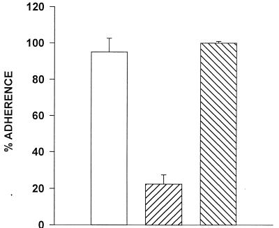
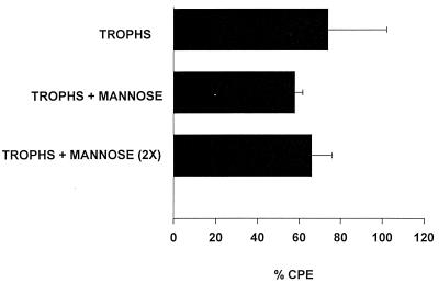
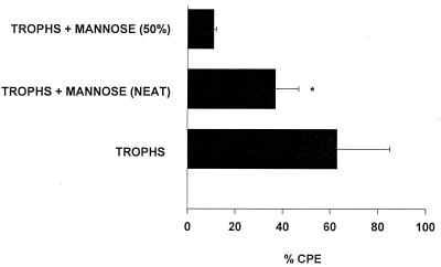
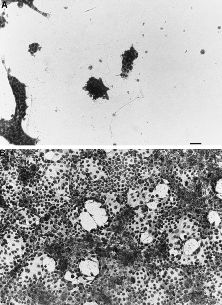
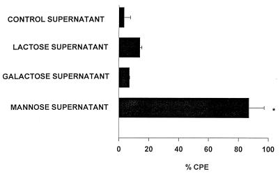
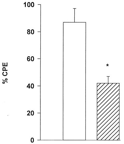
Acanthamoeba keratitis is a chronic inflammatory disease of the cornea which is highly resistant to many antimicrobial agents. The pathogenic mechanisms of this disease are poorly understood. However, it is believed that the initial phases in the pathogenesis of Acanthamoeba keratitis involve parasite binding and lysis of the corneal epithelium. These processes were examined in vitro, using Acanthamoeba castellanii trophozoites. Parasites readily adhered to Chinese hamster corneal epithelial cells in vitro; however, parasite binding was strongly inhibited by mannose but not by lactose. Although mannose prevented trophozoite binding, it did not affect cytolysis of corneal epithelial cells. Moreover, mannose treatment induced trophozoites to release cytolytic factors that lysed corneal epithelial cells in vitro. These factors were uniquely induced by mannose because supernatants collected from either untreated trophozoites or trophozoites treated with other sugars failed to lyse corneal cells. The soluble factors were size fractionated in centrifugal concentrators and found to be > or = 100 kDa. Treatment of the supernatants with the serine protease inhibitor phenylmethylsulfonyl fluoride inhibited most, but not all, of the cytopathic activity. These data suggest that the binding of Acanthamoeba to mannosylated proteins on the corneal epithelium may exacerbate the pathogenic cascade by initiating the release of cytolytic factors.
Figures


Similar articles
-
Effects of mannose on Acanthamoeba castellanii proliferation and cytolytic ability to corneal epithelial cells.Invest Ophthalmol Vis Sci. 2003 Aug;44(8):3424-31. doi: 10.1167/iovs.03-0019. Invest Ophthalmol Vis Sci. 2003. PMID: 12882791
-
Effect of steroids on Acanthamoeba cysts and trophozoites.Invest Ophthalmol Vis Sci. 2001 Nov;42(12):2885-93. Invest Ophthalmol Vis Sci. 2001. PMID: 11687533
-
In vitro pathogenicity of Acanthamoeba is associated with the expression of the mannose-binding protein.Invest Ophthalmol Vis Sci. 2006 Mar;47(3):1056-62. doi: 10.1167/iovs.05-0477. Invest Ophthalmol Vis Sci. 2006. PMID: 16505041
-
The pathogenesis of Acanthamoeba keratitis.Microbes Infect. 1999 May;1(6):437-43. doi: 10.1016/s1286-4579(99)80047-1. Microbes Infect. 1999. PMID: 10602676 Review.
-
The role of the innate and adaptive immune responses in Acanthamoeba keratitis.Arch Immunol Ther Exp (Warsz). 2002;50(1):53-9. Arch Immunol Ther Exp (Warsz). 2002. PMID: 11916309 Review.
Cited by
-
The biology of Acanthamoeba keratitis.Exp Eye Res. 2021 Jan;202:108365. doi: 10.1016/j.exer.2020.108365. Epub 2020 Nov 19. Exp Eye Res. 2021. PMID: 33221372 Free PMC article. Review.
-
Amoebicidal activities of alexidine against 3 pathogenic strains of acanthamoeba.Eye Contact Lens. 2009 Jan;35(1):1-5. doi: 10.1097/ICL.0b013e3181909ae6. Eye Contact Lens. 2009. PMID: 19125040 Free PMC article.
-
Biological characteristics and pathogenicity of Acanthamoeba.Front Microbiol. 2023 Apr 5;14:1147077. doi: 10.3389/fmicb.2023.1147077. eCollection 2023. Front Microbiol. 2023. PMID: 37089530 Free PMC article. Review.
-
ADP and other metabolites released from Acanthamoeba castellanii lead to human monocytic cell death through apoptosis and stimulate the secretion of proinflammatory cytokines.Infect Immun. 2002 Aug;70(8):4424-32. doi: 10.1128/IAI.70.8.4424-4432.2002. Infect Immun. 2002. PMID: 12117953 Free PMC article.
-
Comparison of specific activity and cytopathic effects of purified 33 kDa serine proteinase from Acanthamoeba strains with different degree of virulence.Korean J Parasitol. 2006 Dec;44(4):321-30. doi: 10.3347/kjp.2006.44.4.321. Korean J Parasitol. 2006. PMID: 17170574 Free PMC article.
References
-
- Alizadeh H, Niederkorn J Y, McCulley J P. Acanthamoeba keratitis. In: Pepose J S, Holland G N, Wilhelmus K R, editors. Ocular infection and immunity. St. Louis, Mo: Mosby; 1996. pp. 1062–1071.
-
- Allen P G, Dawidowicz E A. Phagocytosis in Acanthamoeba. I. A mannose receptor is responsible for the binding and phagocytosis of yeast. J Cell Physiol. 1990;145:508–513. - PubMed
-
- Allen P G, Dawidowicz E A. Phagocytosis in Acanthamoeba. II. Soluble and insoluble mannose-rich ligands stimulate phosphoinositide metabolism. J Cell Physiol. 1990;145:514–521. - PubMed
-
- Blaylock W K, Yue B Y, Robin J B. The use of concanavalin A to competitively inhibit Pseudomonas aeruginosa adherence to rabbit corneal epithelium. CLAO J. 1990;16:223–227. - PubMed
Publication types
MeSH terms
Substances
Grants and funding
LinkOut - more resources
Full Text Sources
Other Literature Sources
Research Materials
