. 1998 Jan;66(1):98-106.
doi: 10.1128/IAI.66.1.98-106.1998.

Processing of Mycobacterium tuberculosis bacilli by human monocytes for CD4+ alphabeta and gammadelta T cells: role of particulate antigen

Affiliations

Processing of Mycobacterium tuberculosis bacilli by human monocytes for CD4+ alphabeta and gammadelta T cells: role of particulate antigen

K N Balaji et al. Infect Immun. 1998 Jan.

Abstract

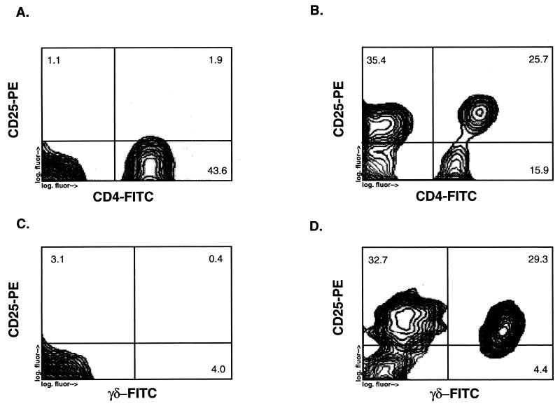
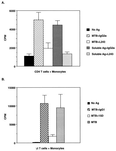
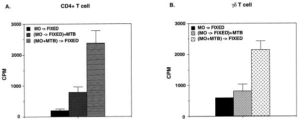
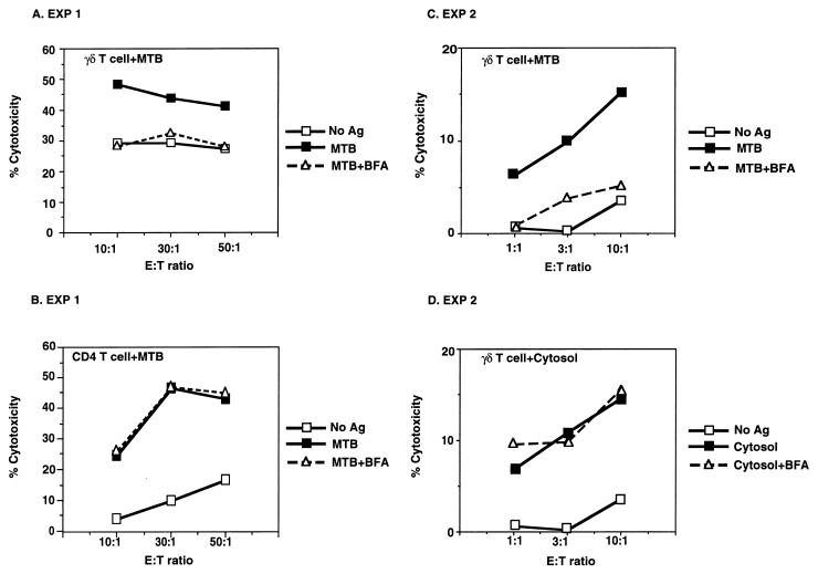
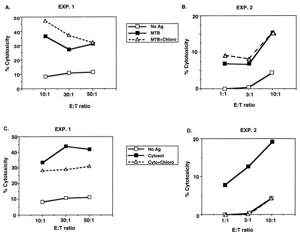
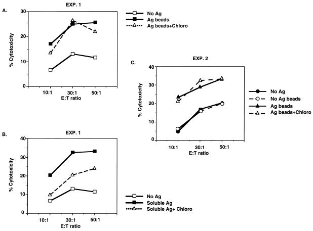
Mycobacterium tuberculosis readily activates both CD4+ and Vdelta2+ gammadelta T cells. Despite similarity in function, these T-cell subsets differ in the antigens they recognize and the manners in which these antigens are presented by M. tuberculosis-infected monocytes. We investigated mechanisms of antigen processing of M. tuberculosis antigens to human CD4 and gammadelta T cells by monocytes. Initial uptake of M. tuberculosis bacilli and subsequent processing were required for efficient presentation not only to CD4 T cells but also to Vdelta2+ gammadelta T cells. For gammadelta T cells, recognition of M. tuberculosis-infected monocytes was dependent on Vdelta2+ T-cell-receptor expression. Recognition of M. tuberculosis antigens by CD4+ T cells was restricted by the class II major histocompatibility complex molecule HLA-DR. Processing of M. tuberculosis bacilli for Vdelta2+ gammadelta T cells was inhibitable by Brefeldin A, whereas processing of soluble mycobacterial antigens for gammadelta T cells was not sensitive to Brefeldin A. Processing of M. tuberculosis bacilli for CD4+ T cells was unaffected by Brefeldin A. Lysosomotropic agents such as chloroquine and ammonium chloride did not affect the processing of M. tuberculosis bacilli for CD4+ and gammadelta T cells. In contrast, both inhibitors blocked processing of soluble mycobacterial antigens for CD4+ T cells. Chloroquine and ammonium chloride insensitivity of processing of M. tuberculosis bacilli was not dependent on the viability of the bacteria, since processing of both formaldehyde-fixed dead bacteria and mycobacterial antigens covalently coupled to latex beads was chloroquine insensitive. Thus, the manner in which mycobacterial antigens were taken up by monocytes (particulate versus soluble) influenced the antigen processing pathway for CD4+ and gammadelta T cells.


Figures

FIG. 1
FIG. 1
Upregulation of IL-2Rα (CD25) expression on peripheral blood CD4+ and γδ T cells by live M. tuberculosis. Shown are the results of an analysis by two-color flow cytometry of CD25 expression on CD4+ and γδ T cells from peripheral blood T cells stimulated for 7 days with either live M. tuberculosis (5 × 10⁶ bacilli per ml) (B and D) or no antigen (A and C). The y axis represents PE fluorescence for CD25, and the x axis represents FITC fluorescence for either CD4+ or γδ T cells.
FIG. 2
FIG. 2
(A) Restriction of CD4 T cells by class II MHC (HLA-DR) expression on M. tuberculosis-infected monocytes. Monocytes were pretreated with either L243 (10 μg/ml) or isotypic control MAb (10 μg/ml) on ice for 120 min before the addition of CD4 T cells (2.5 × 10⁴/well) from a long-term T-cell line with and without M. tuberculosis or soluble mycobacterial antigen in a proliferation assay. The results are the means and standard deviations of triplicate wells and are representative of four experiments. (B) Dependence on Vδ2 TCR expression by γδ T cells for activation by M. tuberculosis-infected monocytes. γδ T cells (2.5 × 10⁴/well) were pretreated with either C448.15D (15D) or isotypic control MAb (2 μg/ml) on ice for 60 min before being added to irradiated heterologous monocytes (5 × 10⁴/well) with and without M. tuberculosis (5 × 10⁶/ml) in a proliferation assay. The results are the means and standard deviations of triplicate wells and are representative of three experiments. Ag, antigen; MTB, M. tuberculosis.
FIG. 3
FIG. 3
Requirement for phagocytosis by monocytes of M. tuberculosis bacilli for activation of CD4+ and γδ T cells. Monocytes (MO) were incubated with M. tuberculosis (MTB) in the presence (CCD-MO+MTB) or absence (MO+MTB) of cytochalasin D (CCD) for 120 min, after which they were washed and irradiated before coculture with CD4+ (A) and γδ (B) T-cell lines in a proliferation assay. Results are means and standard deviations of triplicate wells and are representative of five experiments. Monocyte targets were from the same donor as the CD4+ T-cell line, and the γδ T-cell line was derived from an unrelated donor.
FIG. 4
FIG. 4
Requirement for uptake and processing of M. tuberculosis bacilli for presentation to both CD4+ and γδ T cells. Monocytes (MO) were fixed with paraformaldehyde either before (MO→FIXED) or after [(MO+MTB)→FIXED] a 120-min incubation with live M. tuberculosis (MTB). Fixed monocytes then were added to a proliferation assay with CD4+ (A) and γδ (B) T cells. Results are means and standard deviations of triplicate wells and are representative of four experiments.
FIG. 5
FIG. 5
Differential effects of Brefeldin A on antigen processing of M. tuberculosis bacilli for γδ and CD4+ T cells. Monocytes incubated for 12 h with either M. tuberculosis (MTB) (A, B, and C) or cytosolic mycobacterial antigens (Ag) (D) were treated with Brefeldin A (BFA) for 120 min before serving as targets in a CTL assay with either γδ T-cell lines (A, C, and D) or CD4+ T cell lines (B). In experiment 1 (EXP 1), the same monocyte targets were used for both CD4+ and γδ T-cell lines, with the γδ T-cell lines being derived from an HLA-mismatched donor. Results are representative of four experiments. E:T ratio, effector-to-target ratio.
FIG. 6
FIG. 6
Effect of an inhibitor of lysosomal acidification on antigen processing of M. tuberculosis bacilli by monocytes for CD4+ T-cell lines. Monocytes incubated for 12 h with either M. tuberculosis (MTB) (A and B) or cytosolic antigens (Ag) (C and D) of M. tuberculosis were treated with chloroquine (Chloro) for 120 min before serving as targets in a CTL assay with CD4+ T cells. The long-term CD4+ T-cell line used in experiment 1 (EXP. 1) was generated against total cytosolic proteins of M. tuberculosis, and the long-term CD4+ T-cell line used in experiment 2 was generated against purified 30-kDa (85B) antigen of M. tuberculosis. Results are representative of six experiments. E:T ratio, effector-to-target ratio.
FIG. 7
FIG. 7
Effect of chloroquine on the initial uptake and processing of M. tuberculosis bacilli for CD4+ T cells. Monocytes were preincubated with or without chloroquine (CHLORO) for 30 min, and then M. tuberculosis bacilli (MTB) were added for 120 min and chloroquine treatment was continued. Monocytes were then tested in a CTL assay with a CD4+ T-cell line. In this experiment, chloroquine treatment reduced soluble antigen (Ag) presentation by monocytes by 40%. The long-term CD4+ T-cell line used in this assay is reactive to total cytosolic proteins of M. tuberculosis. Results are representative of three experiments. E:T ratio, effector-to-target ratio.
FIG. 8
FIG. 8
Effect of ammonium chloride (NH4Cl) on the initial uptake and processing of M. tuberculosis bacilli for CD4+ T cells. Monocytes were preincubated with or without ammonium chloride for 30 min, and then M. tuberculosis bacilli (MTB) (A) or cytosolic mycobacterial antigens (CYTOSOL) (B) were added for 120 min on initial uptake and processing of M. tuberculosis bacilli for CD4+ T cells and ammonium chloride treatment was continued. Monocytes then were tested in a CTL assay with a CD4+ T-cell line. Results are representative of three experiments. Ag, antigen; E:T ratio, effector-to-target ratio.
FIG. 9
FIG. 9
Chloroquine insensitivity of antigen processing of cytosolic mycobacterial antigens coupled to latex microspheres for CD4+ T cells. Monocytes were incubated with latex beads coupled with cytosolic mycobacterial antigens (A and C) or with soluble mycobacterial antigens (B) and then treated with chloroquine (Chloro) before use in a CTL assay with a CD4+ T-cell line. Results of two of four representative experiments are shown. Ag, antigen; E:T ratio, effector-to-target ratio.

References

    1. Balaji K N, Schwander S, Rich E A, Boom W H. Alveolar macrophages as accessory cells for human γδ T cells activated by M. tuberculosis. J Immunol. 1995;154:5959–5968. - PubMed
    1. Barnes P F, Mistry S D, Cooper C L, Pirmez C, Rea T H, Modlin R L. Compartmentalization of a CD4+ T lymphocyte subpopulation in tuberculous pleuritis. J Immunol. 1989;142:1114–1119. - PubMed
    1. Bhardwaj V, Colston M J. The processing and presentation of mycobacterial antigens by human monocytes. Eur J Immunol. 1988;18:691–696. - PubMed
    1. Boom W H, Balaji K N, Nayak R, Tsukaguchi K, Chervenak K A. Characterization of a 10- to 14-kilodalton protease-sensitive Mycobacterium tuberculosis H37Ra antigen that stimulates human γδ T cells. Infect Immun. 1994;62:5511–5518. - PMC - PubMed
    1. Boom W H, Chervenak K A, Mincek M A, Ellner J J. Role of the mononuclear phagocyte as an antigen-presenting cell for human gamma delta T cells activated by live Mycobacterium tuberculosis. Infect Immun. 1992;60:3480–3488. - PMC - PubMed

Publication types

MeSH terms