Comparative Study
. 1998 Jan;66(1):161-8.
doi: 10.1128/IAI.66.1.161-168.1998.

Distinct characteristics of resistance to Borrelia burgdorferi-induced arthritis in C57BL/6N mice

Affiliations
Comparative Study

Distinct characteristics of resistance to Borrelia burgdorferi-induced arthritis in C57BL/6N mice

Y Ma et al. Infect Immun. 1998 Jan.

Abstract

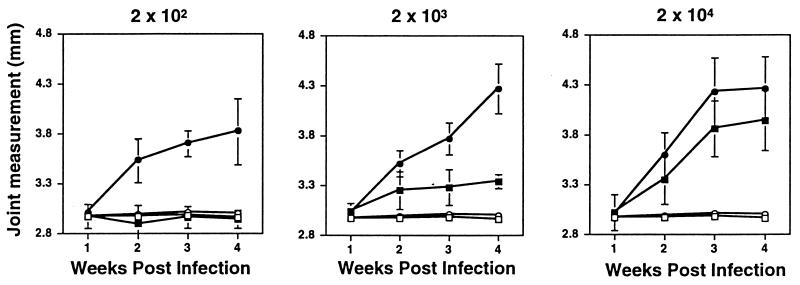
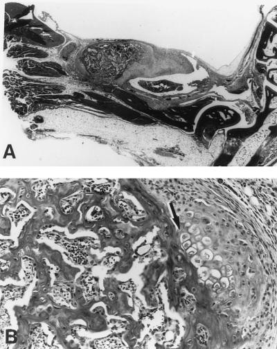
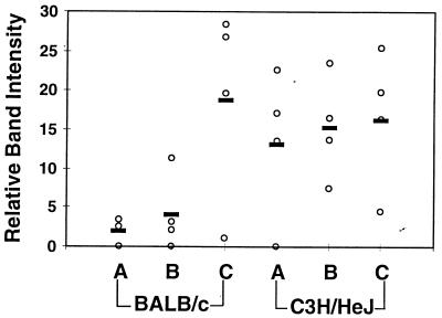
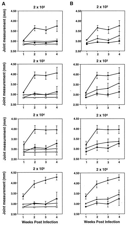
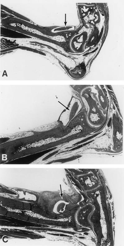
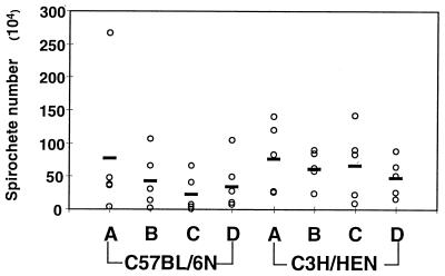
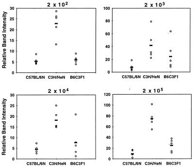
Studies of mice infected with Borrelia burgdorferi have indicated that the severity of arthritis is influenced by the genetic composition of the host: the C3H mouse develops severe arthritis while BALB/c and C57BL/6 mice develop mild arthritis. In this study, the effects of increasing infectious dose on the severity of arthritis were determined in these three mouse strains. C3H/He mice developed severe arthritis at all infectious doses, with 100% infection requiring 200 spirochetes. In BALB/cAnN mice, arthritis severity was dependent on infectious dose; symptoms were mild with infection by 200 B. burgdorferi and progressively more severe with increasing infectious dose. Infection of BALB/cAnN mice with 2 x 10(4) B. burgdorferi resulted in arthritis with severity identical to that in C3H/He mice. Spirochete levels in rear ankle joints of C3H/HeJ and C3H/HeN mice were relatively high, as detected by PCR, and did not increase with infectious dose. Spirochete levels in joints from BALB/cAnN mice increased with increasing infectious dose to levels found in severely arthritic C3H/He mice. Thus, resistance to severe arthritis in BALB/cAnN mice was conditional: it could be overcome by high infectious dose and the arthritis became severe when high levels of B. burgdorferi were present in joints. A unique response to increasing infectious dose was seen in C57BL/6N mice, which displayed mild to moderate arthritis at all doses of B. burgdorferi tested, up to 2 x 10(5). At all infectious doses, the levels of spirochetes in ankle joints of C57BL/6N mice were high, equivalent to those found in the severely arthritic C3H/He mice. The arthritis observed in infected (C57BL/6N x C3H/HeN)F1 mice was of severity intermediate between those of the two parental strains. The finding that resistance to severe arthritis in C57BL/6N mice could not be overcome by high infectious doses and was independent of spirochete levels in joints suggested that it was mediated by a distinct mechanism from that operating in BALB/cAnN mice.


Figures

FIG. 1
FIG. 1
Effects of increasing infectious dose on B. burgdorferi-induced ankle swelling in BALB/cAnN and C3H/HeJN mice. Groups of three to four C3H/HeJN (circles) or BALB/cAnN (squares) mice were infected by intradermal injection with the indicated numbers of the N40 strain of B. burgdorferi (filled symbols). Uninfected control mice were injected with sterile medium (open symbols). Measurements of the most severely swollen ankle were collected weekly for each animal, and the data shown are average values at each time point for each treatment group. Error bars indicate standard deviations within the groups. The same group of mock-infected mice is shown in each panel for comparison. Mice were sacrificed at 4 weeks postinfection and used for the analyses shown in Fig. 2 and 3.
FIG. 2
FIG. 2
Histological analysis of severely arthritic BALB/cAnN mice. (A) Hematoxylin-and-eosin-stained slide from a representative BALB/cAnN mouse from Fig. 1 infected with 2 × 10⁴ B. burgdorferi. Note the thickening of the tendon sheath in the region of the ankle joint. Magnification, ×5.75. (B) Area within the tendon sheath displaying abnormalities of chondrocyte and bone proliferation (arrow). Magnification, ×11.5. For comparison, severe arthritis in C3H/He mice can be seen in Fig. 5C.
FIG. 3
FIG. 3
Detection of spirochete DNA in tissues of mice infected with increasing concentrations of B. burgdorferi. DNA from the mice from Fig. 1 was prepared at sacrifice, and semiquantitative PCR was used to assess the quantity of B. burgdorferi persisting in the rear ankle joint. Samples were amplified with primers for nidogen to ensure loading equivalence and with ospA primers to detect B. burgdorferi DNA, as described in Materials and Methods. The relative intensities of bands were determined by densitometric scanning. Relative densitometric units were determined for ospA products from each infected mouse (three to four mice per group) and are indicated by the open circles while the bold bars indicate the averages for each group. The means and standard deviations for the relative band intensities at the 2 × 10² infection dose were 2.0 ± 1.8 and 13.3 ± 9.6 for the BALB/c and C3H/HeJN mice, respectively. The corresponding results for the other doses were 4.1 ± 4.9 and 15.3 ± 6.6 (2 × 10³) and 18.9 ± 12.5 and 16.48 ± 8.9 (2 × 10⁴) for the BALB/c and C3H/HeJN mice, respectively.
FIG. 4
FIG. 4
Effects of infectious dose on ankle swelling in C57BL/6N and C3H/HeN mice. (A) Groups of four to five C57BL/6N (squares) and C3H/HeN (circles) mice were infected by intradermal injection with the indicated numbers of the N40 strain of B. burgdorferi (filled symbols). Uninfected control mice were injected with sterile medium (open symbols). Weekly measurements of the most severely swollen ankle were made for each animal, and the values were used to determine the means and standard deviations (error bars) within each treatment group at each time point. The same mock-infected mice from each mouse strain are included in each panel for comparison. Mice were sacrificed at 4 weeks postinfection and were used for the analyses shown in Fig. 5 to 7. (B) B6C3F1 mice (triangles) were infected at the same time and with the same doses as the parental mice shown in panel A. Rear ankle measurements from infected B6C3F1 (triangles) are shown in comparison with infected parental strains (same symbols as for panel A). Measurements for uninfected B6C3F1 mice were similar to those for parental mice shown in panel A.
FIG. 5
FIG. 5
Histological analysis of tendonitis development in C57BL/6N, C3H/HeN, and B6C3F1 mice infected with 2 × 10⁴ B. burgdorferi. (A) Representative ankle joint from a C57BL/6N mouse from the experiment shown in Fig. 4. There is very little edema or change in the tendon sheath evident in this section. (B) Ankle joint from a representative B6C3F1 mouse from Fig. 4 showing evidence of arthritis. Note the presence of edema surrounding the tendon crossing the ankle. (C) Ankle joint from a representative C3H/HeN mouse from Fig. 4 showing evidence of severe arthritis. Note the predominant thickening of the tendon sheath in the tibiotarsal region. Arrows identify prominant tendons. Magnification, ×5.75.
FIG. 6
FIG. 6
Effects of B. burgdorferi infectious dose on DNA levels in ankle joints from C57BL/6N and C3H/HeN mice. The concentrations of DNA in the samples were adjusted based on products generated with nidogen primers, as described in Materials and Methods. Samples were amplified with primers for ospB, and products were quantified by phosphor image analysis. The estimation of total B. burgdorferi numbers in each sample of joint DNA was made by comparison with a standard curve, generated as described in Materials and Methods, and calculated from the fraction of total DNA amplified in the PCR. Open circles indicate spirochete numbers from each mouse, and bold bars indicate average numbers for the groups of 4 to 5 mice. Spirochete numbers (means ± standard deviations) per tissue are given for each infectious dose with group A receiving 2 × 10² (C57BL/6, [7.7 ± 10.6] × 105; C3H/HeN, [7.9 ± 5.2] × 105), group B receiving 2 × 10³ (C57BL/6, [4.3 ± 4.2] × 105; C3H/HeN, [6.3 ± 2.6] × 105), group C receiving 2 × 10⁴ (C57BL/6, [2.3 ± 2.9] × 105; C3H/HeN, [6.9 ± 5.4] × 105), and group D receiving 2 × 10⁵ B. burgdorferi (C57BL/6, [3.9 ± 4.0] × 105; C3H/HeN, [4.8 ± 2.9] × 105).
FIG. 7
FIG. 7
Detection of B. burgdorferi DNA in hearts collected from infected C3H/HeN, C57BL/6N, and B6C3F1 mice. Hearts were collected from the mice in Fig. 4, and DNA was prepared for PCR analysis. Samples were determined to contain equivalent amounts of DNA by amplification with nidogen primers, as described in Materials and Methods. Samples for mice infected with each indicated dose of B. burgdorferi were amplified with ospB primers. Phosphor image determination of relative quantities of product were made from samples run on a single gel. Open circles indicate relative band intensities from individual mice and bold bars reflect averages for the groups of 4 to 5 mice. Although means and standard deviations cannot be compared between samples run on different gels, comparisons between C3H/HeN and C57BL/6N mice at each dose of B. burgdorferi were made and indicated significant differences as follows: dose of 200, P = 0.007; dose of 2 × 10³, P = 0.032; dose of 2 × 10⁴, P = 0.001; dose of 2 × 10⁵, P = 0.002.

Similar articles

Cited by

References

    1. Akins D R, Porcella S F, Popova T G, Shevchenko D, Baker S I, Li M, Norgard M V, Radolf J D. Evidence for in vivo but not in vitro expression of a Borrelia burgdorferi outer surface protein F (OspF) homologue. Mol Microbiol. 1995;18:507–520. - PubMed
    1. Anguita J, Persing D H, Rincon M, Barthold S W, Fikrig E. Effect of anti-interleukin 12 treatment on murine lyme borreliosis. J Clin Invest. 1996;97:1028–1034. - PMC - PubMed
    1. Armstrong A L, Barthold S W, Persing D H, Beck D S. Carditis in Lyme disease susceptible and resistant strains of laboratory mice infected with Borrelia burgdorferi. Am J Trop Med Hyg. 1992;47:249–258. - PubMed
    1. Balmelli T, Piffaretti J C. Association between different clinical manifestations of Lyme disease and different species of Borrelia burgdorferi sensu lato. Res Microbiol. 1995;146:329–340. - PubMed
    1. Barbour A G, Tessier S L, Todd W J. Lyme disease spirochetes and ixodid tick spirochetes share a common surface antigenic determinant defined by a monoclonal antibody. Infect Immun. 1983;41:795–804. - PMC - PubMed

Publication types

MeSH terms

LinkOut - more resources