. 1998 Jan;66(1):169-75.
doi: 10.1128/IAI.66.1.169-175.1998.

Identification and characterization of protective T cells in hsp65 DNA-vaccinated and Mycobacterium tuberculosis-infected mice

Affiliations

Identification and characterization of protective T cells in hsp65 DNA-vaccinated and Mycobacterium tuberculosis-infected mice

V L Bonato et al. Infect Immun. 1998 Jan.

Abstract

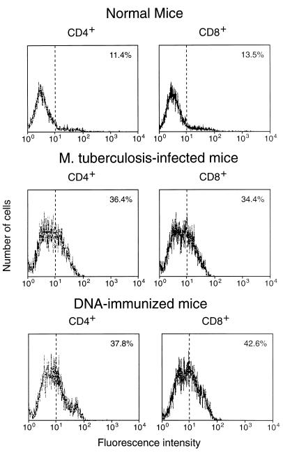
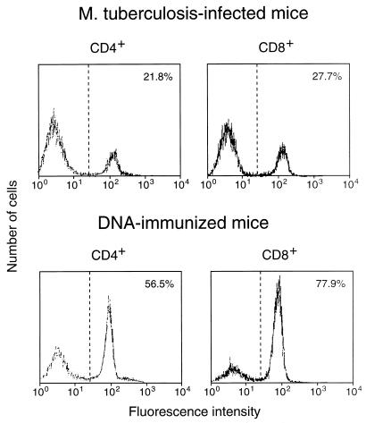
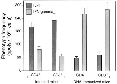
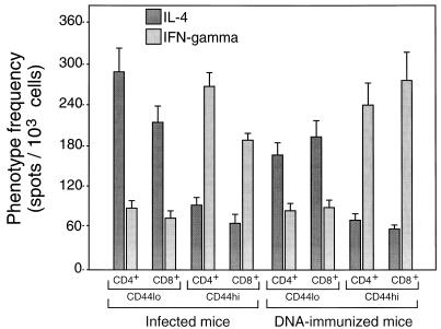
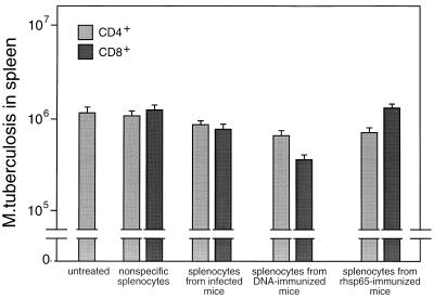
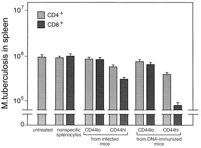
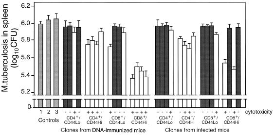
Immunization by intramuscular injection of plasmid DNA expressing mycobacterial 65-kDa heat shock protein (hsp65) protects mice against challenge with virulent Mycobacterium tuberculosis H37Rv. During infection or after immunization, CD4+/CD8- and CD8+/CD4- hsp65-reactive T cells increased equally in spleens. During infection, the majority of these cells were weakly CD44 positive (CD44(lo)) and produced interleukin 4 (IL-4) whereas after immunization the majority were highly CD44 positive (CD44(hi)) and produced gamma interferon (IFN-gamma). In adoptive transfer of protection to naive mice, the total CD8+/CD4- cell population purified from spleens of immunized mice was more protective than that from infected mice. When the cells were separated into CD4+/CD8- and CD8+/CD4- types and then into CD44(hi) and CD44(lo) types, CD44(lo) cells were essentially unable to transfer protection, the most protective CD44(hi) cells were CD8+/CD4-, and those from immunized mice were much more protective than those from infected mice. Thus, whereas the CD44(lo) IL-4-producing phenotype prevailed during infection, protection was associated with the CD8+/CD44(hi) IFN-gamma-producing phenotype that predominated after immunization. This conclusion was confirmed and extended by analysis of 16 hsp65-reactive T-cell clones from infected mice and 16 from immunized mice; the most protective clones, in addition, displayed antigen-specific cytotoxicity.


Figures

FIG. 1
FIG. 1
Expression of CD44 on CD4+/CD8 and CD8+/CD4 splenocytes freshly purified from hsp65 DNA-immunized, M. tuberculosis-infected, or normal control mice. The cells were stained with MAb against CD4 or CD8 (to confirm purity) and against CD44 and then analyzed by FACScan. The percentage of cells showing CD44hi fluorescence is shown in the upper right corner of each panel.
FIG. 2
FIG. 2
Expression of CD44 on CD4+/CD8 and CD8+/CD4 splenocytes when the cells had been amplified by 7-day culture with J774-hsp65 after purification from hsp65 DNA-immunized or M. tuberculosis-infected mice; the cells were stained with MAb against CD44 and analyzed by FACScan. The percentage of cells showing CD44hi fluorescence is shown in the upper right corner of each panel.
FIG. 3
FIG. 3
ELISPOT estimates of the frequencies of cells that produced IL-4 or IFN-γ in response to hsp65 among splenocytes from hsp65 DNA-immunized or M. tuberculosis-infected mice. Purified CD4+/CD8 and CD8+/CD4 cells were amplified by culture for 14 days on J774-hsp65 cells before assay in the presence of J774-hsp65 cells. The results shown are mean estimates (± SD) from triplicate wells at 3 different dilutions after subtraction of estimates obtained by assay in the presence of J774 vector cells (about 10 per 1,000 for CD4+/CD8 cells and 12 per 1,000 for CD8+/CD4 cells).
FIG. 4
FIG. 4
Association of IFN-γ production with CD44hi cells. Purified CD4+/CD8 and CD8+/CD4 cells were amplified by culture for 14 days on J774-hsp65 cells and then separated into CD44hi and CD44lo by FACSort and amplified for a further 12 days before assay in the presence of J774-hsp65 cells as described in the legend to Fig. 3.
FIG. 5
FIG. 5
Adoptive transfer of protective immunity against tuberculosis by bulk transfer of CD4+/CD8 or CD8+/CD4 cells to naive mice. T-cell subsets were obtained by negative and positive selection from spleens of M. tuberculosis-infected or hsp65 DNA-immunized mice. Recipient mice were gamma irradiated and then injected intravenously with 5 × 10⁶ T cells and 1 × 10⁵ M. tuberculosis cells. The numbers of live bacteria in the spleens were determined 4 weeks after infection. Control mice were either untreated or were irradiated and reconstituted with nonspecific splenic T cells enriched from normal mice. Results are shown as mean CFU ± SD from groups of five animals.
FIG. 6
FIG. 6
Association of protection with CD8+/CD44hi hsp65-reactive T cells. Purified CD4+/CD8 and CD8+/CD4 cells were amplified by culture for 14 days on J774-hsp65 cells and then separated into CD44hi and CD44lo types by FACSort and amplified for a further 12 days. The cells were tested for the ability to protect naive recipient mice against challenge with M. tuberculosis as described in the legend to Fig. 5.
FIG. 7
FIG. 7
Protection by T-cell clones. Four strongly growing hsp65-responsive clones of CD4+/CD44lo and four of CD4+/CD44hi, four of CD8+/CD44lo, and four of CD8+/CD44hi phenotypes were selected from spleens of M. tuberculosis-infected and from hsp65 DNA-immunized mice. After characterization for hsp65 antigen-dependent cytotoxicity and IL-4 and IFN-γ production, they were tested for the ability to protect naive mice from challenge with M. tuberculosis as described in the legend to Fig. 5. The numbers of live bacteria in spleens 4 weeks after challenge are shown as mean log10 ± SD for groups of five animals. Dark-shaded and unshaded bars represent data from clones that produced IL-4 and IFN-γ, respectively. Controls were as follows: 1, untreated; 2 and 3, reconstituted, respectively, with CD4+/CD8 and CD8+/CD4 clones having irrelevant antigenic specificity.

References

    1. Bevan M J. Cross-priming for a secondary cytotoxic response to minor H antigens with H-2 congenic cells which do not cross-react in the cytotoxic assay. J Exp Med. 1976;143:1283–1288. - PMC - PubMed
    1. Croft M, Carter L, Swain S L, Dutton R W. Generation of polarized antigen-specific CD8 effector populations: reciprocal action of interleukin (IL)-4 and IL-12 in promoting type 2 versus type 1 cytokine profiles. J Exp Med. 1994;180:1715–1728. - PMC - PubMed
    1. Cron R Q, Gajewski T F, Sharrow S O, Fitch F W, Matis L A, Bluestone J A. Phenotypic and functional analysis of murine CD3+, CD4−, CD8−, TCR-gamma/delta-expressing peripheral T cells. J Immunol. 1989;142:3754–3762. - PubMed
    1. Dutton R W. The regulation of the development of CD8 effector T cells. J Immunol. 1996;157:4287–4292. - PubMed
    1. Ernst D N, Weigle W O, Noonan D J, McQuitty D N, Hobbs M V. The age-associated increase in IFN-gamma synthesis by mouse CD8+ T cells correlates with shifts in the frequencies of cell subsets defined by membrane CD44, CD45RB, 3G11, and MEL-14 expression. J Immunol. 1993;151:575–587. - PubMed

Publication types

MeSH terms