. 1998 Jan;66(1):232-8.
doi: 10.1128/IAI.66.1.232-238.1998.

Listeria monocytogenes virulence factors that stimulate endothelial cells

Affiliations

Listeria monocytogenes virulence factors that stimulate endothelial cells

D A Drevets. Infect Immun. 1998 Jan.

Abstract

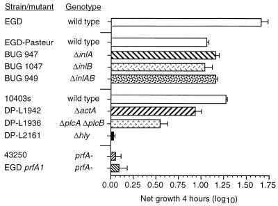
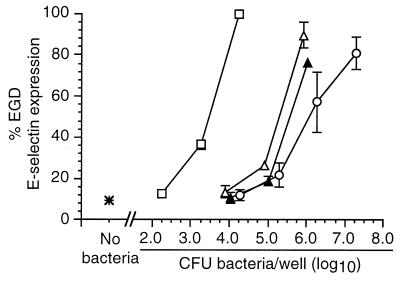
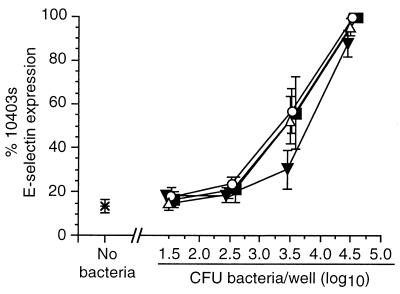
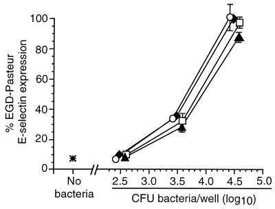
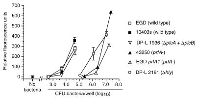
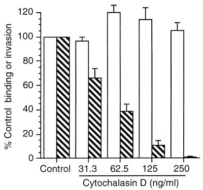
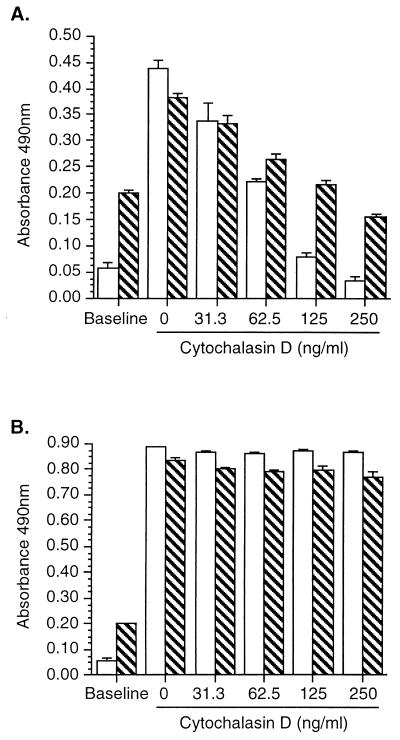
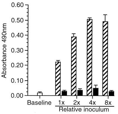
Listeria monocytogenes infection of endothelial cells upregulates surface expression of adhesion molecules and stimulates neutrophil adhesion to infected cell monolayers. The experiments presented here tested the roles of specific bacterial virulence factors as triggers for this inflammatory phenotype and function. Human umbilical vein endothelial cell (HUVEC) monolayers were infected with wild-type L. monocytogenes or L. monocytogenes mutants; then surface expression of E-selectin and neutrophil adhesion were measured. The results showed that delta hly and prfA mutants were the most crippled, requiring 100-fold more mutant bacteria than wild-type bacteria for analogous stimulation. By comparison, L. monocytogenes mutants with deletions of actA, inlA, inlB, inlAB, plcA, and plcB resembled their parent strains, and a delta plcA delta plcB mutant displayed decreased intracellular growth rate but only a minor decrease in stimulation of E-selectin or neutrophil adhesion. Other experiments showed that cytochalasin D-treated HUVEC monolayers bound bacteria, but internalization and increased surface E-selectin and intercellular adhesion molecule-1 expression were profoundly inhibited. However, cytochalasin D had no effect on the HUVEC response to stimulation with lipopolysaccharide or tumor necrosis factor alpha. These data suggest that listeriolysin O production by infecting L. monocytogenes contributes to increased expression of surface E-selectin and intercellular adhesion molecule-1, but neither it nor intracellular replication are directly responsible for this event. Nonetheless it is possible that listeriolysin O potentiates the effect(s) of an other molecule(s) that directly triggers this response. Additionally, cellular invasion by L. monocytogenes appears to be critical for initiating the HUVEC response, potentially by providing a signal which results in upregulation of the necessary bacterial genes.


Figures

FIG. 1
FIG. 1
Growth of wild-type L. monocytogenes and L. monocytogenes mutants within HUVEC. Confluent HUVEC monolayers were infected with 104 CFU. Cells and bacteria were cocultured for 60 min, washed, and then incubated for another 60 min in medium containing gentamicin to kill extracellular bacteria. Triplicate wells from one plate were lysed, and intracellular bacterial CFU at time zero were quantified by serial dilution and plating. The second plate was incubated for another 4 h (time plus 4 h), and CFU were quantified as before. Intracellular growth during the 4-h interval was calculated as follows: log10 CFU at time plus 4 h − log10 CFU at time zero. Results are shown as the mean (±SEM) log10 growth from three experiments.
FIG. 2
FIG. 2
Nonhemolytic L. monocytogenes mutants are crippled in their ability to stimulate increased surface expression of E-selectin on endothelial cells. HUVEC were infected with increasing numbers of wild-type L. monocytogenes (□), prfA mutants 43250 (▴) or EGD prfA1 (▵), or a Δhly mutant (○). Bacterial inocula were measured by serial dilution and plating. After a 4-h incubation, surface E-selectin expression was quantified by whole-cell ELISA. Relative percent E-selectin expression for each bacterium was calculated as follows: absorbance at 490 nm following infection with test bacterium/absorbance at 490 nm following infection with 104 L. monocytogenes EGD organisms. The mean (±SEM) relative percent E-selectin expression from three to seven experiments is shown.
FIG. 3
FIG. 3
Stimulation of HUVEC surface E-selectin expression by L. monocytogenes ΔplcA, ΔplcB, and ΔplcA ΔplcB mutants. HUVEC were infected with increasing numbers of the parent wild-type L. monocytogenes 10403s (▪) or L. monocytogenes ΔplcA (▵), ΔplcB (○), and ΔplcA ΔplcB (▾) mutants. Bacterial inocula were measured by serial dilution and plating. After a 4-h incubation, surface E-selectin expression was quantified by whole-cell ELISA. Relative percent E-selectin expression for each bacterium was calculated as follows: absorbance at 490 nm following infection with test bacterium/absorbance at 490 nm following infection with 104 L. monocytogenes 10403s organisms. The mean (±SEM) relative percent E-selectin expression from three experiments is shown.
FIG. 4
FIG. 4
Stimulation of HUVEC E-selectin expression by L. monocytogenes ΔinlA, ΔinlB, and ΔinlAB mutants. HUVEC were infected with increasing numbers of the parent wild-type L. monocytogenes EGD-Pasteur (⧫) or L. monocytogenes ΔinlA (○), ΔinlB (▴), or ΔinlAB (□) mutants. Bacterial inocula were measured by serial dilution and plating. After a 4-h incubation, surface E-selectin expression was quantified by whole-cell ELISA. Relative percent E-selectin expression for each bacterium was calculated as follows: absorbance at 490 nm following infection with test bacterium/absorbance at 490 nm following infection with 104 L. monocytogenes EGD-Pasteur organisms. The mean (±SEM) relative percent E-selectin expression from three experiments is shown.
FIG. 5
FIG. 5
PMN adhesion to endothelial cells infected by wild-type L. monocytogenes or L. monocytogenes mutants. HUVEC were infected with increasing numbers of wild-type L. monocytogenes or L. monocytogenes mutants and then cultured for another 4 h. Bacterial inocula were measured by serial dilution and plating. Infected monolayers were washed and then incubated for 30 min with 105 calcein-AM-loaded PMNs/well. The monolayers were washed again, and fluorescence emission was measured with a fluorescence microplate reader. PMN adhesion is represented as mean (±SD) relative fluorescence units from quadruplicate groups of wells from one of two experiments with identical results.
FIG. 6
FIG. 6
Cytochalasin D inhibits invasion of endothelial cells by L. monocytogenes. HUVEC were cultured with increasing amounts of cytochalasin D for 60 min and then were infected with 104 L. monocytogenes EGD organisms. Cells and bacteria were incubated for 60 min and washed, and then one set of triplicate wells were lysed and bound CFU were quantified by serial dilution and plating. The remaining cells were incubated with medium containing gentamicin for 60 min, and then intracellular bacterial CFU were quantified as before. Percent bacterial binding (open bars) was calculated as follows: CFU bound/CFU added. Percent invasion (hatched bars) was calculated as follows: intracellular bacterial CFU/CFU bound. Results presented are the mean (±SEM) percent control (no cytochalasin D added) binding and invasion from three experiments.
FIG. 7
FIG. 7
Cytochalasin D inhibits surface E-selectin and ICAM-1 upregulation in response to L. monocytogenes infection but not to stimulation with LPS. HUVEC were incubated with increasing concentrations of cytochalasin D for 60 min; then the cells were infected with 104 L. monocytogenes EGD organisms (A) or stimulated with 100 ng of LPS/ml (B). After 5 h E-selectin (open bars) and ICAM-1 (hatched bars) surface expressions were measured by whole-cell ELISA. Results presented are the mean (±SD) absorbance at 490 nm from quadruplicate wells from one of two experiments with similar results.
FIG. 8
FIG. 8
Increasing the bacterial inoculum does not increase surface E-selectin on cytochalasin D-treated cells. HUVEC were incubated with 250 μg of cytochalasin D/ml for 60 min (black bars) or left untreated (hatched bars) prior to infection with twofold increments of L. monocytogenes EGD. The mean (±SD) inoculum (1×) was 1.72 × 10³ ± 0.23 × 10³ CFU of bacteria/well. Cells were cultured for 60 min, gentamicin was added, and the cells were incubated for another 4 h. E-selectin expression was measured by whole-cell ELISA. Results presented are the mean (±SEM) absorbance at 490 nm from three experiments.

Similar articles

Cited by

References

    1. Beilke M A. Vascular endothelium in immunology and infectious disease. Rev Infect Dis. 1989;11:273–283. - PubMed
    1. Berche P. Bacteremia is required for invasion of the murine central nervous system by Listeria monocytogenes. Microb Pathog. 1995;18:323–336. - PubMed
    1. Bohne J, Sokolovic Z, Goebel W. Transcriptional regulation of prfA and PrfA-regulated virulence genes in Listeria monocytogenes. Mol Microbiol. 1994;11:1141–1150. - PubMed
    1. Bryant A E, Stevens D L. Phospholipase C and perfringolysin O from Clostridium perfringens upregulate endothelial cell-leukocyte adherence molecule 1 and intercellular leukocyte adherence molecule 1 expression and induce interleukin-8 synthesis in cultured umbilical vein endothelial cells. Infect Immun. 1996;64:358–362. - PMC - PubMed
    1. Chakraborty T, Leimeister-Wächter M, Doman E, Hartl M, Goebel W, Nichterlein T, Notermans S. Coordinate regulation of the virulence genes in Listeria monocytogenes requires the product of the prfA gene. J Bacteriol. 1992;174:568–574. - PMC - PubMed

Publication types

MeSH terms

Substances