. 1998 Jan;66(1):336-42.
doi: 10.1128/IAI.66.1.336-342.1998.

Intracellular Staphylococcus aureus escapes the endosome and induces apoptosis in epithelial cells

Affiliations

Intracellular Staphylococcus aureus escapes the endosome and induces apoptosis in epithelial cells

K W Bayles et al. Infect Immun. 1998 Jan.

Abstract

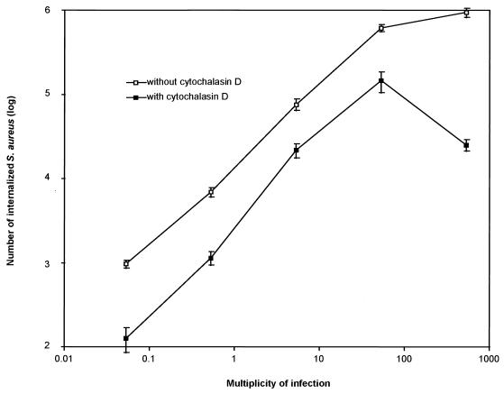
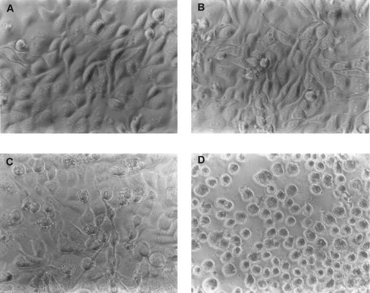
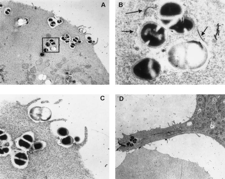
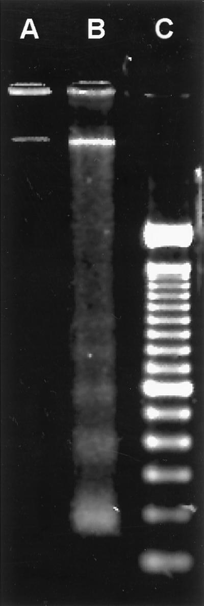
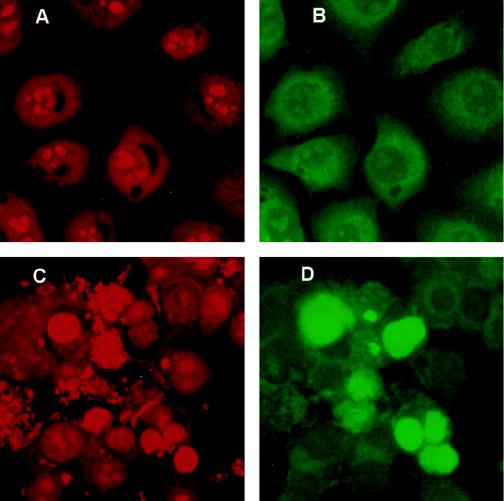
We examined the invasion of an established bovine mammary epithelial cell line (MAC-T) by a Staphylococcus aureus mastitis isolate to study the potential role of intracellular survival in the persistence of staphylococcal infections. S. aureus cells displayed dose-dependent invasion of MAC-T cells and intracellular survival. An electron microscopic examination of infected cells indicated that the bacteria induced internalization via a mechanism involving membrane pseudopod formation and then escaped into the cytoplasm following lysis of the endosomal membrane. Two hours after the internalization of S. aureus, MAC-T cells exhibited detachment from the matrix, rounding, a mottled cell membrane, and vacuolization of the cytoplasm, all of which are indicative of cells undergoing programmed cell death (apoptosis). By 18 h, the majority of the MAC-T cell population exhibited an apoptotic morphology. Other evidence for apoptosis was the generation of MAC-T cell DNA fragments differing in size by increments of approximately 180 bp and terminal deoxynucleotidyl transferase-mediated dUTP nick end labeling of the fragmented nuclear DNA of the infected host cells. These results demonstrate that after internalization S. aureus escapes the endosome and induces apoptosis in nonprofessional phagocytes.


Figures

FIG. 1
FIG. 1
Invasion of MAC-T cells by S. aureus Novel. A dose response invasion assay was performed by exposing MAC-T cell monolayers to various concentrations of S. aureus cells so that the MOI was altered within the range indicated. Culture media were supplemented with either gentamicin alone (open squares) or gentamicin plus cytochalasin D (solid squares). Additional controls showed that overnight incubation of S. aureus cells in the presence of 0.5 μg of cytochalasin D per ml resulted in no loss in viability (data not shown). Data are from a representative experiment repeated four times. Error bars represent the means ± the standard errors of the means.
FIG. 2
FIG. 2
Effect of S. aureus Novel invasion on MAC-T cell morphology. MAC-T cell monolayers were either uninfected (controls) or were incubated in the presence of staphylococci for 2 h prior to the addition of gentamicin (experimental). Photomicrographs were taken at the times indicated below following the addition of gentamicin to uninfected and infected cultures. (A) Control, 2 h; (B) control, 18 h; (C) experimental, 2 h; (D) experimental, 18 h.
FIG. 3
FIG. 3
TEM analysis of MAC-T cells infected with S. aureus Novel. MAC-T cells were grown as described for invasion assays, but gentamicin was never added to the cocultures after inoculation with S. aureus. All panels represent a 3-h coculture of MAC-T cells with S. aureus. (A) Invasion of a MAC-T cell by S. aureus illustrating contact with the MAC-T cell surface, formation of pseudopod-like structures, engulfment of bacteria, phagosome formation, and degradation of the phagosome membrane in the interior of the cell (magnification, ×5,000). (B) Enlargement (magnification, ×30,000) of the boxed area shown in panel A. Arrows indicate fragments of the degraded phagosome membrane. (C) Enlargement (magnification, ×15,000) of pseudopod-like structures and engulfment of bacteria. (D) Cytoplasmic membrane contortion (magnification, ×4,000) associated with many of the infected MAC-T cells.
FIG. 4
FIG. 4
Agarose gel (1.8%) electrophoretic separation of DNA extracted from MAC-T cells. Lane A, uninfected control MAC-T cells treated with gentamicin for 3 h; lane B, MAC-T cells infected with S. aureus Novel for 2 h prior to the addition of gentamicin, then incubated for an additional 3 h; note the ladder of fragmented DNA bands in multiples of 180 bp; lane C, 100-bp DNA ladder. Data are from a representative experiment repeated four times in duplicate.
FIG. 5
FIG. 5
Evaluation of DNA fragmentation in MAC-T cells by the TUNEL method. MAC-T cells were grown in the absence (A and B) or presence (C and D) of S. aureus Novel for 2 h prior to the addition of gentamicin and then for an additional 4 h. Images were captured by confocal microscopy for both propidium iodide (PI; A and C) and fluorescein (B and D) fluorescence. PI was used to visualize all nuclear material. The incorporation of fluorescein-dUTP at the free 3′ hydroxyl ends of fragmented DNA is shown by intense green fluorescence. (A) Uninfected MAC-T cells; (B) the same microscopic field as for panel A; (C) MAC-T cells infected with S. aureus; (D) the same microscopic field as for panel C; note the intense green fluorescence over background. Data are from a representative experiment repeated three times in duplicate. Only those apoptotic bodies that remained adherent during the staining procedure are shown here.

Similar articles

Cited by

References

    1. Almeida R A, Matthews K R, Cifrian E, Guidry A J, Oliver S P. Staphylococcus aureus invasion of bovine mammary epithelial cells. J Dairy Sci. 1996;79:1021–1026. - PubMed
    1. Bielecki J, Youngman P, Connelly P, Portnoy D A. Bacillus subtilis expressing a haemolysin gene from Listeria monocytogenes can grow in mammalian cells. Nature (London) 1990;345:175–176. - PubMed
    1. Blosser T. Economic losses from and the national research program on mastitis in the United States. J Dairy Sci. 1979;62:119–127. - PubMed
    1. Bohach G, Dinges M M, Mitchell D T, Ohlendorf D H, Schlievert P M. Exotoxins. In: Crossley K B, Archer G L, editors. The Staphylococci in human disease. New York, N.Y: Churchill Livingstone; 1997. pp. 83–111.
    1. Chen Y, Smith M R, Thirumalai K, Zychlinsky A. A bacterial invasin induces macrophage apoptosis by binding directly to ICE. EMBO J. 1996;15:3853–3860. - PMC - PubMed

Publication types