. 1998 Jan 15;18(2):625-33.
doi: 10.1523/JNEUROSCI.18-02-00625.1998.

Assembly of proteins to postsynaptic densities after transient cerebral ischemia

Affiliations

Assembly of proteins to postsynaptic densities after transient cerebral ischemia

B R Hu et al. J Neurosci. .

Abstract

Transient ischemia leads to changes in synaptic efficacy and results in selective neuronal damage during the postischemic phase, although the mechanisms are not fully understood. The protein composition and ultrastructure of postsynaptic densities (PSDs) were studied by using a rat transient ischemic model. We found that a brief ischemic episode induced a marked accumulation in PSDs of the protein assembly ATPases, N-ethylmaleimide-sensitive fusion protein, and heat-shock cognate protein-70 as well as the BDNF receptor (trkB) and protein kinases, as determined by protein microsequencing. The changes in PSD composition were accompanied by a 2.5-fold increase in the yield of PSD protein relative to controls. Biochemical modification of PSDs correlated well with an increase in PSD thickness observed in vivo by electron microscopy. We conclude that a brief ischemic episode modifies the molecular composition and ultrastructure of synapses by assembly of proteins to the postsynaptic density, which may underlie observed changes in synaptic function and selective neuronal damage.


Figures
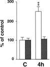
Fig. 1.
Fig. 1.
Yield of postsynaptic densities (PSDs; open bars) and light plasma membranes (LMs; hatched bars) from sham-operated rats (C) and rats subjected to 15 min of ischemia, followed by 4 hr of reperfusion (4h). Data were were expressed as mean ± SD percentage of control. A significant difference in protein yield (**) was observed between the two groups (p < 0.01, Student’s t test).
Fig. 2.
Fig. 2.
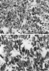
Electron micrographs of isolated cortical PSDs from sham-operated rats (A) and rats subjected to 15 min of ischemia, followed by 4 hr of reperfusion (B). Three preparations of isolated PSDs were used for electron microscopic study. Arrows indicate isolated PSDs in both control and 4 hr reperfusion.Arrowheads point to examples of trilaminar structures observed only in control PSDs, but not in 4 hr reperfused PSDs. Scale bars in A and B, 0.5 μm.
Fig. 3.
Fig. 3.
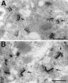
Electron micrographs of E-PTA-stained PSDs (arrows) in neocortical tissue sections of sham-operated control (A) and 4 hr of reperfusion (B). Three tissue sections from three different rats in each group were used for electron microscopic study. Note the increased thickness and fluffier appearance of the PSDs in the postischemic brain, as compared with control. Scale bars inA and B, 0.5 μm.
Fig. 4.
Fig. 4.
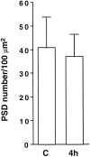
Synaptic density in E-PTA-stained cortical sections. Tissue sections were from sham-operated controls (C) and 4 hr of reperfusion (4h) after 15 min of transient cerebral ischemia. Electron micrographs of E-PTA-stained synapses photographed at a magnification of 8300× were obtained and scanned into a computer. Data were expressed as mean ± SD. No significant difference was observed between these two groups (Student’s t test).
Fig. 5.
Fig. 5.
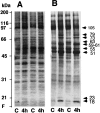
Protein profiles of light membranes (A) and isolated PSDs (B). PSD fractions (20 μg of protein) from control rats (C) and rats subjected to 15 min of ischemia, followed by 4 hr of reperfusion (4h), were used in each lane for SDS-PAGE; gels were stained with Coomassie blue. The gels were dried and photographed. Molecular standards are indicated in kDa on theleft. Molecular sizes of the altered proteins are calculated and labeled in kDa with arrows on theright.
Fig. 6.
Fig. 6.
Immunoblots of PSD proteins in the PSD fractions (PSDs, left panel) and light plasma membrane fractions (LMs, right panel). Samples were from control rats (C) and rats subjected to 15 min of ischemia, followed by 4 hr of reperfusion (4h). The blots were labeled with antibodies against NR1, NR2,TrkB, NSF, and HSC70 (HSC) and visualized with an ECL system. Arrows indicate the bands labeled by the specific antibodies. Molecular standards are indicated in kDa on the right.
Fig. 7.
Fig. 7.
Immunoblots of CaM-kinase II-α (CaMKII), PKC-β (PKC),PSD95, and 5HT2AR in PSD fractions (PSDs, left panel) and light membrane fractions (LMs, right panel). Samples were from control rats (C) and rats subjected to 15 min of ischemia, followed by 4 hr of reperfusion (4h). The blots were labeled with the antibodies (arrows) and visualized with an ECL system. Molecular standards are indicated in kDa on theright.
Fig. 8.
Fig. 8.
Immunoblots of β-tubulin (Tubulin), p97 (P97),Syntaxin, synaptophysin (Synapto-),CREB, and MAPKs in the PSD fractions (PSDs, left panel) and light membrane fractions (LMs, right panel), or nuclear fractions (N,right panel) or cytosolic fractions (S3, right panel). Samples were from control rats (C) and rats subjected to 15 min of ischemia, followed by 4 hr of reperfusion (4h). The blots were labeled with the antibodies (arrows) and visualized with an ECL system. Molecular standards are indicated in kDa on the right.

References

    1. Acharya U, Jacobs R, Peters JM, Watson N, Farquhar MG, Malhotra V. The formation of Golgi stacks from vesiculated Golgi membranes requires two distinct fusion events. Cell. 1995;82:895–904. - PubMed
    1. Andiné P, Jacobson I, Hagberg H. Enhanced calcium uptake by CA1 pyramidal cell dendrites in the post-ischemic phase despite subnormal evoked field potentials: excitatory amino acid receptor dependency and relationship to neuronal damage. J Cereb Blood Flow Metab. 1992;12:773–783. - PubMed
    1. Aronowski J, Grotta JC, Waxham MN. Ischemia-induced translocation of Ca2+/calmodulin-dependent protein kinase II: potential role in neuronal damage. J Neurochem. 1992;58:1743–1753. - PubMed
    1. Bloom FE, Aghajanian GK. Cytochemistry of synapses: a selective staining method for electron microscopy. Science. 1966;154:1575–1577. - PubMed
    1. Bloom FE, Aghajanian GK. Fine structural and cytochemical analysis of staining of synaptic junctions with phosphotungstic acid. J Ultrastruct Res. 1968;22:361–375. - PubMed

Publication types

MeSH terms

Substances