

Temporal patterns of gonadotropin-releasing hormone (GnRH), c-fos, and galanin gene expression in GnRH neurons relative to the luteinizing hormone surge in the rat
- PMID: 9425013
- PMCID: PMC6792541
- DOI: 10.1523/JNEUROSCI.18-02-00713.1998
Temporal patterns of gonadotropin-releasing hormone (GnRH), c-fos, and galanin gene expression in GnRH neurons relative to the luteinizing hormone surge in the rat
Abstract
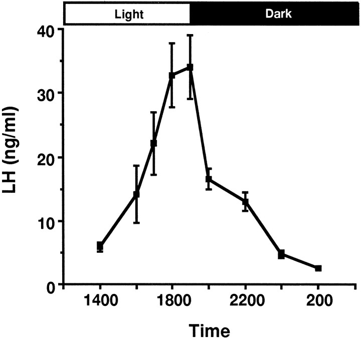
Gonadotropin-releasing hormone (GnRH) neurons increase their expression of Fos and galanin coincident with the luteinizing hormone (LH) surge in the female rat. To define the temporal relationships between the expression of these genes and the GnRH gene itself and to gain insight about the possible functional interactions of these processes, we compared levels of c-fos, galanin, and GnRH mRNA in GnRH neurons and plasma levels of LH in the rat, beginning 6 hr before and continuing for 24 hr after a sex steroid-induced LH surge. LH levels were increased significantly by 1600 hr. They increased twofold further by 1800 hr and then returned to baseline by 2400 hr. Using in situ hybridization, we determined that levels of c-fos mRNA in GnRH neurons were elevated significantly at 1600 hr only, whereas levels of galanin mRNA in GnRH neurons first increased twofold by 1800 hr, increased an additional twofold by 2400 hr, and remained elevated at all time points sampled thereafter. There were no significant changes in cellular levels of GnRH mRNA over the time points sampled. These results are consistent with the hypothesis that the induction of c-fos gene expression in GnRH neurons leads to an increase in galanin gene expression, and that the sustained increase in galanin mRNA levels in GnRH neurons reflects either the need to replenish galanin stores that are depleted at the time of the LH surge or the involvement of galanin with physiological events that occur on the day of estrus.
Figures


Similar articles
-
The regulation of galanin gene expression in gonadotropin-releasing hormone neurons.Mol Cell Endocrinol. 1998 May 25;140(1-2):137-42. doi: 10.1016/s0303-7207(98)00037-9. Mol Cell Endocrinol. 1998. PMID: 9722181 Review.
-
Activation-dependent regulation of galanin gene expression in gonadotropin-releasing hormone neurons in the female rat.Endocrinology. 1994 May;134(5):1991-8. doi: 10.1210/endo.134.5.7512492. Endocrinology. 1994. PMID: 7512492
-
Sexual differentiation of galanin gene expression in gonadotropin-releasing hormone neurons.Endocrinology. 1996 Nov;137(11):4767-72. doi: 10.1210/endo.137.11.8895345. Endocrinology. 1996. PMID: 8895345
-
Regulation of galanin gene expression in gonadotropin-releasing hormone neurons during the estrous cycle of the rat.Endocrinology. 1993 Apr;132(4):1836-44. doi: 10.1210/endo.132.4.7681766. Endocrinology. 1993. PMID: 7681766
-
Galanin: analysis of its coexpression in gonadotropin-releasing hormone and growth hormone-releasing hormone neurons.Ann N Y Acad Sci. 1998 Dec 21;863:221-35. doi: 10.1111/j.1749-6632.1998.tb10697.x. Ann N Y Acad Sci. 1998. PMID: 9928173 Review.
Cited by
-
Kisspeptin neurones do not directly signal to RFRP-3 neurones but RFRP-3 may directly modulate a subset of hypothalamic kisspeptin cells in mice.J Neuroendocrinol. 2013 Oct;25(10):876-86. doi: 10.1111/jne.12084. J Neuroendocrinol. 2013. PMID: 23927071 Free PMC article.
-
Gonadal Cycle-Dependent Expression of Genes Encoding Peptide-, Growth Factor-, and Orphan G-Protein-Coupled Receptors in Gonadotropin- Releasing Hormone Neurons of Mice.Front Mol Neurosci. 2021 Jan 18;13:594119. doi: 10.3389/fnmol.2020.594119. eCollection 2020. Front Mol Neurosci. 2021. PMID: 33551743 Free PMC article.
-
Influence of ERβ selective agonism during the neonatal period on the sexual differentiation of the rat hypothalamic-pituitary-gonadal (HPG) axis.Biol Sex Differ. 2012 Jan 19;3:2. doi: 10.1186/2042-6410-3-2. Biol Sex Differ. 2012. PMID: 22260364 Free PMC article.
-
Vasoactive intestinal peptide modulation of the steroid-induced LH surge involves kisspeptin signaling in young but not in middle-aged female rats.Endocrinology. 2014 Jun;155(6):2222-32. doi: 10.1210/en.2013-1793. Epub 2014 Mar 21. Endocrinology. 2014. PMID: 24654782 Free PMC article.
-
Sexually dimorphic testosterone secretion in prenatal and neonatal mice is independent of kisspeptin-Kiss1r and GnRH signaling.Endocrinology. 2012 Feb;153(2):782-93. doi: 10.1210/en.2011-1838. Epub 2011 Dec 27. Endocrinology. 2012. PMID: 22202164 Free PMC article.
References
-
- Anouar Y, MacArthur L, Cohen J, Iacangelo AL, Eiden LE. Identification of a TPA-responsive element mediating preferential transactivation of the galanin gene promoter in chromaffin cells. J Biol Chem. 1994;269:6823–6831. - PubMed
-
- Berriman SJ, Wade GN, Blaustein JD. Expression of Fos-like proteins in gonadotropin-releasing hormone neurons of Syrian hamsters: effects of estrous cycles and metabolic fuels. Endocrinology. 1992;131:2222–2228. - PubMed
-
- Bond CT, Hayflick JS, Seeburg PH, Adelman JP. The rat gonadotropin-releasing hormone: SH locus: structure and hypothalamic expression. Mol Endocrinol. 1989;3:1257–1262. - PubMed
-
- Burton KA, Kabigting EB, Steiner RA, Clifton DK. Identification of target cells for growth hormone’s action in the arcuate nucleus. Am J Physiol. 1995;269:E716–E722. - PubMed
Publication types
MeSH terms
Substances
Grants and funding
LinkOut - more resources
Full Text Sources