. 1998 Jan 15;18(2):731-40.
doi: 10.1523/JNEUROSCI.18-02-00731.1998.

Overloaded endoplasmic reticulum-Golgi compartments, a possible pathomechanism of peripheral neuropathies caused by mutations of the peripheral myelin protein PMP22

Affiliations

Overloaded endoplasmic reticulum-Golgi compartments, a possible pathomechanism of peripheral neuropathies caused by mutations of the peripheral myelin protein PMP22

D D'Urso et al. J Neurosci. .

Abstract

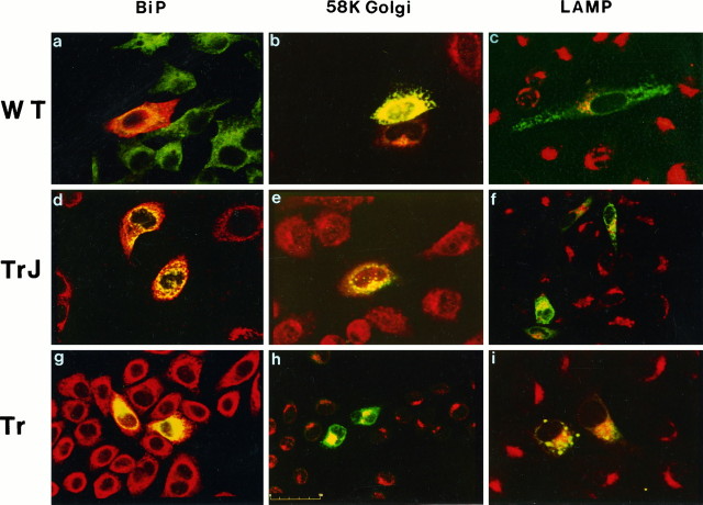
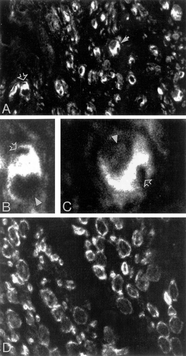
Nonconservative point mutations of the peripheral myelin protein 22 (PMP22) are associated with Charcot-Marie-Tooth type 1A disease, the most common inherited peripheral neuropathy in humans, and with the Trembler J (TrJ) and Trembler (Tr) alleles in mice. We investigated the intracellular transport of wild-type PMP22 and its TrJ and Tr mutant forms in Schwann cells and in a non-neuronal cell line. In contrast to wild type, mutant proteins were not inserted into the plasma membrane and accumulated in the endoplasmic reticulum and Golgi compartments. Coexpression of each mutant with wild-type PMP22 confirmed the different intracellular distribution of the mutant forms, indicating that neither the TrJ nor Tr protein has a dominant-negative effect on the cellular distribution of wild-type PMP22. Accumulation of PMP22 immunoreactivity in the cell body of myelinating Schwann cells was also observed in nerve biopsies obtained from CMT1A patients carrying the TrJ point mutation. We propose that impaired trafficking of mutated PMP22 affects Schwann cell physiology leading to myelin instability and loss.


Figures

Fig. 1.
Fig. 1.
Expression of wild-type, TrJ-, and Tr-PMP22 in transfected HeLa cells. Mutants are not inserted in the plasma membrane and accumulate in the perinuclear regions. PMP22 immunostaining of permeabilized cultures expressing wild type (A), TrJ (C), and Tr (E) PMP22. In nonpermeabilized transfectants (B, D, F), a PMP22 antibody directed against an extracellular domain of the protein recognizes only the wild-type form on the membrane (B). No signal was obtained in cultures transfected with TrJ-PMP22 (D) or Tr-PMP22 (F). Scale bar: 10 μm in A,B, C, E; 20 μm inD, F.
Fig. 2.
Fig. 2.
Cellular sorting of wild-type, TrJ-, and Tr-MP22 proteins in transfected HeLa cells. Merged confocal images of transfected cultures immunostained for PMP22 (green; red only ina) and the ER protein BiP (a, d, g, red;green only in a), the 58 kDa Golgi protein (b, e, h, red), and the lysosomal associated protein LAMP2 (c, f, i, red). Colocalization of the antigens appears yellow. Scale bar, 50 μm.
Fig. 3.
Fig. 3.
Stable transfection of PMP22 in HeLa cells. wt-PMP22 expression in clonal lines was analyzed by (A) Northern blotting. PMP22 mRNA was present only in cells transfected with the expression vector (lane 3). No signal was detectable in the untransfected HeLa cells (lane 1) or cells transfected with the neomycin resistance gene (lane 2). B, In PMP22 expressors the protein was localized in the plasma membrane and in the perinuclear regions, as detected by immunofluorescence and confocal microscopy. Scale bar, 10 μm.
Fig. 4.
Fig. 4.
Membrane targeting of wt-PMP22 is not impaired by coexpression of mutant PMP22. A clonal line of HeLa cells that stably expressed wt-PMP22 were transiently transfected with tagged Flag-TrJ and Flag-Tr PMP22, and the localization of PMP22 was determined by immunofluorescence using PMP22 and anti-Flag antibodies. PMP22 mutants were identified by their C-terminal Flag-epitope (B, D). The membrane targeting of the wt-PMP22 (A, C) is not affected in cells that coexpress TrJ (B) or Tr (D) mutants. Examples of coexpressing cells are marked by arrows.
Fig. 5.
Fig. 5.
TrJ and Tr proteins concentrate in the perinuclear regions of transfected Schwann cells and do not reach the plasma membrane. PMP22 immunostaining of permeabilized Schwann cells transfected with wt- (a), TrJ- (c), Tr-PMP22 (e). When cells were not permeabilized, a weak signal was obtained for PMP22 immunoreactivity on the surface of cells expressing wild-type protein (b, arrows). No membrane staining was visible in TrJ (d) and Tr (f) transfectants. Scale bar, 10 μm.
Fig. 6.
Fig. 6.
TrJ and Tr proteins accumulate in the ER and Golgi compartments of transfected Schwann cells and do not show a dominant-negative effect on the cellular distribution of wt-PMP22. Confocal optical sections of transfected Schwann cells double-immunostained for PMP22 (red) and the ER marker BiP (a, c, d, green), or the 58 kDa Golgi protein (b, d, f, green). In merged images, the yellow signals show colocalization of the antigens. gand h are superimposed confocal micrographs of Schwann cells cotransfected with wt-PMP22 (red) and Flag-TrJ (g, green) or Flag-Tr (h, green). Anti-flag and anti-PMP22 immunostaining colocalize (yellow) only in the perinuclear regions. Mutants do not interfere with the sorting of wt-PMP22.
Fig. 7.
Fig. 7.
PMP22 is abundant in the cytoplasm of myelinating Schwann cells in CMT1A sural nerve. A, PMP22 immunostaining in CMT1A tissue section highlights cell bodies of Schwann cells. Two examples are enlarged in B(open arrow in A) and C(full arrow in A). InB and C, open arrowsindicate the position of dislocated axons, andarrowheads the position of the nuclei. D, PMP22 immunoreactivity in control sural nerve. Magnification 60× inA and D; 100× in B andC.

References

    1. Adlkofer K, Martini R, Aguzzi A, Zielasek J, Toyka KV, Suter U. Hypermyelination and demyelinating peripheral neuropathy in PMP22-deficient mice. Nat Genet. 1995;11:274–280. - PubMed
    1. Baechner D, Liehr T, Hameister H, Altenberger H, Grehel H, Suter U, Rautenstrauss B. Widespread expression of the peripheral myelin protein-22 gene (PMP22) in neural and non-neural tissues during murine development. J Neurosci Res. 1995;42:733–741. - PubMed
    1. Cheng SH, Gregory RJ, Marshall J, Paul S, Souza DW, White GA, O’Riordan CR, Smith AE. Defective intracellular transport and processing of CFTR is the molecular basis of most cystic fibrosis. Cell. 1990;63:827–834. - PubMed
    1. Chomczynski P, Sacchi N. Single-step method of RNA isolation by acid guanidinium thiocyanate-phenol-chloroform extraction. Anal Biochem. 1987;162:156–159. - PubMed
    1. De Jonghe P, Timmerman V, Nelis E, Martin J-J, Van Broeckhoven C. Charcot-Marie-Tooth disease and related peripheral neuropathies. J Periph Nerve System. 1997;2:370–387. - PubMed

Publication types