. 1998 Jan 15;18(2):779-93.
doi: 10.1523/JNEUROSCI.18-02-00779.1998.

Fetal spinal cord transplants support growth of supraspinal and segmental projections after cervical spinal cord hemisection in the neonatal rat

Affiliations

Fetal spinal cord transplants support growth of supraspinal and segmental projections after cervical spinal cord hemisection in the neonatal rat

P S Diener et al. J Neurosci. .

Abstract

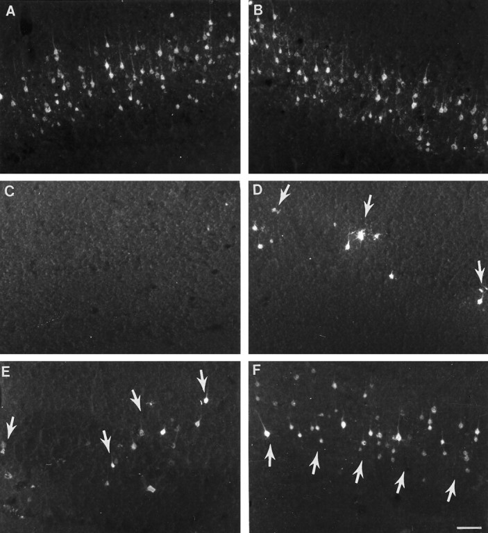
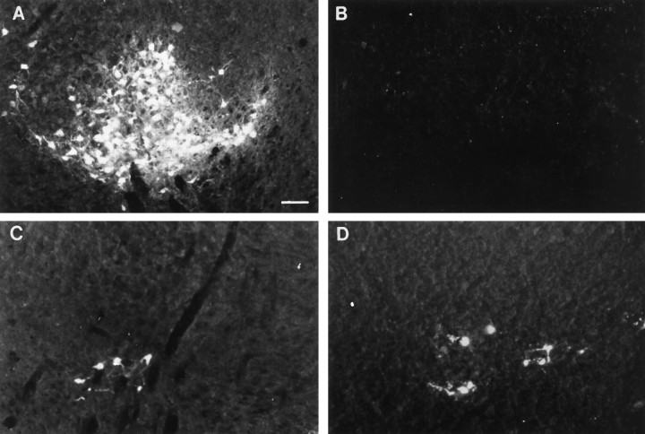
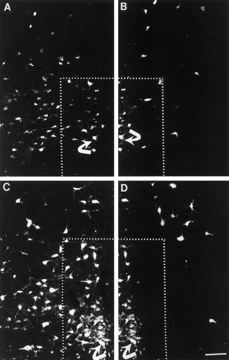
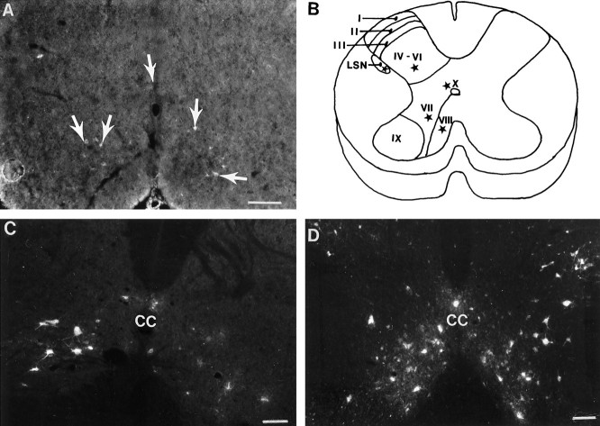
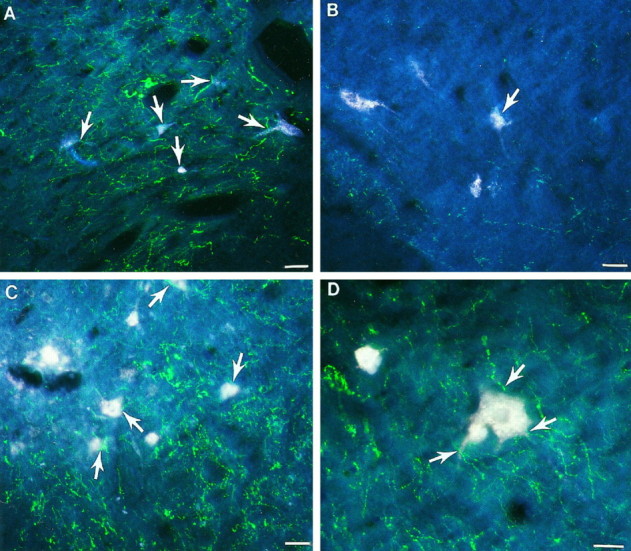
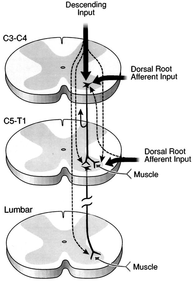
Cervical spinal cord injury at birth permanently disrupts forelimb function in goal-directed reaching. Transplants of fetal spinal cord tissue permit the development of skilled forelimb use and associated postural adjustments (, companion article). The aim of this study was to determine whether transplants of fetal spinal cord tissue support the remodeling of supraspinal and segmental pathways that may underlie recovery of postural reflexes and forelimb movements. Although brainstem-spinal and segmental projections to the cervical spinal cord are present at birth, skilled forelimb reaching has not yet developed. Three-day-old rats received a cervical spinal cord overhemisection with or without transplantation of fetal spinal cord tissue (embryonic day 14); unoperated pups served as normal controls. Neuroanatomical tracing techniques were used to examine the organization of CNS pathways that may influence target-directed reaching. In animals with hemisections only, corticospinal, brainstem-spinal, and dorsal root projections within the spinal cord were decreased in number and extent. In contrast, animals receiving hemisections plus transplants exhibited growth of these projections throughout the transplant and over long distances within the host spinal cord caudal to the transplant. Raphespinal axons were apposed to numerous propriospinal neurons in control and transplant animals; these associations were greatly reduced in the lesion-only animals. These observations suggest that after neonatal cervical spinal cord injury, embryonic transplants support axonal growth of CNS pathways and specifically supraspinal input to propriospinal neurons. We suggest that after neonatal spinal injury in the rat, the transplant-mediated reestablishment of supraspinal input to spinal circuitry is the mechanism underlying the development of target-directed reaching and associated postural adjustments.


Figures

Fig. 1.
Fig. 1.
Segments of cervical spinal cord through the lesion and lesion plus transplant site. A, C, D, Representative spinal cord cross sections stained with cresyl violet. These sections demonstrate the transverse extent of the lesion (or lesion plus transplant) in animals meeting the inclusion criteria for this study (see Materials and Methods). Spinal cords of both HX and HX + TP animals were completely overhemisected, as shown inA and C (i.e., typical overhemisections included damage to the right side of the cord and bilateral ablation of the dorsal funiculus). B, The tracings are representative sections through the lesion site of different HX animals to depict the minimal and maximal extent of injury acceptable for this study, as described in the criteria for lesion section (see text).C, Photomicrograph of a representative section through the lesion plus transplant region demonstrating the transverse extent of the apposition of the transplant to the host cord (small arrowheads). D, High-power view of the interface between host and transplant to show (1) that the transplant grew to fill the lesion cavity and (2) the close apposition (large arrowheads) of transplant and host tissue without an intervening cellular barrier. Scale bars: A, C, 100 μm; D, 50 μm. DH, Dorsal horn;TP, transplant; wm, white matter;gm, gray matter; VH, ventral horn.
Fig. 2.
Fig. 2.
Effect of neonatal cervical spinal cord injury on the motoneuron pool in the brachial spinal cord segment C5.A, B, High-power view of both ventral horns from a representative cresyl violet-stained cross section through a C5 segment from an HX animal. The photomicrographs show that a healthy-appearing motoneuron pool in laminae IX (delineated byopen arrows) is present. Scale bar, 50 μm.C, Histogram showing that the total number of motoneurons present in laminae IX of each group’s left and right ventral horn is similar (CON, n = 3;HX, n = 3; HX + TP,n = 3). Error bars represent SD;p > 0.05.
Fig. 3.
Fig. 3.
Axonal projections penetrate into and beyond the transplant. A, Photomicrograph of FluoroRuby-labeled corticospinal axons (CS) within the transplant. Many CS axons extend throughout the transverse extent of the transplant. B, Photomicrograph of a horizontal section through a transplant labeled with antibodies against CGRP. Immunoreactive CGRP dorsal root axons are visualized projecting along the length of the transplant. Individual fibers contain multiple varicosities. C, Low-power photomicrograph at the apposition site of the host cord and fetal spinal cord transplant. The interface of the transplant to the host tissue is depicted by arrowheads. Serotonergic axons project robustly throughout the host tissue adjacent to the transplant and within the transplant. D, High-power photomicrograph of the transplant at the interface (arrowheads) with the host tissue. Axons immunoreactive for serotonin (arrows) grew within the transplant. Individual axons have many varicosities along their length. E, Low-power photomicrograph of neurons intrinsic to the spinal cord transplant. Neurons were retrogradely labeled with FluoroGold from the lumbar spinal cord. Numerous neurons within the transplant (arrows) are labeled, indicative of their long-distance growth into the host cord. Host–transplant interface is marked with a dashed line.F, High-power photomicrograph of the identified neurons (arrows) from E. Scale bars: A, B, C, E, 50 μm; D, F, 100 μm.
Fig. 4.
Fig. 4.
Retrograde tracing of corticospinal neurons from the lumbar spinal cord. In control animals (A, B), retrogradely labeled corticospinal neurons were located in layer V of the sensorimotor cortex bilaterally. After neonatal hemisection, the number of neurons labeled is reduced in both the contralateral (C; devoid of labeled cells) and ipsilateral (D; arrow indicating labeled cell) cortices. E, F, Transplantation spares some of the neurons in both the contralateral (E; 3–4 small cells denoted by arrows) and ipsilateral (F; numerous white cells) cortex. The cortex ipsilateral to the spinal cord lesion (F) has more labeled cells than the contralateral cortex (E). Scale bar (shown inF for A–F): 100 μm.
Fig. 5.
Fig. 5.
Retrograde tracing of corticospinal neurons from the cervical cord. A, B, CON cortices showing the normal distribution in layer V of corticospinal neurons retrogradely traced from cervical spinal cord injections. The mediolateral distribution and the dense packing of cells in laminae V is greater than after lumbar injections (compare with Fig.4A,B). After neonatal HX, the number of retrogradely labeled neurons is dramatically reduced in both the contralateral (C) and ipsilateral (D) sensorimotor cortices. In contrast, in all animals with lesion plus transplant, many retrogradely labeled corticospinal neurons are present in the sensorimotor cortex both contralateral (E) and ipsilateral (F) to the spinal cord lesion. There are substantially more retrogradely labeled corticospinal neurons in lesion plus transplant animals (E, F) than in lesion-only animals (C, D). Arrowsindicate representative corticospinal neurons labeled retrogradely with FluoroGold. Scale bar (shown in F forA–F): 100 μm.
Fig. 6.
Fig. 6.
Retrograde tracing of rubrospinal neurons after FluoroGold application. The normal pattern of labeling in unlesioned animals is shown in A. The retrograde labeling in the intact RN in HX and HX + TP animals was similar to that in normal animals (data not shown). In contrast, the axotomized RN from HX rats (B) is essentially devoid of labeled neurons after either lumbar (B) or cervical (data not shown) spinal cord lesions. In all animals with transplants at the lesion site, RN neurons were retrogradely labeled from either the lumbar (C) or cervical (D) spinal cord. Scale bar (shown in A forA–D): 100 μm.
Fig. 7.
Fig. 7.
Raphespinal neurons labeled with the retrograde tracer FluoroGold. In each set of panels, the corresponding neuron is identified by a curved arrow for orientation.A, B, Photomicrographs of a section from the caudal medulla from a representative HX animal. Diminished labeling is noted in the raphe obscurus, magnus, and pallidus (outlined area, A–D) on the axotomized (B) side in comparison to the side contralateral (A) to the overhemisection. C, D, Photomicrographs of a caudal medullary section from a representative HX + TP animal. Labeling varies among transplant animals, but typically a greater number of neurons are labeled in the raphe obscurus and magnus both ipsilateral (D) and contralateral (C) to the spinal cord injury than in HX animals. Scale bar (shown in D for A–D): 100 μm.
Fig. 8.
Fig. 8.
Retrograde labeling of propriospinal neurons. A, In normal animals at P3, propriospinal neurons (arrows) labeled with Fast Blue are distributed throughout laminae VII–VIII and X. This indicates that at the time of the neonatal spinal cord injury, at least some of the propriospinal neurons are axotomized directly. B, Schematic diagram representing the general distribution of the propriospinal neurons (represented as stars in LSN, laminae IV–VI, VII–VIII, and X) in the brachial cord of a normal adult rat. C, HX group, spinal cord segment caudal to the injury site. Compared with the normal distribution of propriospinal neurons in adult rats (data not shown), there is a decrease in the number of propriospinal neurons after neonatal hemisection (C). The decrease in labeling is greatest on the side ipsilateral to the lesion but is evident bilaterally. D, HX + TP group, spinal cord segment caudal to the lesion plus transplant. In contrast to lesion-only animals, animals with transplants had many retrogradely labeled propriospinal neurons present bilaterally. These neurons were located in all of the appropriate laminae (IV–VIII, X, and LSN). Thus, the transplants rescue a substantial proportion of the propriospinal neurons. Scale bars, 50 μm. LSN, Lateral spinal nucleus; CC, central canal.
Fig. 9.
Fig. 9.
Effect of neonatal cervical spinal cord injury on the supraspinal projections to propriospinal neurons. Fluorescent photomicrographs of laminae VIII of double-labeled spinal cord sections from the C5 spinal cord segment. A, Representative section from a normal animal. Numerous propriospinal neurons (white cells) are labeled, and serotonergic axons (green fibers) are widely distributed throughout the ventral horn. Serotonergic axons are closely associated with the propriospinal neurons (arrows).B, In the HX group, although propriospinal neurons are retrogradely labeled (white cells), the serotonergic axons (green fibers) are substantially decreased in the ventral horn and are rarely associated with the propriospinal neurons (arrow depicts one cell with an adjacent axon).C, The HX + TP group has both labeled propriospinal neurons (white cells) and serotonergic axons (green fibers) distributed throughout the ventral horn. Many serotonergic axons are closely associated with the propriospinal neurons (arrows). D, High-powered view of a representative propriospinal neuron (large white cell) with adjacent serotonergic fibers ( arrows) in a transplant animal. The presence of a transplant at the lesion site not only preserves more propriospinal neurons than in lesion-only animals, but the descending input to these neurons also seems to be reestablished. Scale bars, 50 μm.
Fig. 10.
Fig. 10.
Summary diagram of a proposed mechanism that guides target reaching and postural adjustments after neonatal cervical spinal cord injury and transplantation. [Spinal cord levels are noted. Propriospinal neurons are represented as dark black neurons schematically located in the C3–C4 (short propriospinal neurons) and C5–T1 (long propriospinal neurons) segments. Supraspinal (descending) and segmental afferent input are labeled.] Before skilled movement emerges, neonatal high cervical spinal cord injury at C3 disrupts (1) the C3–C4 propriospinal network and (2) the supraspinal and segmental input directed to the C3–C4 propriospinal neurons, the forelimb and hindlimb segment motoneurons, and the interneurons. This schematic diagram represents some of the normal input that contributes to the propriospinal network, which we suggest is reestablished in the presence of a transplant growing within the C3 spinal cord segment. Because target reaching and postural adjustments develop postnatally, the proposed pathway is suggested also to develop after birth in the normal and transplant animals but not in lesion-only animals. The aberrant motor patterns exhibited by lesion-only rats (Diener and Bregman, 1998) are suggested to be commensurate with failed remodeling of the pathways identified in the schematic (refer to other photomicrographs in this paper). We suggest that through a transplant-mediated response, the descending and segmental projections to multiple spinal levels are reestablished (dashed arrows emanating from the descending andsegmental arrows). The transplant may also mediate the extension of collaterals that normally would provide feedback to higher centers (indicated by the collateral with arrowheadcurving upward from the C3–C4 propriospinal axon). After the converging input is integrated, the short (C3–C4) propriospinal neurons may project to the brachial segments (possibly using the transplant as a bridge or a relay; refer to Figs. 3, 4, 5, 7, 8). All of these projections may influence forelimb motoneurons and long propriospinal neurons, which in turn may guide lower spinal cord segments. This schematic suggests a neural mechanism that may be reestablished after high spinal injury and transplantation to influence the development of postural adjustments (e.g., axial and hindlimb movements) in coordination with the development of forelimb movements (e.g., reaching), thereby furnishing an explanation for the skilled movement observed in the transplant rats (Diener and Bregman, 1998).
Fig. 10.
Fig. 10.
Summary diagram of a proposed mechanism that guides target reaching and postural adjustments after neonatal cervical spinal cord injury and transplantation. [Spinal cord levels are noted. Propriospinal neurons are represented as dark black neurons schematically located in the C3–C4 (short propriospinal neurons) and C5–T1 (long propriospinal neurons) segments. Supraspinal (descending) and segmental afferent input are labeled.] Before skilled movement emerges, neonatal high cervical spinal cord injury at C3 disrupts (1) the C3–C4 propriospinal network and (2) the supraspinal and segmental input directed to the C3–C4 propriospinal neurons, the forelimb and hindlimb segment motoneurons, and the interneurons. This schematic diagram represents some of the normal input that contributes to the propriospinal network, which we suggest is reestablished in the presence of a transplant growing within the C3 spinal cord segment. Because target reaching and postural adjustments develop postnatally, the proposed pathway is suggested also to develop after birth in the normal and transplant animals but not in lesion-only animals. The aberrant motor patterns exhibited by lesion-only rats (Diener and Bregman, 1998) are suggested to be commensurate with failed remodeling of the pathways identified in the schematic (refer to other photomicrographs in this paper). We suggest that through a transplant-mediated response, the descending and segmental projections to multiple spinal levels are reestablished (dashed arrows emanating from the descending andsegmental arrows). The transplant may also mediate the extension of collaterals that normally would provide feedback to higher centers (indicated by the collateral with arrowheadcurving upward from the C3–C4 propriospinal axon). After the converging input is integrated, the short (C3–C4) propriospinal neurons may project to the brachial segments (possibly using the transplant as a bridge or a relay; refer to Figs. 3, 4, 5, 7, 8). All of these projections may influence forelimb motoneurons and long propriospinal neurons, which in turn may guide lower spinal cord segments. This schematic suggests a neural mechanism that may be reestablished after high spinal injury and transplantation to influence the development of postural adjustments (e.g., axial and hindlimb movements) in coordination with the development of forelimb movements (e.g., reaching), thereby furnishing an explanation for the skilled movement observed in the transplant rats (Diener and Bregman, 1998).

Similar articles

Cited by

References

    1. Alstermark B, Sasaki S. Integration in descending motor pathways controlling the forelimb in the cat. 13. Corticospinal effects in shoulder: elbow, wrist, and digit motoneurones. Exp Brain Res. 1985;59:353–364. - PubMed
    1. Alstermark B, Wessberg J. Timing of postural adjustment in relation to forelimb target-reaching in cats. Acta Physiol Scand. 1985;125:337–340. - PubMed
    1. Alstermark B, Lindstrom S, Lundberg A, Sybirska E. Integration in descending motor pathways controlling the forelimb in the cat. 8. Ascending projection to the lateral reticular nucleus from C3–C4 propriospinal neurones also projecting to forelimb motoneurones. Exp Brain Res. 1981a;42:282–298. - PubMed
    1. Alstermark B, Lundberg A, Norrsell U, Sybirska E. Integration in descending motor pathways controlling the forelimb in the cat. 9. Differential behavioural defects after spinal cord lesions interrupting defined pathways from higher centres to motoneurones. Exp Brain Res. 1981b;42:299–318. - PubMed
    1. Alstermark B, Lundberg A, Sasaki S. Integration in descending motor pathways controlling the forelimb in the cat. 11. Inhibitory pathways from higher motor centres and forelimb afferents to C3–C4 propriospinal neurones. Exp Brain Res. 1984a;56:293–307. - PubMed

Publication types