. 1998 Jan 12;140(1):1-15.
doi: 10.1083/jcb.140.1.1.

Retrograde transport of Golgi-localized proteins to the ER

Affiliations

Retrograde transport of Golgi-localized proteins to the ER

N B Cole et al. J Cell Biol. .

Abstract

The ER is uniquely enriched in chaperones and folding enzymes that facilitate folding and unfolding reactions and ensure that only correctly folded and assembled proteins leave this compartment. Here we address the extent to which proteins that leave the ER and localize to distal sites in the secretory pathway are able to return to the ER folding environment during their lifetime. Retrieval of proteins back to the ER was studied using an assay based on the capacity of the ER to retain misfolded proteins. The lumenal domain of the temperature-sensitive viral glycoprotein VSVGtsO45 was fused to Golgi or plasma membrane targeting domains. At the nonpermissive temperature, newly synthesized fusion proteins misfolded and were retained in the ER, indicating the VSVGtsO45 ectodomain was sufficient for their retention within the ER. At the permissive temperature, the fusion proteins were correctly delivered to the Golgi complex or plasma membrane, indicating the lumenal epitope of VSVGtsO45 also did not interfere with proper targeting of these molecules. Strikingly, Golgi-localized fusion proteins, but not VSVGtsO45 itself, were found to redistribute back to the ER upon a shift to the nonpermissive temperature, where they misfolded and were retained. This occurred over a time period of 15 min-2 h depending on the chimera, and did not require new protein synthesis. Significantly, recycling did not appear to be induced by misfolding of the chimeras within the Golgi complex. This suggested these proteins normally cycle between the Golgi and ER, and while passing through the ER at 40 degrees C become misfolded and retained. The attachment of the thermosensitive VSVGtsO45 lumenal domain to proteins promises to be a useful tool for studying the molecular mechanisms and specificity of retrograde traffic to the ER.


Figures

Figure 1
Figure 1
Schematic representation of VSVGtsO45 chimeras. The temperature-sensitive lumenal domain of VSVGtsO45 was fused in-frame with (left to right): a full-length human homologue of the KDELR (VSVG–KDELR; Hsu et al., 1992); a full-length mutant form of the KDELR, which contains a single amino acid change (Asp to Asn at position 195) that fails to redistribute into the ER upon overexpression of KDEL-containing ligands, and is presumably recycling defective (analogous to Townsley et al., 1993); the transmembrane domain and cytoplasmic tail of TGN38 (Luzio et al., 1990), which contain nonoverlapping domains sufficient to confer steady-state TGN localization (Humphrey et al., 1993; Ponnambalam et al., 1994); the transmembrane domain and cytoplasmic tail of the cell surface protein, the α chain of the IL2R (Leonard et al., 1984); and a construct in which the transmembrane domain of VSVGtsO45 was replaced by a synthetic transmembrane domain composed of 15 leucines. This has been shown to result in accumulation within the Golgi complex of proteins normally destined for the plasma membrane (Munro, 1995). The quaternary structures of these constructs have not been analyzed, yet we assume that VSVG–TGN38, VSVG–IL2R, and VSVG–Leu15 form trimers, since it is the lumenal domain of VSVGtsO45 that is responsible for trimer formation (Doms et al., 1988). The oligomeric nature of KDELR is not known.
Figure 2
Figure 2
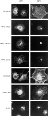
Localization of VSVGtsO45 chimeras at nonpermissive (40°C) and permissive (32°C) temperatures. COS cells were transiently transfected with the indicated constructs and maintained at either 40° or 32°C. After 40 h, cells were fixed, permeabilized, and prepared for indirect immunofluorescence using polyclonal anti-VSV antibodies followed by rhodamine anti–rabbit secondary antibodies. At 40°C, VSVGtsO45, as well as each of the chimeras, were retained in the ER (left), whereas at 32°C, the constructs were distributed to the Golgi complex and/or the plasma membrane (right). β-COP staining (bottom) indicates the distribution of the Golgi complex, and was from cells double labeled for VSVG–Leu15. Bar, 10 μm.
Figure 3
Figure 3
Processing of VSVG chimeras at nonpermissive and permissive temperatures. COS cells were pulse labeled with [35S]methionine for 20 min at 40°C, and then chased at 32°C in unlabeled medium for 2 h, or maintained at 40°C for 4 h. The cells were detergent solubilized and immunoprecipitated with anti-VSVG antibodies. Immunoprecipitates were treated with or without endo H or a combination of endo H and neuraminidase (endo H/NA) before analysis by SDS-PAGE under reducing conditions. Note the increase in mobility for tsO45 and several of the chimeras when treated with endo H/neuraminidase. Complete processing for the Leu15 and IL2R chimeras occurred during longer chase periods.
Figure 4
Figure 4
Colocalization of VSVG chimeras with Golgi markers by immunofluorescence microscopy. COS cells transfected at 32°C with the indicated chimeras and indicated Golgi marker proteins (GM130 shows endogenous protein) were treated with cycloheximide for 3 h, fixed, and then double labeled with antibodies to VSVG (left) and Golgi proteins (top) followed by FITC-coupled (VSVG) and rhodamine-coupled (Golgi marker) secondary antibodies. Each panel shows an overlay image, with yellow indicating the region of overlap. Bar, 5 μm.
Figure 5
Figure 5
Membrane cycling dynamics of VSVG chimeras. COS cells transfected at 40°C were shifted to either 18°C for 3 h (VSVGtsO45, IL2R, and LEU15 chimeras), or to 32°C for 2 h (KDELR wild type, KDELRm, and TGN 38 chimeras; left), and then shifted to 40°C for 2 h (middle column), before shifting back to 32°C for an additional 2 h (right). Cells were fixed, permeabilized, and prepared for immunofluorescence microscopy using anti-VSV antibodies. The Golgi complex was visualized with antibodies to cotransfected α-mannosidase II (Man II), followed by FITC-labeled anti–mouse secondary antibodies (bottom). The panels showing mannosidase II staining correspond to cells double labeled for VSVG–TGN38. Whereas VSVGtsO45, VSVG-IL2R and some VSVG-Leu15 were delivered to the plasma membrane upon a shift to 40°C (middle column), the KDELR wild type, KDELRm, and TGN38 chimeras had redistributed into the ER. Mannosidase II staining was generally unaffected by the temperature shift, although a slight increase in ER staining can often be seen. Notice the presence of Golgi staining with the Leu15 and IL2R chimeras, but not VSVGtsO45, after a return to 32°C (right). Bar, 10 μm.
Figure 9
Figure 9
The relationship between misfolding and recycling in intact cells. COS cells expressing VSVG–TGN38 were pulse labeled at 40°C, and then chased in unlabeled medium at 32°C for either 5 min or 2 h. After each chase point, equal aliquots were shifted back to 40°C for an additional 10 min. Cells were solubilized and immunoprecipitated with polyclonal anti-VSV or monoclonal I14 antibodies. Whereas labeled material within the ER is sensitive to misfolding, material chased into the Golgi becomes resistant. Continued incubation at 40°C leads to a decrease in the percentage of folded but not total VSVG–TGN38. A subsequent shift back to 32°C permits refolding of VSVG–TGN38. Notice that a wild-type VSVG–TGN38 chimera is totally resistant to misfolding at 40°C. Quantitation reflects the average of two independent experiments.
Figure 6
Figure 6
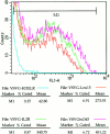
Surface staining of VSVG chimeras by flow cytometry. COS cells were transiently transfected with 5 μg VSVGtsO45, or the LEU15, IL2R, or KDELR wild-type chimeric plasmids, and then maintained at 40°C. At 40 h posttransfection, cycloheximide was added, and cells were placed at 18°C for 3 h, and then returned to 40°C for an additional 2 h. Intact cells were harvested and stained on ice with anti-VSVG monoclonal antibodies, and then with secondary FITC-conjugated anti–mouse IgG. Shown is the surface-staining intensity (x-axis) measured for cells gated for positive VSVGtsO45 surface expression (M1). Cell number is plotted on the y-axis. Statistical analysis indicates significantly higher mean fluorescence intensity for VSVGtsO45 (mean value ∼451) than for the Leu15 (∼274) and IL2R (∼341) chimeras, with the KDELR wild-type chimera (∼43) serving as a negative cell surface control. Parallel samples analyzed in the presence of 0.2% saponin determined total cellular staining and showed equivalent expression levels for each construct.
Figure 7
Figure 7
Analysis of cycling using conformation-sensitive antibodies. CHO cells stably expressing VSVG–TGN38 were treated as indicated and stained with conformation-sensitive I14 antibodies to VSVG (a, c, e, g, i, and k), and antibodies to the Golgi enzyme, mannosidase II (b, d, f, h, j, and l). Images were acquired on a Zeiss laser scanning confocal microscope. Notice the complete lack of I14 staining in cells treated at 40°C (e), but not when recycling was inhibited (c). The brightness of the cells in e and k was artificially enhanced to show the cell outline. Bar, 10 μm.
Figure 7
Figure 7
Analysis of cycling using conformation-sensitive antibodies. CHO cells stably expressing VSVG–TGN38 were treated as indicated and stained with conformation-sensitive I14 antibodies to VSVG (a, c, e, g, i, and k), and antibodies to the Golgi enzyme, mannosidase II (b, d, f, h, j, and l). Images were acquired on a Zeiss laser scanning confocal microscope. Notice the complete lack of I14 staining in cells treated at 40°C (e), but not when recycling was inhibited (c). The brightness of the cells in e and k was artificially enhanced to show the cell outline. Bar, 10 μm.
Figure 8
Figure 8
Fractionation of ER and Golgi membranes and misfolding in vitro. (A) COS cells transfected with VSVG–TGN38 were labeled with [35S]methionine, chased to generate labeled molecules in both the ER and Golgi complex, and then homogenized and fractionated on a 0–26% Optiprep gradient, as described in Materials and Methods. Fractions were analyzed for the presence of specific markers; ribophorin (ER), and galactosyltransferase activity (Golgi). (B) Pooled ER and Golgi fractions were aliquoted into equal volumes and incubated at either 32° or 40°C for 60 min. Membranes were lysed at the indicated temperatures, placed on ice, and immunoprecipitated with polyclonal anti-VSV antiserum or conformation-sensitive anti-VSVG I14 antibodies. Precipitates were analyzed by SDS-PAGE and scanning densitometry. Notice the relative ability of I14 antibodies to recognize VSVG–TGN38 from Golgi, but not from ER, fractions at 40°C.

Similar articles

Cited by

References

    1. Akagi S, Yamamoto A, Yoshimori T, Masaki R, Ogawa R, Tashiro Y. Distribution of protein disulfide isomerase in rat epiphyseal chondrocytes. J Histochem Cytochem. 1989;37:1835–1844. - PubMed
    1. Arar C, Carpentier V, Le Caer JP, Monsigny M, Legrand A, Roche AC. ERGIC-53, a membrane protein of the endoplasmic reticulum-Golgi intermediate compartment, is identical to MR60, an intracellular mannose-specific lectin of myelomonocytic cells. J Biol Chem. 1995;270:3551–3553. - PubMed
    1. Balch WE, Keller DS. ATP-coupled transport of vesicular stomatitis virus G protein. Functional boundaries of secretory compartments. J Biol Chem. 1986;261:14690–14696. - PubMed
    1. Balch WE, Elliott MM, Keller DS. ATP-coupled transport of vesicular stomatitis virus G protein between the endoplasmic reticulum and the Golgi. J Biol Chem. 1986;261:14681–14689. - PubMed
    1. Blum R, Feick P, Puype M, Vandekerckhove J, Klengel R, Nastainczyk W, Schulz I. Tmp21 and p24A, two type I proteins enriched in pancreatic microsomal membranes, are members of a protein family involved in vesicular trafficking. J Biol Chem. 1996;271:17183–17189. - PubMed

MeSH terms