. 1998 Jan 12;140(1):29-37.
doi: 10.1083/jcb.140.1.29.

Prospore membrane formation defines a developmentally regulated branch of the secretory pathway in yeast

Affiliations

Prospore membrane formation defines a developmentally regulated branch of the secretory pathway in yeast

A M Neiman. J Cell Biol. .

Abstract

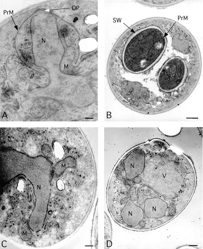
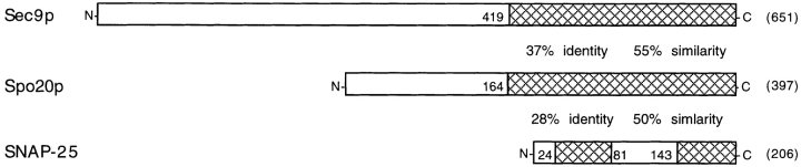
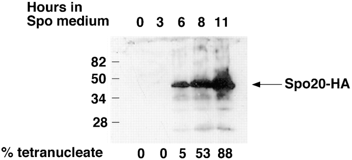
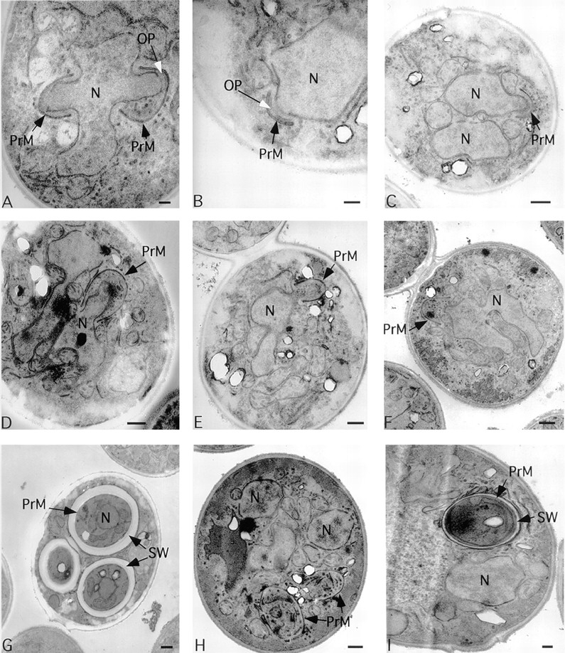
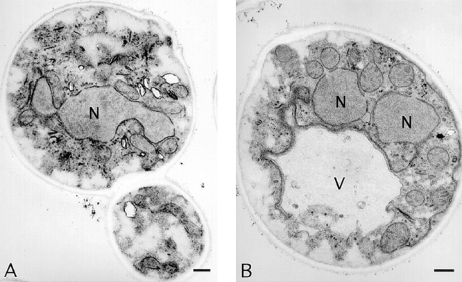
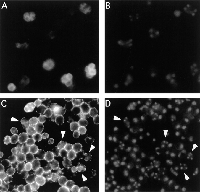
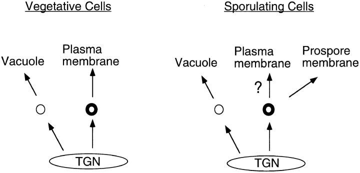
Spore formation in yeast is an unusual form of cell division in which the daughter cells are formed within the mother cell cytoplasm. This division requires the de novo synthesis of a membrane compartment, termed the prospore membrane, which engulfs the daughter nuclei. The effect of mutations in late-acting genes on sporulation was investigated. Mutation of SEC1, SEC4, or SEC8 blocked spore formation, and electron microscopic analysis of the sec4-8 mutant indicated that this inability to produce spores was caused by a failure to form the prospore membrane. The soluble NSF attachment protein 25 (SNAP-25) homologue SEC9, by contrast, was not required for sporulation. The absence of a requirement for SEC9 was shown to be due to the sporulation-specific induction of a second, previously undescribed, SNAP-25 homologue, termed SPO20. These results define a developmentally regulated branch of the secretory pathway and suggest that spore morphogenesis in yeast proceeds by the targeting and fusion of secretory vesicles to form new plasma membranes in the interior of the mother cell. Consistent with this model, the extracellular proteins Gas1p and Cts1p were localized to an internal compartment in sporulating cells. Spore formation in yeast may be a useful model for understanding secretion-driven cell division events in a variety of plant and animal systems.


Figures

Figure 1
Figure 1
Prospore membranes are absent in a sec4/sec4 mutant. (A) SEC4 cell (NH144) late in prospore membrane formation. The gap in the nuclear envelope underneath the outer plaque is the position of the spindle pole body. (B) NH144, after closure. (C and D) Cells of strain AN81 (sec4-8/sec4-8) at stages comparable to those in A and B, respectively. N, nucleus; M, mitochondrion; V, vacuole; OP, outer plaque; PrM, prospore membrane; SW, spore wall. All cells were sporulated at 23°C for 3 h and then shifted to the restrictive temperature for sec4-8 (34°C). Bars: (A and C) 200 nm; (B and D) 500 nm.
Figure 2
Figure 2
SPO20 encodes a second yeast SNAP-25 homologue. Alignment of domains in Sec9p, Spo20p, and mouse SNAP-25. Amino acid numbers are indicated adjacent to hatched areas, and numbers in parentheses are total amino acids. These sequence data are available from GenBank/EMBL/DDBJ under accession numbers Z49211 (SPO20), L34336 (SEC9), and M22012 (SNAP-25).
Figure 3
Figure 3
Time course of Spo20p induction. At the times indicated, samples of strain AN67 carrying pRS425-SPO20HA were removed from sporulation medium, and extracts were made and analyzed by Western blot using anti-HA antibodies. Equal cell equivalents were loaded in each lane. Marks at left indicate the position of molecular size standards. The fraction of cells at each time point (at least 200 cells counted) that were tetranucleate, as determined by DAPI staining, is shown below each lane. The predicted molecular mass for the Spo20-HA fusion protein is 49,800 D.
Figure 4
Figure 4
spo20 mutants fail to package daughter nuclei into spores. (A, D, and G) Isogenic SPO20 (NH144) or (B, C, E, F, H, and I) spo20ΔURA3/spo20ΔUR A3 (AN67) cells at different stages of prospore membrane formation: early (A– C); middle (D–F); or after closure (G–I). (A and B) The gap in the nuclear envelope underneath the outer plaque is the position of the spindle pole body. N, nucleus; OP, outer plaque; PrM, prospore membrane; SW, spore wall. Bars: (A, B, and I) 200 nm; (C–H) 500 nm.
Figure 5
Figure 5
Prospore membranes are absent in a spo20/spo20 sec9/ sec9 double mutant. Cells of strain AN80 (spo20ΔURA3/spo20ΔURA3sec9-4/sec9-4) at times corresponding to middle (A) and postclosure stages (B) of prospore membrane formation. N, nucleus; V, vacuole. Cells were sporulated at the restrictive temperature for sec9-4 (34°C). Bars, 500 nm.
Figure 6
Figure 6
Gas1p and Cts1p localization in sporulating cells. (A) Indirect immunofluorescence of Gas1p in sporulating cells of strain NH144. (B) DAPI staining of cells in A. (C) Indirect immunofluorescence of Cts1p in sporulating cells of strain NH144. (D) DAPI staining of cells in C. Arrowheads (C and D) indicate tetranucleate cells.
Figure 7
Figure 7
A sporulation-specific branch of the yeast secretory pathway. Routes for vesicular traffic out of the TGN in vegetative and sporulating cells are shown. Thick circles indicate vesicles dependent on SEC4 function for fusion with their target membrane. In both situations, SEC4-independent vesicles (thin circles) are targeted to the vacuole. “?” indicates that it is unclear whether trafficking to the plasma membrane continues in cells developing a prospore membrane (see text).

Similar articles

Cited by

References

    1. Aalto MK, Ronne H, Keranen S. Yeast syntaxins Sso1p and Sso2p belong to a family of related membrane proteins that function in vesicular transport. EMBO (Eur Mol Biol Organ) J. 1993;12:4095–4104. - PMC - PubMed
    1. Angold RE. The formation of the generative cell in the pollen grain of Emdmyion non-scriptus(L) J Cell Sci. 1968;3:573–578. - PubMed
    1. Brennwald P, Kearns B, Champion K, Keranen S, Bankaitis V, Novick P. Sec9 is a SNAP-25-like component of a yeast SNARE complex that may be the effector of Sec4 function in exocytosis. Cell. 1994;79:245–258. - PubMed
    1. Briza P, Breitenbach M, Ellinger A, Segall J. Isolation of two developmentally regulated genes involved in spore wall maturation in Saccharomyces cerevisiae. . Genes Dev. 1990;4:1775–1789. - PubMed
    1. Byers, B. 1981. Cytology of the yeast life cycle. In The Molecular Biology of the Yeast Saccharomyces: Life Cycle and Inheritance. J.N. Strathern, E.W. Jones, and J.R. Broach, editors. Cold Spring Harbor Laboratory, Cold Spring Harbor, NY. 59–96.

Publication types

MeSH terms