. 1998 Jan 12;140(1):39-47.
doi: 10.1083/jcb.140.1.39.

Sphingomyelinase treatment induces ATP-independent endocytosis

Affiliations

Sphingomyelinase treatment induces ATP-independent endocytosis

X Zha et al. J Cell Biol. .

Abstract

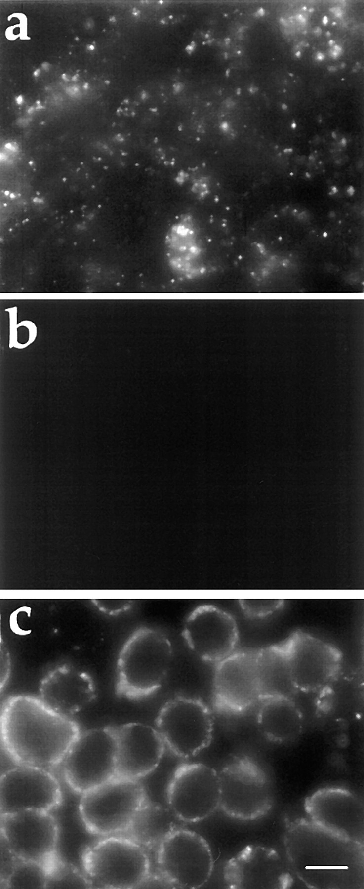
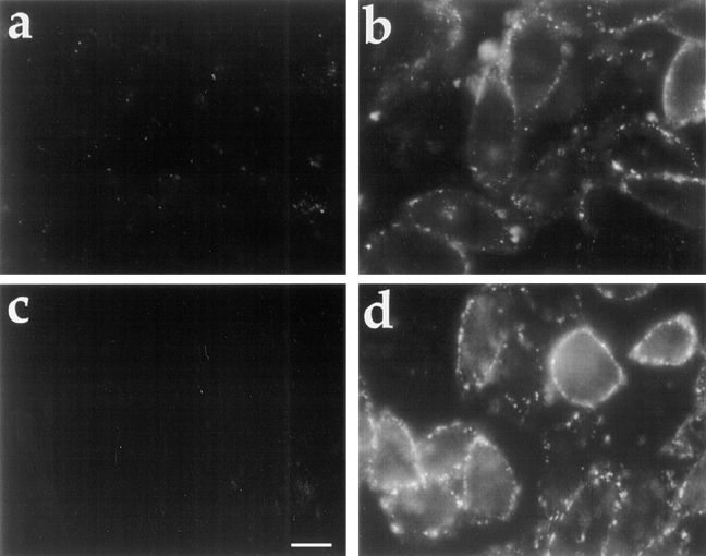
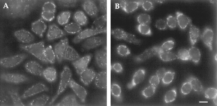
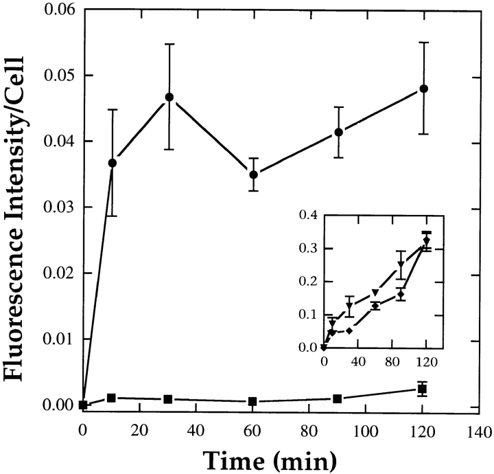
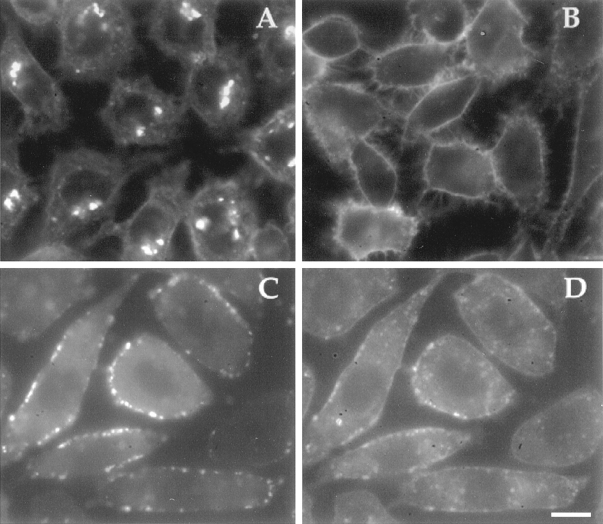
ATP hydrolysis has been regarded as a general requirement for internalization processes in mammalian cells. We found, however, that treatment of ATP-depleted macrophages and fibroblasts with exogenous sphingomyelinase (SMase) rapidly induces formation of numerous vesicles that pinch off from the plasma membrane; the process is complete within 10 min after adding SMase. By electron microscopy, the SMase-induced vesicles are approximately 400 nm in diameter and lack discernible coats. 15-30% of plasma membrane is internalized by SMase treatment, and there is no detectable enrichment of either clathrin or caveolin in these vesicles. When ATP is restored to the cells, the SMase-induced vesicles are able to deliver fluid-phase markers to late endosomes/lysosomes and return recycling receptors, such as transferrin receptors, back to the plasma membrane. We speculate that hydrolysis of sphingomyelin on the plasma membrane causes inward curvature and subsequent fusion to form sealed vesicles. Many cell types express a SMase that can be secreted or delivered to endosomes and lysosomes. The hydrolysis of sphingomyelin by these enzymes is activated by several signaling pathways, and this may lead to formation of vesicles by the process described here.


Figures

Figure 8
Figure 8
Sorting of transferrin receptors in SMase-induced vesicles. J774 macrophages were preincubated for 15 min in 0.2% BSA/DME plus 100 mM 2-deoxyglucose/sodium azide, followed by a 10-min incubation in the same medium containing Cy3-Tf (20 μg/ml). Because of the energy depletion, Cy3-Tf was only bound to the cell surface at this stage. The cells were then rinsed and incubated in DME containing energy poisons, FITC-dextran, and SMase for 10 min. At the end of this 10-min incubation, a mild acid wash was performed to strip Cy3-Tf that still remained on the cell surface. Cells were then either viewed immediately by fluorescence microscopy (a and b) or chased in DME for 20 min (c and d). Without any chase, internalized Cy3-Tf (b) was colocalized with FITC-dextran (a) in the peripheral vesicles. After a 20-min chase in complete medium without energy poison, FITC-dextran–containing vesicles moved into the cells (c), and Tf was mostly returned to the cell surface (d). Bar, 20 μm.
Figure 1
Figure 1
Fluorescence microscopy of FITC-dextran endocytosis in J774 macrophages. J774 macrophages were preincubated for 5 min in 0.2% BSA/DME alone (a), or containing 100 mM 2-deoxyglucose and 100 mM sodium azide (b and c). The cells were then incubated in the same medium containing 5 mg/ml FITC-dextran alone (a and b), or plus 50 mU/ml SMase (c) for 10 min. Cells were fixed and examined by fluorescence microscopy as described in Materials and Methods. Bar, 10 μm.
Figure 2
Figure 2
Electron microscopy of HRP endocytosis. J774 macrophages were preincubated with sodium azide/2-deoxyglucose (100 mM) for 5 min and then incubated in the same medium plus SMase (50 mU/ml) and HRP (10 mg/ml) for 10 min. The cells were fixed, incubated with diaminobenzidine and H2O2, and processed for electron microscopy as described in Materials and Methods. HRP-containing vesicles typically exhibited precipitated diaminobenzidine along the lumenal edge of the compartment (filled arrowheads). Lumenal diaminobenzidine precipitation often formed a horseshoe shape (open arrowheads). A lack of precipitated diaminobenzidine in the center of the compartment indicated a relatively low concentration of soluble protein. The SMase-induced compartments exhibited diameters ranging from 50 to 500 nm. In the presence of energy poisons, SMase-generated compartments exhibited a peripheral distribution in the cytoplasm. Limiting membranes of SMase-generated compartments did not contain discernible coat structures and were not continuous with membranes of any other organelles. Bar, 1 μm.
Figure 3
Figure 3
Fluorescence microscopy of FITC-dextran uptake in TRVb-1 cells. Cells were preincubated for 5 min in 0.2% BSA/ HF-12 medium (a and b), or glucose-free 0.2% BSA/HF-12 plus 5 mM 2-deoxyglucose and 5 mM sodium azide (c and d). The cells were then incubated in the same medium with 5 mg/ml FITC-dextran (a and c) or FITC-dextran plus SMase (50 mU/ml; b and d) for 2.5 min. Cells were then fixed and viewed by fluorescence microscopy. Bar, 10 μm.
Figure 4
Figure 4
Fluorescence microscopy of Lucifer yellow uptake and retention in SL-O–permeabilized TRVb-1 cells. Cells were preincubated for 10 min in 0.2% BSA/glucose-free HF-12 plus 10 mM 2-deoxyglucose and 10 mM sodium azide. The cells were then chilled to 0°C, treated with SL-O (0.4 U/ml) for 3 min, and followed by three washes with cold PBS. After returning to 37°C for 10 min, cells were exposed to Lucifer yellow (10 mg/ml) with SMase (50 mU/ml) for another 10 min. The cells were either viewed right away (a) or transferred into a medium free of Lucifer yellow for an additional 2 h before viewed by fluorescence microscopy (b). Bar, 10 μm.
Figure 5
Figure 5
Kinetics of dextran uptake in J774 macrophages. Cells were preincubated for 5 min in 0.2% BSA/DME containing 100 mM azide and 100 mM 2-deoxyglucose, or in the same medium without energy poisons (inset). Cells were then incubated with 5 mg/ml FITC-dextran (▪, ♦) or FITC-dextran plus 50 mU/ml SMase (•, ▾) for times ⩽2 h. The uptake of FITC-dextran was then quantified by fluorescence microscopy as described previously (Dunn and Maxfield, 1990). Each data point presents an average fluorescence intensity from four or five fields of cells (50–70 cells/fields) ± SD. SMase treatment of energy-poisoned cells led to uptake of FITC-dextran that was comparable to the cumulative uptake in control cells after ∼20 min. In energy-replete cells (inset), SMase treatment caused an increase in the initial dextran uptake with a rate of sustained uptake similar to control cells.
Figure 6
Figure 6
SMase-induced endovesiculation of the plasma membrane. The plasma membrane of TRVb-1 cells was labeled with the fluorescent lipid analogue C6-NBD-gal. Cells were then energy depleted by incubation with sodium azide/deoxyglucose (5 mM) for 10 min at 37°C. After a subsequent incubation in the same medium with (c and d) or without (a and b) SMase (50 mU/ml), the lipid label was backexchanged by washing with ice-cold medium containing 5% BSA (b and d) or washed with ice-cold medium alone (a and c). In the absence of SMase (a and b), almost all of the C6-NBD-gal is backexchanged, whereas in the presence of SMase, C6-NBD-gal located in peripheral vesicles is not available for backexchange. Bar, 10 μm.
Figure 7
Figure 7
Fusion of SMase-induced vesicles with late endosomes/ lysosomes. J774 macrophages were incubated with TRITC-dextran for 1 h and chased in dextran-free medium for 30 min. The cells were then incubated for 5 min in 0.2% BSA/DME plus 100 mM 2-deoxyglucose/sodium azide, followed by a 10-min incubation in the same medium containing FITC-dextran (5 mg/ml) and SMase (50 mU/ml). After rinsing, the cells were chased for 1 h either in the same medium containing energy poisons (a and b) or in complete medium without energy poisons (c and d). The cells were fixed, and then a pair of fluorescence images of FITC-dextran (a and c) and TRITC-dextran (b and d) were acquired from each field of cells by confocal microscopy. In the continuous presence of energy poisons, the FITC-containing compartments remain in the periphery (a) and do not fuse with the TRITC-dextran– labeled endosomes and lysosomes (b; arrows). In cells chased in complete medium that allows restoration of ATP, there is extensive colocalization between FITC-dextran–containing compartments (c) and TRITC-dextran compartments (d; arrows). Bar, 10 μm.
Figure 9
Figure 9
SMase treatment of BODIPY-C12-SM–labeled cells. TRVb-1 cells were labeled with BODIPY-C12-SM on ice for 30 min. Cells were incubated with SMase (50 mU/ml) (A) or without (B) for 5 min. Some of the cells were incubated with SL-O (0.4 U/ml) for 5 min on ice (C and D), washed with cold PBS and transferred to 37°C for 3 min, and then incubated with SMase (50 mU/ml)/ rhodamine-dextran (5 mg/ml) for 5 min. Cells were washed and transferred into HF-12 medium for ∼20 min at room temperature before being viewed by fluorescence microscopy. Rhodamine-dextran–containing vesicles (C) colocalized with vesicles labeled with BODIPY fluorescence (D). Bar, 10 μm.

Similar articles

Cited by

References

    1. Allan D, Walklin CM. Endovesiculation of human erythrocytes exposed to sphingomyelinase C: a possible explanation for the enzyme-resistant pool of sphingomyelin. Biochim Biophys Acta. 1988;938:403–410. - PubMed
    1. Araki N, Johnson MT, Swanson JA. A role for phosphoinositide 3-kinase in the completion of macropinocytosis and phagocytosis by macrophages. J Cell Biol. 1996;135:1249–1260. - PMC - PubMed
    1. Bednarek SY, Orci L, Schekman R. Traffic COPs and the formation of vesicle coats. Trends Cell Biol. 1996;6:468–473. - PubMed
    1. Callahan JW, Jones CS, Davidson DJ, Shankaran P. The active site of lysosomal sphingomyelinase: evidence for the involvement of hydrophobic and ionic groups. J Neurosci Res. 1983;10:151–163. - PubMed
    1. Chatterjee S. Neutral sphingomyelinase. Adv Lipid Res. 1993;26:25–48. - PubMed

Publication types

MeSH terms

Substances