. 1998 Jan 12;140(1):101-10.
doi: 10.1083/jcb.140.1.101.

Low density membranes are associated with RNA-binding proteins and thylakoids in the chloroplast of Chlamydomonas reinhardtii

Affiliations

Low density membranes are associated with RNA-binding proteins and thylakoids in the chloroplast of Chlamydomonas reinhardtii

W Zerges et al. J Cell Biol. .

Abstract

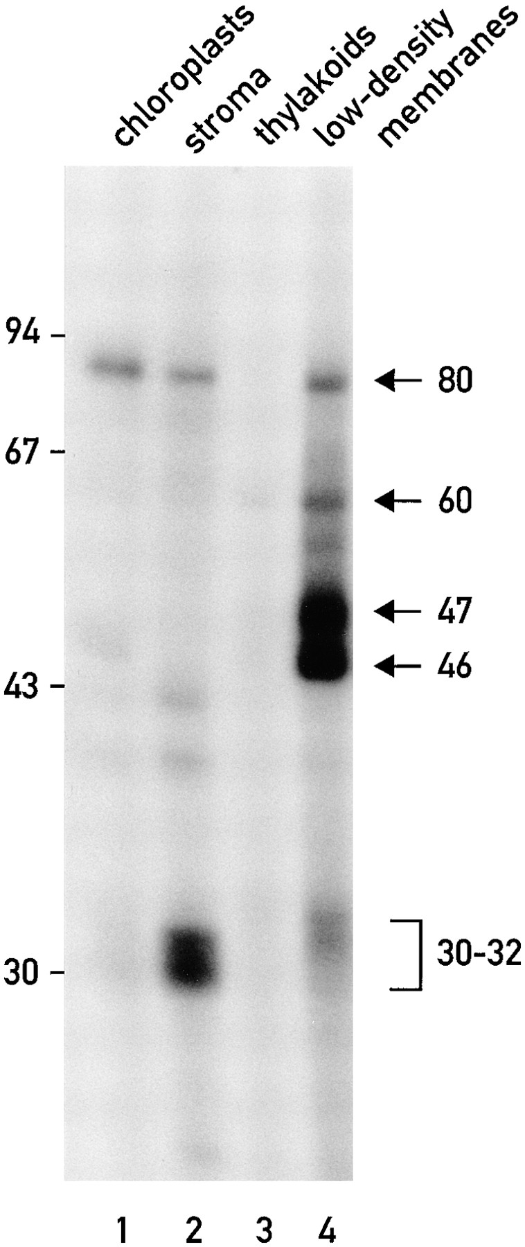
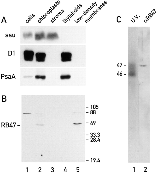
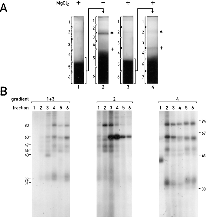
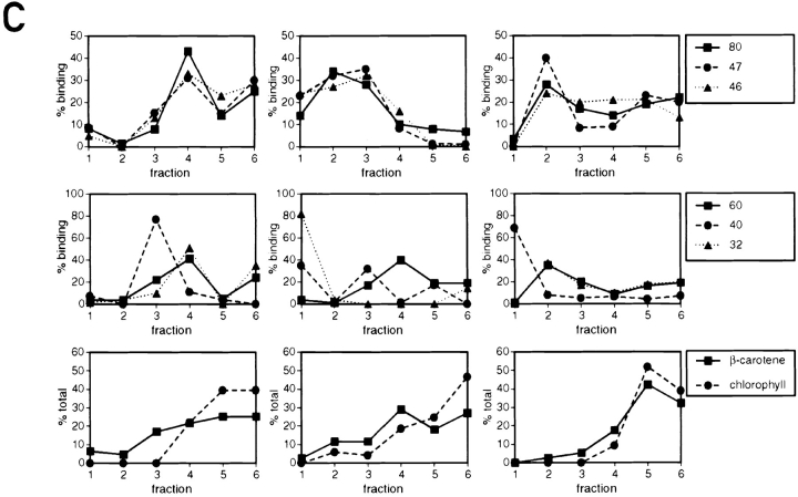
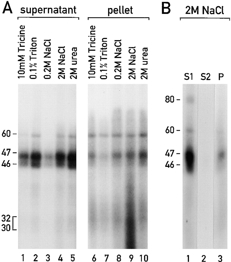
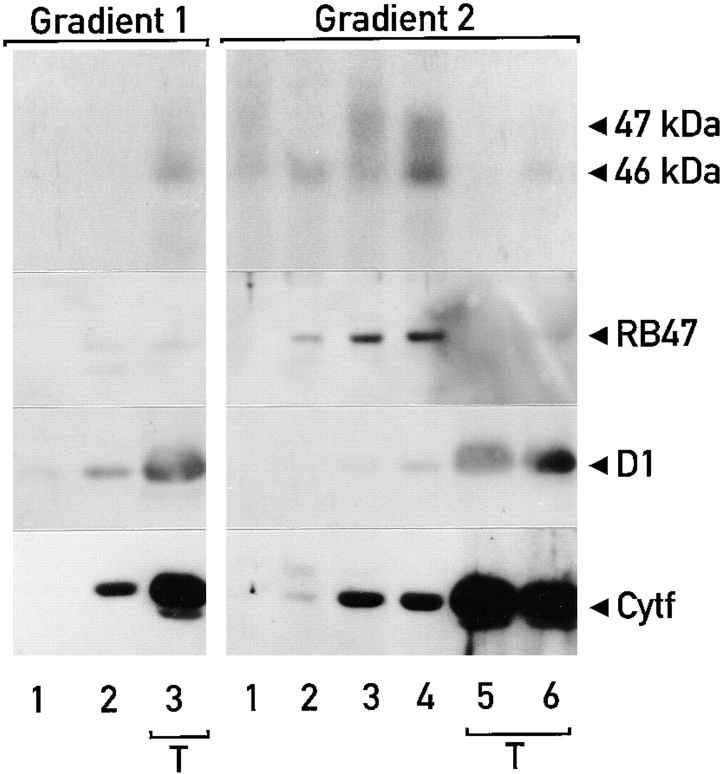
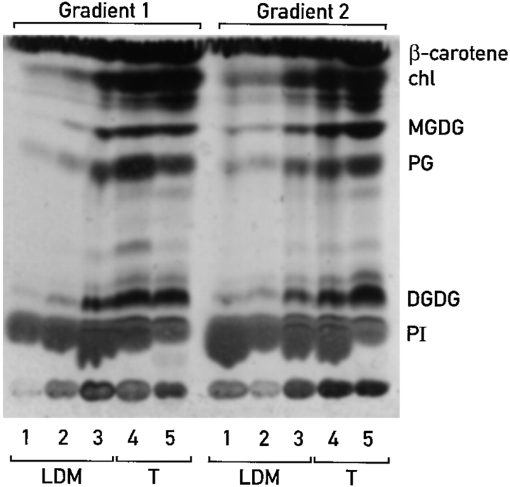
Chloroplast subfractions were tested with a UV cross-linking assay for proteins that bind to the 5' untranslated region of the chloroplast psbC mRNA of the green alga Chlamydomonas reinhardtii. These analyses revealed that RNA-binding proteins of 30-32, 46, 47, 60, and 80 kD are associated with chloroplast membranes. The buoyant density and the acyl lipid composition of these membranes are compatible with their origin being the inner chloroplast envelope membrane. However, unlike previously characterized inner envelope membranes, these membranes are associated with thylakoids. One of the membrane-associated RNA-binding proteins appears to be RB47, which has been reported to be a specific activator of psbA mRNA translation. These results suggest that translation of chloroplast mRNAs encoding thylakoid proteins occurs at either a subfraction of the chloroplast inner envelope membrane or a previously uncharacterized intra-chloroplast compartment, which is physically associated with thylakoids.


Figures

Figure 1
Figure 1
RNA-binding proteins (of ∼30, 32, 46, 47, 60, and 80 kD) cofractionate with the chloroplast low density membranes. UV light was used to cross-link the 32P-RNA probe derived from the psbC 5′ UTR to proteins in extracts from isolated chloroplasts (lane 1), chloroplast stroma (lane 2), thylakoids (lane 3), and the low density chloroplast membranes (lane 4). Each sample contained 20 μg of protein.
Figure 2
Figure 2
The RNA-binding protein, RB47, cofractionates with low density chloroplast membranes. (A and B) The immunoblots analyze 20-μg samples of protein extracts from total cells (lane 1) isolated chloroplasts (lane 2), chloroplast stroma (lane 3), thylakoids (lane 4), and the low density chloroplast membranes (lane 5). The immunoblot in A was reacted with antisera against marker proteins for the chloroplast stroma, the small subunit of Rubisco (ssu); for the stacked (appressed) thylakoid membranes, the D1 subunit of PSII (D1), and for the nonstacked or stroma lamellae of thylakoid membranes, the PsaA subunit of PSI (PsaA). The immunoblot in B was reacted with antisera against the RNA-binding protein RB47 (Danon and Mayfield, 1991, 1994). (C) The 47-kD RNA-binding protein (radiolabeled by UV cross-linked to the 32P-RNA probe ) comigrates during SDS-PAGE (and in the same lane) with RB47 detected with the antiserum against it.
Figure 4
Figure 4
The low density membranes (with the RNA-binding proteins) are associated with thylakoid membranes. (A) Sequential sucrose density gradients were used to fractionate chloroplast membranes. On gradient 1 total chloroplast membranes were fractionated in the presence of 5 mM MgCl2. Thylakoids from this gradient (the dark band at the bottom) and the low density membranes associated with them were separated on gradient 2, which lacked MgCl2. The band of orange membranes observed on gradient 2 is indicated with an asterisk. When both gradients contained MgCl2 (gradients 3 and 4) considerably fewer low density membranes were observed above the thylakoid membranes. The lower density of the thylakoids in the absence of Mg2+ (indicated by their upward shift in gradient 2 and the +) reflects the destacking of the grana. (B) Binding of proteins to the 32P-RNA derived from the psbC 5′ UTR in the fractions from the gradients shown in A was measured with the UV cross-linking assay. Samples represented equivalent proportions of each fraction (i.e., were analyzed on a per chloroplast basis) and contained between 8 and 30 μg protein. (C) The binding signals for each protein were determined by phosphorimaging and graphed as percentage of the total signal for that protein in each gradient. Chlorophyll concentrations throughout each gradient are also graphed to indicate the distribution of thylakoid membranes.
Figure 4
Figure 4
The low density membranes (with the RNA-binding proteins) are associated with thylakoid membranes. (A) Sequential sucrose density gradients were used to fractionate chloroplast membranes. On gradient 1 total chloroplast membranes were fractionated in the presence of 5 mM MgCl2. Thylakoids from this gradient (the dark band at the bottom) and the low density membranes associated with them were separated on gradient 2, which lacked MgCl2. The band of orange membranes observed on gradient 2 is indicated with an asterisk. When both gradients contained MgCl2 (gradients 3 and 4) considerably fewer low density membranes were observed above the thylakoid membranes. The lower density of the thylakoids in the absence of Mg2+ (indicated by their upward shift in gradient 2 and the +) reflects the destacking of the grana. (B) Binding of proteins to the 32P-RNA derived from the psbC 5′ UTR in the fractions from the gradients shown in A was measured with the UV cross-linking assay. Samples represented equivalent proportions of each fraction (i.e., were analyzed on a per chloroplast basis) and contained between 8 and 30 μg protein. (C) The binding signals for each protein were determined by phosphorimaging and graphed as percentage of the total signal for that protein in each gradient. Chlorophyll concentrations throughout each gradient are also graphed to indicate the distribution of thylakoid membranes.
Figure 3
Figure 3
Membrane association of the RNA-binding proteins. (A) Samples (1.0 μg protein) of low density chloroplast membranes containing the RNA-binding proteins (which had been radiolabeled by the UV cross-linking to the 32P-RNA probe) were treated with 10 mM tricine, pH 7.8, 0.1% Triton X-100, 0.2 M NaCl, 2 M NaCl and 2 M urea. The samples were incubated 30 min at 0°C. After centrifugation of the extracts at 105 g for 60 min, supernatant and pellet fractions were analyzed by SDS-PAGE and autoradiography. (B) A sample of low density membranes with the 32P-RNA–binding proteins was treated for 30 min at 0°C with 2.0 M NaCl, and then centrifuged at 105 g for 60 min. The membrane pellet was resuspended in 2.0 M NaCl, and again incubated for 30 min at 0°C and then centrifuged at 105 g for 60 min. The first and second supernatant fractions (S1 and S2) and the final membrane pellet fraction (P) were analyzed by SDS-PAGE and autoradiography. The UV crosslinking of the 80-kD protein was often not detected for unknown reasons (see A and B). However, in other experiments, extraction of this protein paralleled the extraction of the 46-, 47- and 60-kD proteins.
Figure 5
Figure 5
Immunoblot and UV cross-linking analyses of fractions from gradients 1 and 2. The 46-, 47-, and 60-kD RNA-binding proteins, which were found to bind to the 32P-RNA probe in the UV cross-linking assay, cofractionate with thylakoids (T) in the presence of magnesium ions (Gradient 1). In the absence of magnesium ions (Gradient 2), these proteins and the low density membranes associated with them were separated from thylakoid membranes (T). Thylakoid membranes were followed by their enrichment for the PSII reaction center subunit D1, and cytochrome f detected by immunoblot analysis. RB47 (detected by immunoblot analysis) also cofractionates with the low density membranes that are associated with thylakoid membranes in the presence of magnesium ions. Samples contained 20 μg of protein.
Figure 6
Figure 6
The low density membranes that are associated with thylakoids in the presence of magnesium have acyl lipid compositions that are similar to the chloroplast envelope membranes. The thin layer chromatogram resolves acyl lipid classes in fractions from gradients similar to those shown in Fig. 4. Indicated are the pigments β-carotene and chlorophyll (chl), the galactolipids monogalactosyldiacylglycerol (MGDG) and digalactosyldiacylglycerol (DGDG), and the phospholipids phosphatidylglycerol (PG) and phosphatidylinositol (PI). LDM, the low density membranes. T, thylakoid membranes. Samples represent equivalent fractions of each fraction (i.e., were analyzed on a per chloroplast basis).

Similar articles

Cited by

References

    1. Andreasson E, Svensson P, Weibull C, Albertsson P-A. Separation and characterization of stromal and grana membranes -evidence for heterogeneity in antenna size of both photosystem I and photosystem II. Biochim Biophys Acta. 1988;936:339–350.
    1. Barber J. Membrane surface charges and potentials in relation to photosynthesis. Biochim Biophys Acta. 1980;594:253–308. - PubMed
    1. Block, M.A., A-J. Dorne, J. Joyard, and R. Douce. 1983. Preparation and characterization of membrane fractions enriched in outer and inner envelope membranes from spinach chloroplasts. J. Biol. Chem. 258. 13273–13280. - PubMed
    1. Chen Q, Adams CC, Usack L, Yang J, Monde R-A, Stern DB. An AU-rich element in the 3′ untranslated region of the spinach chloroplast petDgene participates in sequence-specific RNA-protein complex formation. Mol Cell Biol. 1995;15:2010–2018. - PMC - PubMed
    1. Clemetson JM, Boschetti A, Clemetson KJ. Chloroplast envelope proteins are encoded by the chloroplast genome of Chlamydomonas reinhardtii. . J Biol Chem. 1992;267:19773–19779. - PubMed

Publication types

MeSH terms

Substances