. 1998 Jan 12;140(1):159-69.
doi: 10.1083/jcb.140.1.159.

Epimorphin functions as a key morphoregulator for mammary epithelial cells

Affiliations

Epimorphin functions as a key morphoregulator for mammary epithelial cells

Y Hirai et al. J Cell Biol. .

Abstract

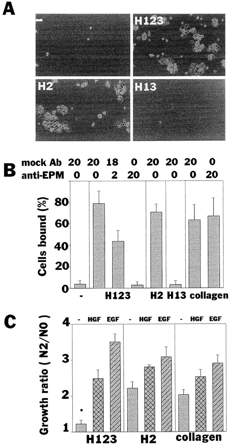
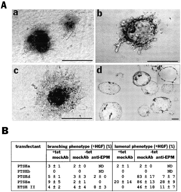
Hepatocyte growth factor (HGF) and EGF have been reported to promote branching morphogenesis of mammary epithelial cells. We now show that it is epimorphin that is primarily responsible for this phenomenon. In vivo, epimorphin was detected in the stromal compartment but not in lumenal epithelial cells of the mammary gland; in culture, however, a subpopulation of mammary epithelial cells produced significant amounts of epimorphin. When epimorphin-expressing epithelial cell clones were cultured in collagen gels they displayed branching morphogenesis in the presence of HGF, EGF, keratinocyte growth factor, or fibroblast growth factor, a process that was inhibited by anti-epimorphin but not anti-HGF antibodies. The branch length, however, was roughly proportional to the ability of the factors to induce growth. Accordingly, epimorphin-negative epithelial cells simply grew in a cluster in response to the growth factors and failed to branch. When recombinant epimorphin was added to these collagen gels, epimorphin-negative cells underwent branching morphogenesis. The mode of action of epimorphin on morphogenesis of the gland, however, was dependent on how it was presented to the mammary cells. If epimorphin was overexpressed in epimorphin-negative epithelial cells under regulation of an inducible promoter or was allowed to coat the surface of each epithelial cell in a nonpolar fashion, the cells formed globular, alveoli-like structures with a large central lumen instead of branching ducts. This process was enhanced also by addition of HGF, EGF, or other growth factors and was inhibited by epimorphin antibodies. These results suggest that epimorphin is the primary morphogen in the mammary gland but that growth factors are necessary to achieve the appropriate cell numbers for the resulting morphogenesis to be visualized.


Figures

Figure 3
Figure 3
Preparation of recombinant epimorphin. (A) Schematic diagram of epimorphin isoforms and the recombinant epimorphin fragments used in this study. There are three epimorphin isoforms produced by alternative splicing. Isoforms I and II (34-kD products) have a putative membrane-spanning region at the COOH terminus, whereas isoform III (31-kD products) has not. The recombinant epimorphin fragment H123 represents the epimorphin sequence shared by all the isoforms. H13 is a fusion peptide of the NH2- and COOH-terminal coiled-coil domains of H123, and H2 is a peptide of only the cellular recognition domain inserted between the coiled-coil domains. *, NH2-terminally tagged histidine residues. (B) Coomassie brilliant blue–stained epimorphin fragments in SDS-PAGE gel.
Figure 7
Figure 7
Nonpolar presence of recombinant epimorphin around each cell of the cluster leads to “lumen” formation. (A) Immunoblot detection of recombinant epimorphin in the clusters of SCp2 cells. (Right lane) Clusters of SCp2 cells precultured on H123 for 4 d (SCp2H123IN). (Left lane) Purified H123 (5 ng). (B) Detection of recombinant epimorphin (H123) in the cell clusters. Sections of the clusters were stained with anti-epimorphin antibodies (a) and with DAPI (b). (C) Appearance of the clusters of H123-containing SCp2 cells (SCp2 H123IN) in collagen gels, cultured for 8 d in the absence (a) or presence (b) of 50 ng/ml HGF. (D) Quantification of the branching and “lumenal” phenotype of the clusters of SCp2. Clusters of cells precultured on H123, H2, or H13 are shown as H123IN, H2IN, or H13IN, respectively. Note that H123 but not H2 and H13 fragments in the cluster induced “lumen” formation. EGF as well as HGF dramatically enhanced the effect of H123. Bars: (B) 120 μm; (C) 200 μm.
Figure 1
Figure 1
Expression of epimorphin in the mammary gland. (A) Localization of epimorphin in the sections of glands from virgin (a and b), mid-pregnant (c), and lactating (d) CD-1 mice. The area boxed in black in a is shown at higher magnification in b. (B) Detection of epimorphin-producing cell types. Frozen sections of mammary glands from mid-pregnant mice were immunostained with anti-epimorphin antibodies and rhodamine-labeled secondary antibodies (red). Sections were also stained for E-cadherin (a), α-smooth muscle actin (b), or vimentin (c) using their specific antibodies and FITC-labeled secondary antibodies (green). Nuclei were visualized with DAPI (blue). (d) Living cells were treated with a mixture of anti-epimorphin and anti-vimentin antibodies, and labeled antibodies were visualized with rhodamine- (for epimorphin) and FITC- (for vimentin) labeled secondary antibodies. Note that both anti-epimorphin and vimentin antibodies stained sections (c), but only the former labeled the living cells, indicating epimorphin is localized also at the cell surface (d). Bars, 120 μm.
Figure 2
Figure 2
Expression of epimorphin in epithelial model cells. Sections of cell clusters of CID-9 (a–c), EpH4 (d–f), and SCp2 (g–i) were stained simultaneously for epimorphin (a, d, and g), vimentin (b, e, and h), and DAPI (c, f, and i). *, epimorphin-negative subpopulation of EpH4. Note that 30–40% of CID-9 cells and 60–70% of EpH4 cells expressed epimorphin. Almost all SCp2 cells were negative for epimorphin but a minor subpopulation (∼5%) expressed epimorphin (g′ and i′). Bar, 120 μm.
Figure 4
Figure 4
Attachment and proliferation of an epithelial cell line on recombinant epimorphin. (A and B) Attachment of SCp2 cells to substrate-coated epimorphin fragments. Cells bound to each epimorphin fragment in 8 h were photographed (A) and quantified (B). Uncoated (−) and collagen type I–coated wells were used as negative and positive controls, respectively. Medium contained mock antibodies and anti-epimorphin antibodies at a total concentration of 20 μg/ml as indicated. P values between − and H123 (mock Ab20), and H123 (mock Ab20) and H123 (anti-EPM20) were <0.0001. (C) The growth ratios (cell number in 2 d plus 8 h divided by cell numbers 8 h after plating) of SCp2 cells cultured on type I collagen, H123, and H2 fragments. HGF and EGF were tested at 50 ng/ml. Significant inhibition of cell growth was observed in cells cultured on H123 in the absence of additional growth factors. *, P values versus H2 (−) and collagen (−) were <0.001.
Figure 5
Figure 5
Effect of recombinant epimorphin and growth factors on branching morphogenesis of SCp2 cells. (A) Morphology of SCp2 cell clusters cultured for 8 d in collagen gels mixed with (a and b) or without (c and d) recombinant epimorphin H123. Presence of H123 in substrate and outside the clusters is indicated as H123EX. HGF (50 ng/ml) was added to the medium in b and d. b′ and d′ are sections of b and d stained with hematoxylin, respectively. (B) Quantification of the branching phenotype of SCp2 cells depicted in A. Cell clusters were surrounded by H123, H2, or H13 in collagen (indicated as H123EX, H2EX, or H13EX, respectively). Note that H123, but not H2 and H13 fragments induced branching morphogenesis in collagen gels. HGF dramatically enhanced the effect of H123 and functional blocking antibodies to HGF (100 μg/ml) completely neutralized HGF. (C) Effect of other growth factors on morphology of SCp2 cell clusters. EGF (a, d, and g), KGF (b and e), and FGF (c and f) can also induce branching morphogenesis in the presence (a–c and g) but not in the absence (a–c) of epimorphin H123 (H123EX). All of the factors were added to medium at 50 ng/ml. Note that EGF induced epimorphin-dependent branching morphogenesis even in the presence of function-blocking anti-HGF antibodies. (D) Summary of above data. For growth in collagen, when the average diameter of >50 clusters was 1–2× the control (size of cluster without the factor addition) and P value versus control was <0.1, it was denoted +; 2–3× the control is ++, and 3× or larger, +++. Branching was scored as described in Materials and Methods. Bars: (A and C) 200 μm.
Figure 6
Figure 6
Branching morphogenesis of clonal derivatives of SCp2 cells. (A) The level of endogenous epimorphin in parental SCp2 and an epimorphin-positive clonal population (D6). Note that only very faint bands of endogenous epimorphin were detected in the SCp2 cells, as would be expected from the immunofluorescence studies (only ∼5% express epimorphin). (B) Detection of endogenous epimorphin in the clusters of SCp2 and D6 cells. The sections of SCp2 (a and c) and D6 (b and d) cells were stained for epimorphin (a and b) and DAPI (c and d). The expression of epimorphin was still heterogenous in D6 cells and only the peripheral portion of the clusters were epimorphin-positive. (C) Appearance of cell clusters of D6 cultured for 8 d in collagen gels with (c) or without (a and b) additional recombinant epimorphin H123, in the presence of 50 ng/ml HGF. Mock antibodies (a and c) or anti-epimorphin antibodies (b) were added to medium at 100 μg/ ml. c′ is a section of c stained with hematoxilin. Note that D6 demonstrated branching morphogenesis in collagen, but exogenous epimorphin (H123EX) enhanced, and anti-epimorphin antibodies perturbed the phenotypic appearance. (D) Effect of H123 and anti-epimorphin antibodies on the appearance of branching phenotype of several subclones derived from SCp2. About 60% of D6 and 40% of I6 cells were epimorphin positive, whereas PTSEa and PTSEb (refer to Fig. 8) were epimorphin negative. The values indicate mean ± SD of three separate experiments. Bars: (B) 120 μm; (C) 200 μm.
Figure 8
Figure 8
Epimorphin expression and cellular growth in the transfectants. (A) Characterization of transfectant clones isolated and used in this study: PTSEd, PTSEe (from SCp2), and ETSEII (from EpH4) expressed epimorphin transgene after removal of tetracycline. PTSEa and PTSEb cells were isolated from SCp2 as controls. Note that ETSEII was from an epimorphin-negative subpopulation of EpH4 cells. (B) Immunoblot analysis of epimorphin in the transfectants. The molecular mass of introduced extracellular epimorphin was 37 kD, which was reduced to that of endogenous epimorphin isoform I (34 kD) when medium contained 0.02 and 0.1 μg/ml tunicamycin. (C) The growth ratios of PTSEa and PTSEd cultured with or without tetracycline for 2 d. Media containing 50 ng/ml HGF and EGF were also tested. Note that the growth of transfected cells was severely suppressed when epimorphin transgene was induced (*).
Figure 9
Figure 9
Epimorphin production by transfectants in clonal cultures leads to lumen formation. (A) The appearances of clusters of PTSEd cultured in the presence (a) or absence (b–d) of 5 μg/ml tetracycline, either for 8 d (a–c) or 14 d (d). Anti-epimorphin antibodies (100 μg/ml) were added to medium in c. d′ is a section of d stained with hematoxylin. Medium contained 50 ng/ml HGF in each case. The appearance in c was often slightly different from a, probably due to nonhomogeneous blocking of epimorphin transgene by the antibodies, leading to some focal, polar presentation of epimorphin outside the cluster. This is in turn would allow some fine branching from the cluster mass (which nevertheless did not resemble a branching phenotype). (B) Quantification of the appearance of branching and lumenal phenotype of the transfectants (The values indicate mean ± SD of three separate experiments). The concentration of the anti-epimorphin or mock antibodies added to medium was 100 μg/ml. Bars, 200 μm.

References

    1. Bennett MK, Garcia-Arraras JE, Elferink LA, Peterson K, Fleming AM, Hazuka CD, Sheller RH. The syntaxin family of vesicular transport receptors. Cell. 1993;74:863–873. - PubMed
    1. Brinkmann V, Foroutan H, Sachs M, Weidner KM, Birchmeier W. Hepatocyte growth factor/scatter factor induces a variety of tissue-specific morphogenic programs in epithelial cells. J Cell Biol. 1995;131:1573–1586. - PMC - PubMed
    1. Butt KI, Manabe M, Yaguchi H, Tsuboi R, Ogawa H. Immunolocalization of epimorphin in skin. J Dermatol Sci. 1996;13:193–201. - PubMed
    1. Coleman S, Silberstein GB, Daniel CW. Ductal morphogenesis in the mouse mammary gland: evidence supporting a role for epidermal growth factor. Dev Biol. 1988;127:304–315. - PubMed
    1. Daniel CW, Strickland P, Friedmann Y. Expression and functional role of E- and P-cadherins in mouse mammary ductal morphogenesis and growth. Dev Biol. 1995;169:511–519. - PubMed

Publication types

MeSH terms