. 1998 Jan 12;140(1):171-82.
doi: 10.1083/jcb.140.1.171.

Ultraviolet light induces apoptosis via direct activation of CD95 (Fas/APO-1) independently of its ligand CD95L

Affiliations

Ultraviolet light induces apoptosis via direct activation of CD95 (Fas/APO-1) independently of its ligand CD95L

Y Aragane et al. J Cell Biol. .

Abstract

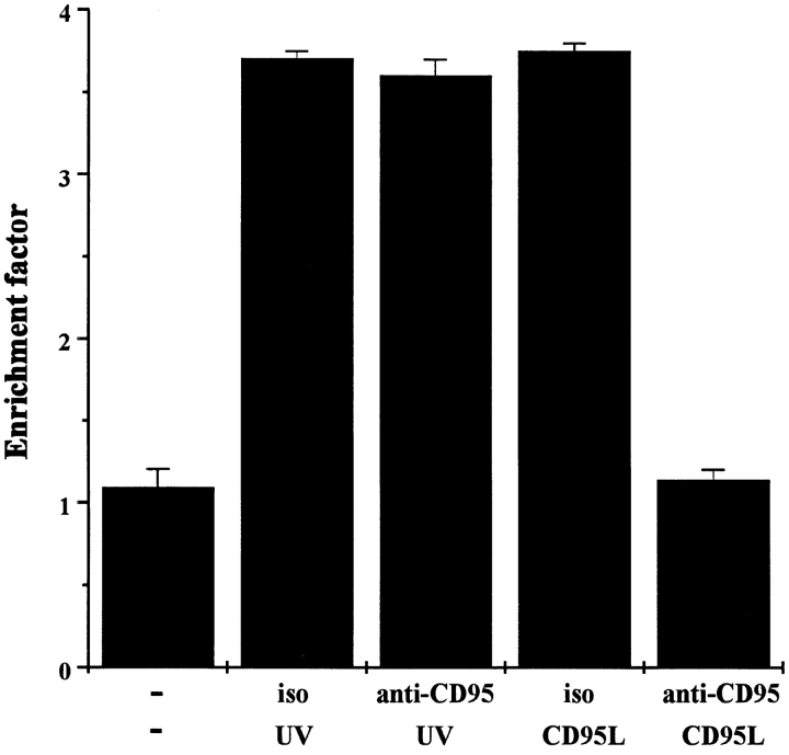
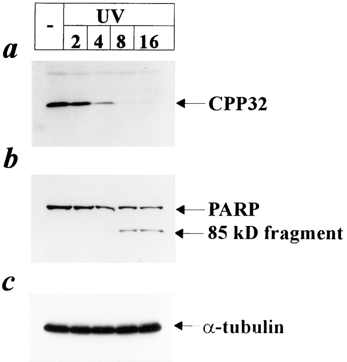
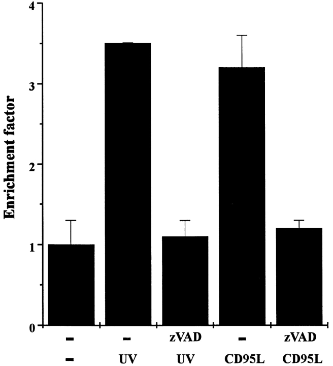
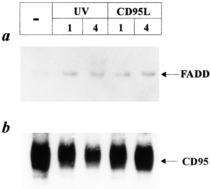
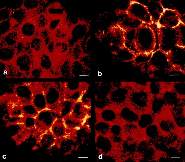
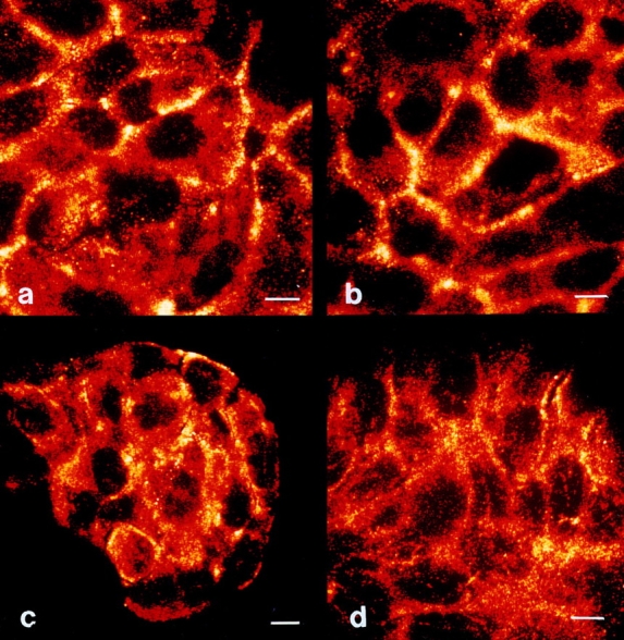
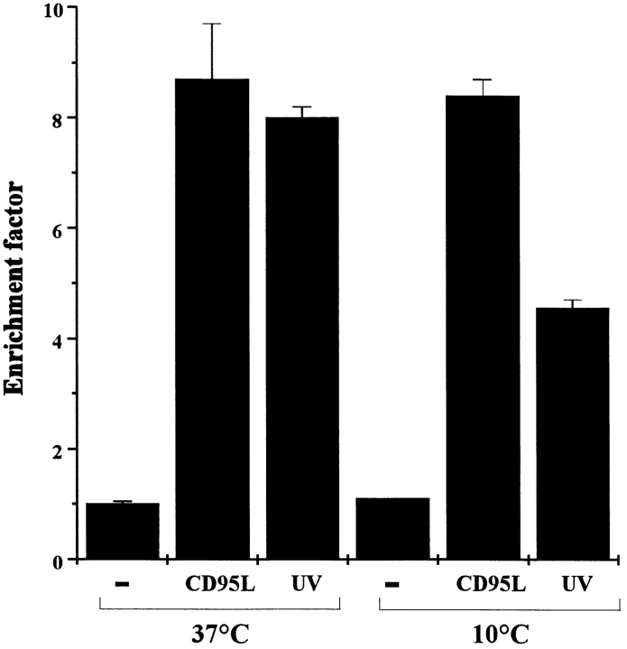
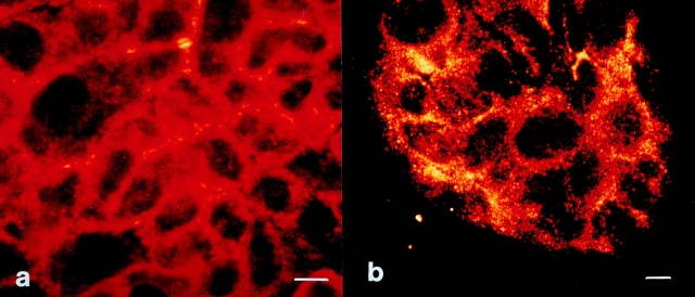
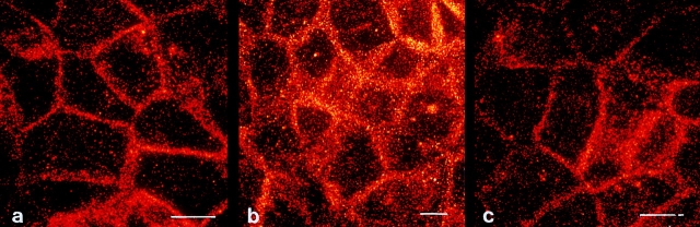
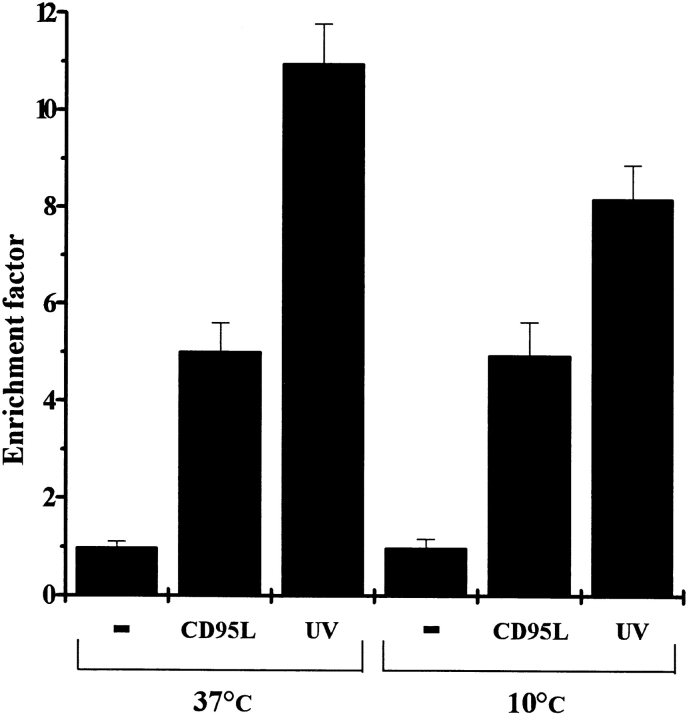
Induction of apoptosis in keratinocytes by UV light is a critical event in photocarcinogenesis. Although p53 is of importance in this process, evidence exists that other pathways play a role as well. Therefore, we studied whether the apoptosis-related surface molecule CD95 (Fas/APO-1) is involved. The human keratinocyte cell line HaCaT expresses CD95 and undergoes apoptosis after treatment with UV light or with the ligand of CD95 (CD95L). Incubation with a neutralizing CD95 antibody completely prevented CD95L-induced apoptosis but not UV-induced apoptosis, initially suggesting that the CD95 pathway may not be involved. However, the protease CPP32, a downstream molecule of the CD95 pathway, was activated in UV-exposed HaCaT cells, and UV-induced apoptosis was blocked by the ICE protease inhibitor zVAD, implying that at least similar downstream events are involved in CD95- and UV-induced apoptosis. Activation of CD95 results in recruitment of the Fas-associated protein with death domain (FADD) that activates ICE proteases. Immunoprecipitation of UV-exposed HaCaT cells revealed that UV light also induces recruitment of FADD to CD95. Since neutralizing anti-CD95 antibodies failed to prevent UV-induced apoptosis, this suggested that UV light directly activates CD95 independently of the ligand CD95L. Confocal laser scanning microscopy showed that UV light induced clustering of CD95 in the same fashion as CD95L. Prevention of UV-induced CD95 clustering by irradiating cells at 10 degrees C was associated with a significantly reduced death rate. Together, these data indicate that UV light directly stimulates CD95 and thereby activates the CD95 pathway to induce apoptosis independently of the natural ligand CD95L. These findings further support the concept that UV light can affect targets at the plasma membrane, thereby even inducing apoptosis.


Figures

Figure 1
Figure 1
Anti-CD95 antibody prevents CD95L- but not UV- induced apoptosis. HaCaT cells (2 × 10⁵/ml) were pretreated with a neutralizing anti-CD95 antibody (anti-CD95) or with an isotype control (iso). 30 min later, cells were either exposed to 300 J/m2 UV light or stimulated with 100 ng/ml recombinant CD95L. For control purposes cells were left untreated (−). 16 h later, apoptosis was examined by determining nucleosomal DNA fragmentation using an apoptosis determination kit. Rate of apoptosis is reflected by the enrichment of nucleosomes in the cytoplasm shown on the y-axis. Data presented show one representative of three independently performed experiments.
Figure 2
Figure 2
Cleavage of CPP32 and PARP by UV light. HaCaT cells were irradiated with 300 J/m2 UV or left untreated. 2, 4, 8, and 16 h after stimulation proteins were extracted, and Western blot analysis was performed using antibodies directed against CPP32 (a) or PARP (b). To monitor equal loading of protein samples, Western blot using an antibody directed against α-tubulin was performed (c).
Figure 3
Figure 3
The ICE inhibitor zVAD inhibits both UV- and CD95L-induced cell death. HaCaT cells were either pretreated with 20 μM zVAD or left untreated. 30 min later, cells were either exposed to 300 J/m2 UV light, stimulated with 100 ng/ml recombinant CD95L, or left untreated (−). 16 h later, apoptosis was examined by determining nucleosomal DNA fragmentation using an apoptosis determination kit. Rate of apoptosis is reflected by the enrichment of nucleosomes in the cytoplasm shown on the y-axis. Data presented show one representative of three independently performed experiments.
Figure 4
Figure 4
UV light induces recruitment of FADD to CD95. HaCaT cells were either stimulated with 100 ng/ml recombinant CD95L, irradiated with 300 J/m2 UV light, or left untreated. 1 and 4 h later, proteins were extracted and immunoprecipitated with an antibody directed against CD95. After blotting to nitrocellulose membranes, Western blot analysis was performed using an antibody directed against FADD (a). To monitor loading of protein samples, the same membranes were reprobed with an anti-CD95 antibody (b).
Figure 5
Figure 5
UV light induces CD95 clustering on HaCaT cells. HaCaT cells were either left untreated (a), stimulated with 100 ng/ml recombinant CD95L (b), irradiated with 300 J/m2 UV light (c), or treated with 10 ng/ml TNF-α (d). 30 min later cells were fixed, incubated with an antibody directed against CD95, incubated with an FITC-conjugated secondary antibody, and subjected to confocal laser scanning microscopy. Bars, 5 μm.
Figure 6
Figure 6
Kinetics of UV- induced CD95 clustering. HaCaT cells were irradiated with 300 J/m2 UV light. Cells were fixed after 0.5 (a), 2 (b), 6 (c), or 10 h (d) of incubation with an antibody directed against CD95. They were then incubated with an FITC-conjugated secondary antibody and subjected to confocal laser scanning microscopy. Bars, 5 μm.
Figure 7
Figure 7
Prevention of CD95 aggregation reduces UV-induced apoptosis. HaCaT cells were kept at 10°C, and 30 min later cells were either exposed to 300 J/m2 UV light, stimulated with 100 ng/ ml recombinant CD95L, or left untreated (−). 10 min after stimulation, cells were incubated at 37°C. 16 h later apoptosis was examined by determining nucleosomal DNA fragmentation using an apoptosis determination kit. For control purposes, cells were kept all the time at 37°C but were otherwise treated in an identical way. Rate of apoptosis is reflected by the enrichment of nucleosomes in the cytoplasm shown on the y-axis. Data presented show one representative of three independently performed experiments.
Figure 8
Figure 8
Low temperature prevents UV-induced clustering of CD95. HaCaT cells were kept at 10°C, and 30 min later cells were exposed to 300 J/m2 UV light (a) or stimulated with 100 ng/ml recombinant CD95L (b). 10 min after stimulation, cells were incubated at 37°C. 2 h later cells were fixed, incubated with an antibody directed against CD95, incubated with an FITC-conjugated secondary antibody, and subjected to confocal laser scanning microscopy. Bars, 5 μm.
Figure 9
Figure 9
UV light induces CD95 clustering on SCL-1 cells. SCL-1 cells were either left untreated (a) or irradiated with 300 J/m2 UV light at 37°C (b) or at 10°C (c). 30 min later, cells were fixed, incubated with an antibody directed against CD95, incubated with an FITC-conjugated secondary antibody, and subjected to confocal laser scanning microscopy. Bars, 5 μm.
Figure 10
Figure 10
Prevention of CD95 aggregation reduces UV-induced apoptosis in SCL-1 cells. SCL-1 cells were kept at 10°C, and 30 min later cells were either exposed to 300 J/m2 UV light, stimulated with 100 ng/ml recombinant CD95L, or left untreated (−). 10 min after stimulation, cells were incubated at 37°C. 16 h later apoptosis was examined by determining nucleosomal DNA fragmentation using an apoptosis determination kit. For control purposes, cells were kept all the time at 37°C but were otherwise treated in an identical way. Rate of apoptosis is reflected by the enrichment of nucleosomes in the cytoplasm shown on the y-axis. Data presented show one representative of three independently performed experiments.

Similar articles

Cited by

References

    1. Alnemri ES, Livingston DJ, Nicholson DW, Salyesen G, Thornberry NA, Wong WW, Yuan J. Human ICE/CED-3 protease nomenclature. Cell. 1996;87:171. - PubMed
    1. Ameisen JC. Programmed cell death (apoptosis) and cell survival regulation: relevance to AIDS and cancer. AIDS. 1994;8:1197–1213. - PubMed
    1. Barr PJ, Tomei LD. Apoptosis and its role in human disease. Biotechnology NY. 1994;12:487–493. - PubMed
    1. Bellgrau D, Gold D, Selawry H, Moore J, Franzusoff A, Duke RC. A role for CD95 ligand in preventing graft rejection. Nature. 1995;377:630–632. - PubMed
    1. Benassi L, Ottani D, Fantini F, Marconi A, Chiodino C, Gianetti A, Pincelli C. 1,25-Dihydroxyvitamin D3, transforming growth factor β1, calcium and ultraviolet B radiation induce apoptosis in cultured human keratinocytes. J Invest Dermatol. 1997;109:276–282. - PubMed

Publication types

MeSH terms