. 1998 Jan 12;140(1):197-210.
doi: 10.1083/jcb.140.1.197.

Direct and regulated interaction of integrin alphaEbeta7 with E-cadherin

Affiliations

Direct and regulated interaction of integrin alphaEbeta7 with E-cadherin

J M Higgins et al. J Cell Biol. .

Abstract

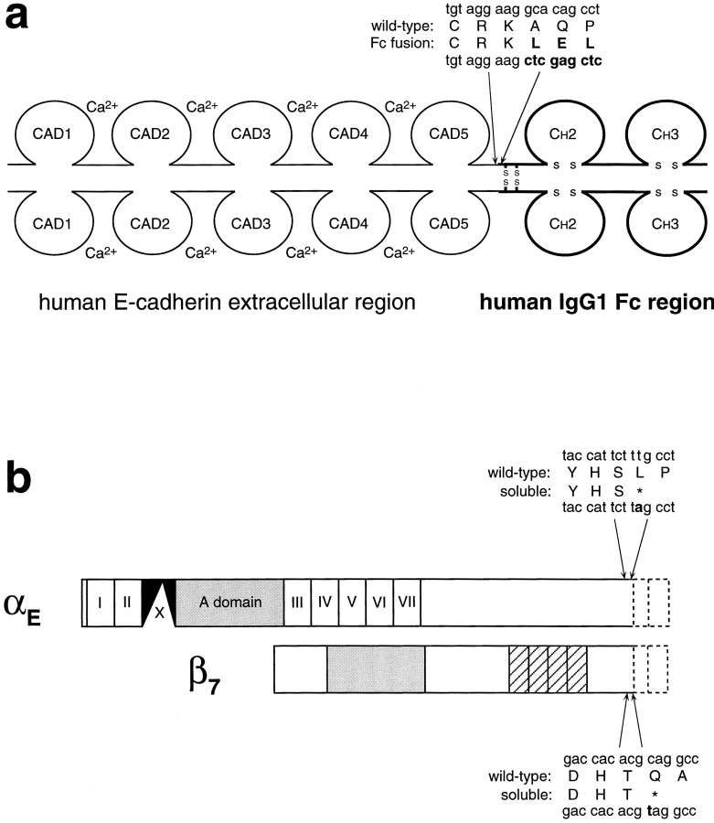
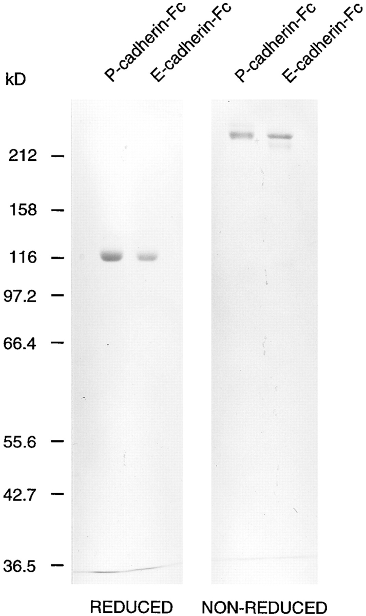
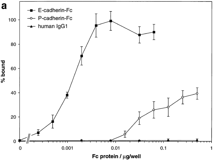
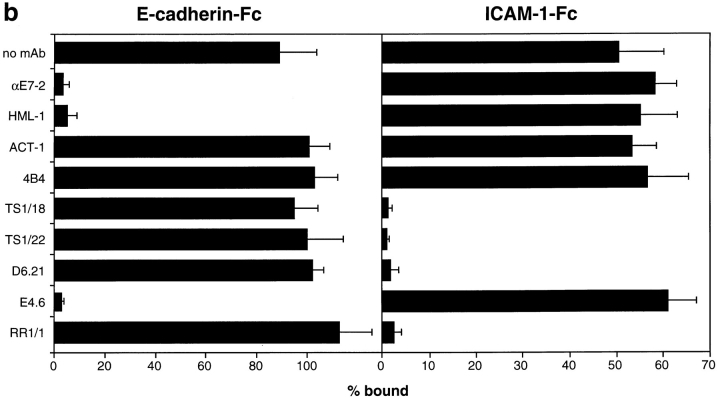
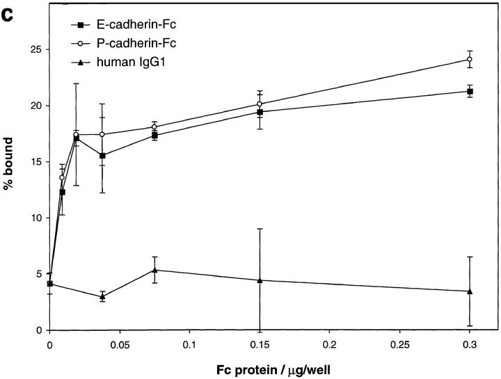
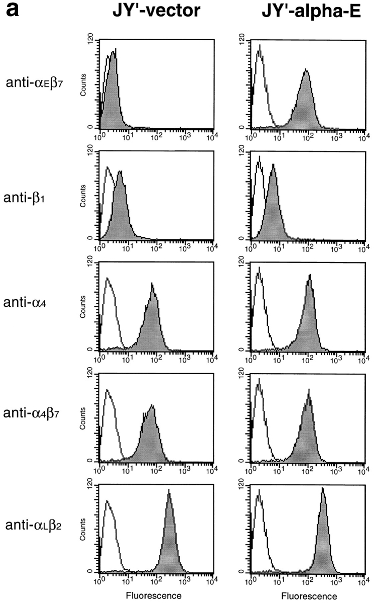
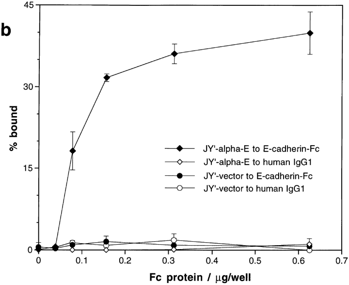
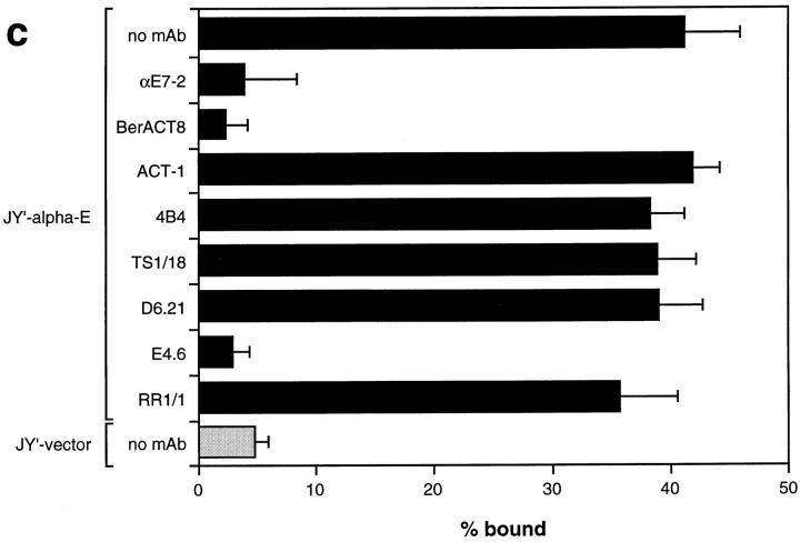
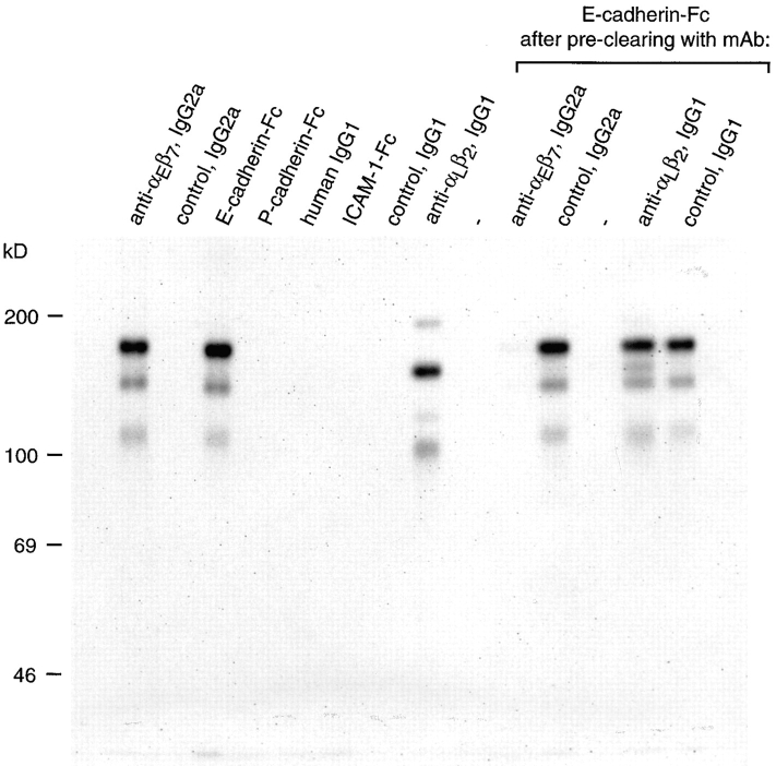
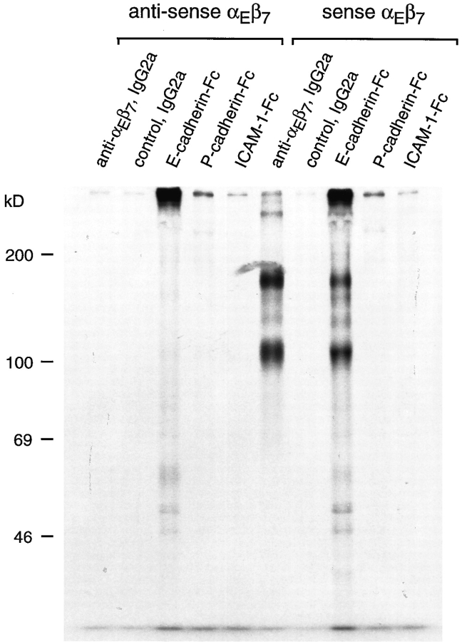
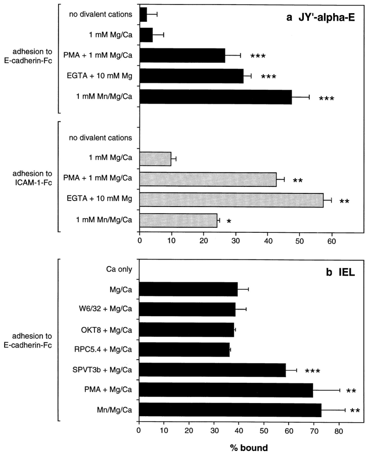
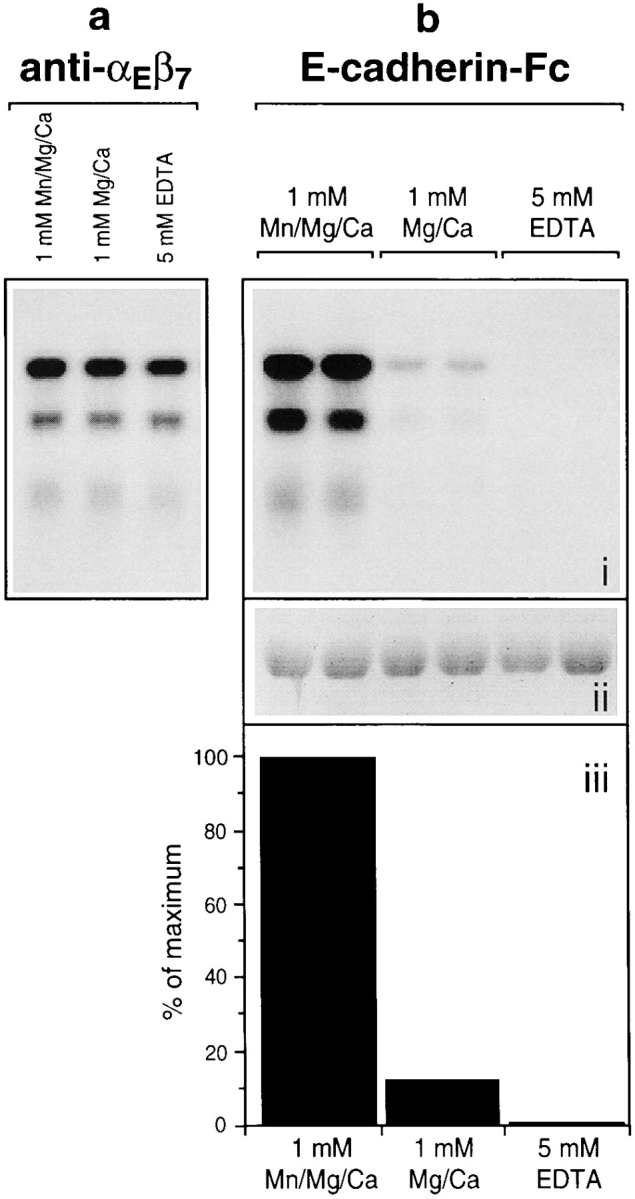
The cadherins are a family of homophilic adhesion molecules that play a vital role in the formation of cellular junctions and in tissue morphogenesis. Members of the integrin family are also involved in cell to cell adhesion, but bind heterophilically to immunoglobulin superfamily molecules such as intracellular adhesion molecule (ICAM)-1, vascular cell adhesion molecule (VCAM)-1, or mucosal addressin cell adhesion molecule (MadCAM)-1. Recently, an interaction between epithelial (E-) cadherin and the mucosal lymphocyte integrin, alphaEbeta7, has been proposed. Here, we demonstrate that a human E-cadherin-Fc fusion protein binds directly to soluble recombinant alphaEbeta7, and to alphaEbeta7 solubilized from intraepithelial T lymphocytes. Furthermore, intraepithelial lymphocytes or transfected JY' cells expressing the alphaEbeta7 integrin adhere strongly to purified E-cadherin-Fc coated on plastic, and the adhesion can be inhibited by antibodies to alphaEbeta7 or E-cadherin. The binding of alphaEbeta7 integrin to cadherins is selective since cell adhesion to P-cadherin-Fc through alphaEbeta7 requires >100-fold more fusion protein than to E-cadherin-Fc. Although the structure of the alphaE-chain is unique among integrins, the avidity of alphaEbeta7 for E-cadherin can be regulated by divalent cations or phorbol myristate acetate. Cross-linking of the T cell receptor complex on intraepithelial lymphocytes increases the avidity of alphaEbeta7 for E-cadherin, and may provide a mechanism for the adherence and activation of lymphocytes within the epithelium in the presence of specific foreign antigen. Thus, despite its dissimilarity to known integrin ligands, the specific molecular interaction demonstrated here indicates that E-cadherin is a direct counter receptor for the alphaEbeta7 integrin.


Figures

Figure 1
Figure 1
Structure of soluble recombinant E-cadherin–Fc and truncated αEβ7. (a) Structure of the human E-cadherin–Fc fusion protein. The sequence of the extracellular juxtamembrane region of wild-type E-cadherin and the alterations resulting from fusion with the human Fc region are shown. Regions corresponding to the Fc portion are shown in bold. The corresponding sequences in P-cadherin, tgc cct gga ccc tgg aaa (encoding CPGPWK), become tgc cct gga ctc gag ctc (encoding CPGLEL) in the P-cadherin–Fc fusion protein. (b) Structure of soluble truncated αEβ7. In the αE chain, the EF hand–like repeats are labeled I to VII, the extra “X” domain is shown in black, and the A domain in grey. In the β7 chain, the β integrin conserved region that may resemble an A domain (Lee et al., 1995) is shown in grey, and the cysteine-rich repeats are hatched. The transmembrane and cytoplasmic regions that were removed from each chain are shown as dotted lines. The change made in the cDNA sequence of each chain to introduce a stop codon immediately before the transmembrane region is shown in bold.
Figure 2
Figure 2
SDS-PAGE of purified recombinant P- and E-cadherin–Fc fusion proteins. Approximately 2 μg of P-cadherin–Fc and 1.5 μg of E-cadherin–Fc protein were subjected to 7.5% SDS-PAGE in reducing and nonreducing conditions and Coomassie blue staining.
Figure 3
Figure 3
Adhesion of cultured intestinal IEL and epithelial cells to E- and P-cadherin–Fc. (a) Dose response of IEL adhesion to E- and P-cadherin–Fc. Serial dilutions of purified E-cadherin– Fc, P-cadherin–Fc, and human IgG1 were immobilized on microtiter plate wells coated with polyclonal goat anti– human IgG antibody and blocked with BSA. The adhesion of cultured IEL was determined in the presence of 1 mM MnCl2, 1 mM MgCl2, 1 mM CaCl2, 0.1% BSA, HBS, pH 7.4, as described in Materials and Methods. Adhesion was determined in two independent experiments, each in triplicate, and the results are expressed as the mean percent bound ± 1 SD (n = 6). (b) Inhibition of adhesion of IEL to E-cadherin–Fc and ICAM-1–Fc by mAbs. Microtiter plate wells were coated directly with 0.06 μg/well of human E-cadherin–Fc or 0.6 μg/well ICAM-1–Fc protein, and blocked with BSA. The adhesion of cultured IEL was determined in the presence of 1 mM MnCl2, 1 mM MgCl2, 1 mM CaCl2, 0.1% BSA, HBS, pH 7.4, and various mAbs. The purified mAbs αE7-2 (anti-αEβ7), D6.21 (anti-αLβ2), 4B4 (anti-β1), ACT-1 (anti-α4β7), E4.6 (anti–E-cadherin), and RR1/1 (anti–ICAM-1) were used at 10 μg/ml. Ascites fluid containing HML-1 (anti-αEβ7), TS1/22 (anti-αLβ2), and TS1/18 (anti-β2) were used at a dilution of 1:50. All antibodies were preincubated with IEL for 10 min on ice before addition to the plate except for RR1/1 and E4.6, which were preincubated in the microtiter plate. All mAbs were mouse IgG1 except HML-1 (mouse IgG2a). The results are expressed as the mean percent bound + 1 SD (n = 5). (c) Dose response of 16E6.A5 cell adhesion to E- and P-cadherin–Fc. Microtiter plates were coated as described for a. Adhesion was determined as described in Materials and Methods and the results are expressed as the mean percent bound ± 1 SD (n = 3). The adhesion of 16E6.A5 cells to E-cadherin–Fc is inhibited by >95% with the anti–E-cadherin mAb HECD-1, but not with the anti–P-cadherin mAb NCC-CAD299. In contrast, the adhesion of 16E6.A5 cells to P-cadherin–Fc is inhibited by 80% with the anti–P-cadherin mAb NCC-CAD299, but not with the anti– E-cadherin mAb HECD-1 (data not shown).
Figure 3
Figure 3
Adhesion of cultured intestinal IEL and epithelial cells to E- and P-cadherin–Fc. (a) Dose response of IEL adhesion to E- and P-cadherin–Fc. Serial dilutions of purified E-cadherin– Fc, P-cadherin–Fc, and human IgG1 were immobilized on microtiter plate wells coated with polyclonal goat anti– human IgG antibody and blocked with BSA. The adhesion of cultured IEL was determined in the presence of 1 mM MnCl2, 1 mM MgCl2, 1 mM CaCl2, 0.1% BSA, HBS, pH 7.4, as described in Materials and Methods. Adhesion was determined in two independent experiments, each in triplicate, and the results are expressed as the mean percent bound ± 1 SD (n = 6). (b) Inhibition of adhesion of IEL to E-cadherin–Fc and ICAM-1–Fc by mAbs. Microtiter plate wells were coated directly with 0.06 μg/well of human E-cadherin–Fc or 0.6 μg/well ICAM-1–Fc protein, and blocked with BSA. The adhesion of cultured IEL was determined in the presence of 1 mM MnCl2, 1 mM MgCl2, 1 mM CaCl2, 0.1% BSA, HBS, pH 7.4, and various mAbs. The purified mAbs αE7-2 (anti-αEβ7), D6.21 (anti-αLβ2), 4B4 (anti-β1), ACT-1 (anti-α4β7), E4.6 (anti–E-cadherin), and RR1/1 (anti–ICAM-1) were used at 10 μg/ml. Ascites fluid containing HML-1 (anti-αEβ7), TS1/22 (anti-αLβ2), and TS1/18 (anti-β2) were used at a dilution of 1:50. All antibodies were preincubated with IEL for 10 min on ice before addition to the plate except for RR1/1 and E4.6, which were preincubated in the microtiter plate. All mAbs were mouse IgG1 except HML-1 (mouse IgG2a). The results are expressed as the mean percent bound + 1 SD (n = 5). (c) Dose response of 16E6.A5 cell adhesion to E- and P-cadherin–Fc. Microtiter plates were coated as described for a. Adhesion was determined as described in Materials and Methods and the results are expressed as the mean percent bound ± 1 SD (n = 3). The adhesion of 16E6.A5 cells to E-cadherin–Fc is inhibited by >95% with the anti–E-cadherin mAb HECD-1, but not with the anti–P-cadherin mAb NCC-CAD299. In contrast, the adhesion of 16E6.A5 cells to P-cadherin–Fc is inhibited by 80% with the anti–P-cadherin mAb NCC-CAD299, but not with the anti– E-cadherin mAb HECD-1 (data not shown).
Figure 4
Figure 4
Analysis of JY′ cells transfected with αE cDNA. (a) Flow cytometric analysis of the cell surface expression of integrins on JY′ cells transfected with pSRα-neo vector alone (JY′-vector) or with pSRα-neo/αE (JY′-αE). The staining with control mAb P3 is shown unshaded. Staining with αE7-2 (anti-αEβ7), 4B4 (anti-β1), B5G10 (anti-α4), ACT-1 (anti-α4β7), and TS1/22 (anti-αLβ2) is shown shaded. All mAbs were mouse IgG1. (b) Adhesion of transfected JY′ cells to E-cadherin–Fc. Microtiter plate wells were coated directly with serial dilutions of E-cadherin–Fc or human IgG1, and blocked with BSA. The adhesion of JY′-vector and JY′-αE cells was determined in the presence of 1 mM MnCl2, 1 mM MgCl2, 1 mM CaCl2, 0.1% BSA, HBS, pH 7.4, as described in Materials and Methods. The results are expressed as the mean percent bound ± 1 SD (n = 3). (c) Inhibition of adhesion of JY′-αE cells to E-cadherin–Fc by mAbs. Microtiter plate wells were coated directly with 0.63 μg/well of E-cadherin–Fc or human IgG1, and blocked with BSA. The adhesion of transfected JY′ cells in the presence of various mAbs was determined as described for IEL in Fig. 3 b. The purified mAbs αE7-2, and BerACT8 (anti-αEβ7), ACT-1 (anti-α4β7), 4B4 (anti-β1), D6.21 (anti-αLβ2), E4.6 (anti–E-cadherin), and RR1/1 (anti–ICAM-1) were used at 10 μg/ml. Ascites fluid containing TS1/18 (anti-β2) was used at a dilution of 1:100. All mAbs were mouse IgG1. The results are expressed as the mean percent bound + 1 SD (n = 4). Adhesion of JY′-vector and JY′-αE cells to human IgG1 in this experiment was <3%.
Figure 4
Figure 4
Analysis of JY′ cells transfected with αE cDNA. (a) Flow cytometric analysis of the cell surface expression of integrins on JY′ cells transfected with pSRα-neo vector alone (JY′-vector) or with pSRα-neo/αE (JY′-αE). The staining with control mAb P3 is shown unshaded. Staining with αE7-2 (anti-αEβ7), 4B4 (anti-β1), B5G10 (anti-α4), ACT-1 (anti-α4β7), and TS1/22 (anti-αLβ2) is shown shaded. All mAbs were mouse IgG1. (b) Adhesion of transfected JY′ cells to E-cadherin–Fc. Microtiter plate wells were coated directly with serial dilutions of E-cadherin–Fc or human IgG1, and blocked with BSA. The adhesion of JY′-vector and JY′-αE cells was determined in the presence of 1 mM MnCl2, 1 mM MgCl2, 1 mM CaCl2, 0.1% BSA, HBS, pH 7.4, as described in Materials and Methods. The results are expressed as the mean percent bound ± 1 SD (n = 3). (c) Inhibition of adhesion of JY′-αE cells to E-cadherin–Fc by mAbs. Microtiter plate wells were coated directly with 0.63 μg/well of E-cadherin–Fc or human IgG1, and blocked with BSA. The adhesion of transfected JY′ cells in the presence of various mAbs was determined as described for IEL in Fig. 3 b. The purified mAbs αE7-2, and BerACT8 (anti-αEβ7), ACT-1 (anti-α4β7), 4B4 (anti-β1), D6.21 (anti-αLβ2), E4.6 (anti–E-cadherin), and RR1/1 (anti–ICAM-1) were used at 10 μg/ml. Ascites fluid containing TS1/18 (anti-β2) was used at a dilution of 1:100. All mAbs were mouse IgG1. The results are expressed as the mean percent bound + 1 SD (n = 4). Adhesion of JY′-vector and JY′-αE cells to human IgG1 in this experiment was <3%.
Figure 5
Figure 5
Binding of αEβ7 from an IEL lysate to E-cadherin–Fc. Cultured intestinal IEL were surface labeled with 125I and solubilized as described in the text. Batches of lysate were subjected to immunoadsorption with mAbs or human IgG1 Fc containing proteins in the presence of 1 mM MnCl2, 1 mM MgCl2, 1 mM CaCl2. Samples were resolved by 7.5% SDS-PAGE in nonreducing conditions, and visualized by autoradiography. The precipitations with αE7-1 and RPC5.4 mAbs (both mouse IgG2a) represent the material obtained using 0.1 μl ascites from 7 × 10⁴ cell equivalents, for TS1/22 and P3 mAbs (both mouse IgG1, with rabbit anti–mouse IgG) using 0.5 μl ascites from 7 × 10⁵ cell equivalents, and for E-cadherin–Fc, P-cadherin–Fc, ICAM-1–Fc and human IgG1 using 5 μg fusion protein from 3 × 10⁶ cell equivalents. For the preclearing experiments, batches of 1.5 × 10⁶ cell equivalents were preabsorbed with the stated antibodies and protein A–Sepharose, before immunoadsorption with 2.5 μg E-cadherin–Fc.
Figure 6
Figure 6
Direct binding of soluble recombinant αEβ7 to E-cadherin–Fc. COS-7 cells transiently transfected with either αE and β7 constructs in the sense or in the antisense orientation were metabolically labeled with 35S amino acids as described in the text. The medium was then made 1 mM with respect to MnCl2 and subjected to immunoadsorption with antibodies or Fc-fusion proteins. Samples were resolved on 7.5% SDS-PAGE in nonreducing conditions, and visualized by autoradiography. The precipitations with αE7-1 and RPC5.4 mAbs (both mouse IgG2a) represent the material obtained using 0.25 ml medium and 0.5 μl ascites, and for E-cadherin–Fc, P-cadherin–Fc, and ICAM-1–Fc, using 1.0 ml medium and 5 μg fusion protein. Aggregated material is present in the both control and test E-cadherin–Fc adsorptions.
Figure 7
Figure 7
Regulation of JY′-αE cell and IEL adhesion to E-cadherin–Fc and ICAM-1–Fc. (a) The adhesion of JY′-αE cells to 0.6 μg/well directly coated E-cadherin–Fc and ICAM-1–Fc under various conditions. EDTA (10 mM) was present during coating and blocking of the microtiter plates, and was washed out with HBS before adding cells. The adhesion buffer was 0.1% BSA/ HBS, pH 7.4, containing divalent cations and 50 ng/ml PMA or 1 mM EGTA where appropriate. The results are expressed as mean percent adhesion + 1 SD (n = 6 for adhesion to E-cadherin–Fc, n = 3 for adhesion to ICAM-1–Fc). The percent adhesion to human IgG1 under the each condition was subtracted from the results (<5% in each case). (b) The adhesion of IEL to 0.1 μg/well E-cadherin–Fc under various conditions. The adhesion buffer was 0.05 mM MgCl2, 1 mM CaCl2, 0.1% BSA, HBS, pH 7.4, unless otherwise stated. Ascites containing antibodies (all mouse IgG2a) to MHC I (W6/32), CD8α (OKT8), nonbinding control (RPC5.4), and CD3 (SPVT3b) were used at a dilution of 1:100 and cross-linked with 10 μg/ml rabbit anti–mouse IgG polyclonal antibody. Where appropriate, 1 mM MnCl2 or 50 ng/ml PMA was also included. The results are expressed as mean percent adhesion + 1 SD (n = 6, except for experiments with OKT8 and RPC5.4 where n = 3). In both a and b, P values calculated by Welch's alternate t test compared with the Mg/Ca alone are as follows: ***P < 0.0001, **P < 0.001, *P < 0.01. In addition, for adhesion to E-cadherin–Fc, P values by the Mann-Whitney test compared with Mg/Ca alone are: ***P = 0.002, **P < 0.005 (see Materials and Methods).
Figure 8
Figure 8
Direct binding of αEβ7 to E-cadherin–Fc is modulated by divalent cations. (a) Immunoadsorption of αEβ7 by an anti– αEβ7 mAb is unaffected by divalent cations. Batches of 125I-labeled IEL lysate were subjected to immunoadsorption with the αE7-1 mAb in the presence of divalent cations or EDTA as shown. Samples were resolved by 7.5% SDS-PAGE in nonreducing conditions, and αEβ7 was visualized by autoradiography. (b) Immunoadsorption of αEβ7 by E-cadherin–Fc is modulated by divalent cations. (i) In duplicate, batches of 125I-labeled IEL lysate were subjected to immunoadsorption with E-cadherin–Fc in the conditions shown in a. (ii) The presence of an equal quantity of E-cadherin–Fc in the immunoadsorbed material was confirmed by 7.5% SDS-PAGE in reducing conditions and Coomassie blue staining. (iii) Quantitation of αEβ7 bound to E-cadherin–Fc in each condition. Phosphorimage quantitation of αEβ7 was based upon the 175- and 135-kD bands corresponding to the αE-chain (Shaw et al., 1994). The averages of duplicates for each band were combined. The precipitations with αE7-1 represent the material obtained using 0.05-μl ascites from 6 × 10⁴ cell equivalents and for E-cadherin–Fc using 5 μg fusion protein from 2 × 10⁶ cell equivalents.

References

    1. Band H, Hochstenbach F, McLean J, Hata S, Krangel MS, Brenner MB. Immunochemical proof that a novel rearranging gene encodes the T cell receptor δ subunit. Science. 1987;238:682–684. - PubMed
    1. Barnstable CJ, Bodmer WF, Brown G, Galfre G, Milstein C, Williams AF, Ziegler A. Production of monoclonal antibodies to group A erythrocytes, HLA and other human cell surface antigens-new tools for genetic analysis. Cell. 1978;14:9–20. - PubMed
    1. Ben-Ze'ev A. Cytoskeletal and adhesion proteins as tumor suppressors. Curr Opin Cell Biol. 1997;9:99–108. - PubMed
    1. Bergelson JM, Hemler ME. Integrin-ligand binding. Do integrins use a ‘MIDAS touch' to grasp an Asp? . Curr Biol. 1995;5:615–617. - PubMed
    1. Blaschuk OW, Sullivan R, David S, Pouliot Y. Identification of a cadherin cell adhesion recognition sequence. Dev Biol. 1990;139:227–229. - PubMed

Publication types

MeSH terms