. 1998 Jan 12;140(1):211-21.
doi: 10.1083/jcb.140.1.211.

Identification of p130Cas as a mediator of focal adhesion kinase-promoted cell migration

Affiliations

Identification of p130Cas as a mediator of focal adhesion kinase-promoted cell migration

L A Cary et al. J Cell Biol. .

Abstract

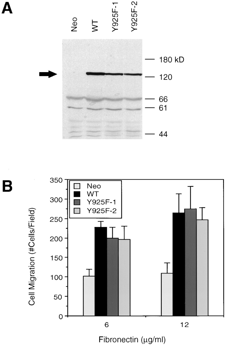
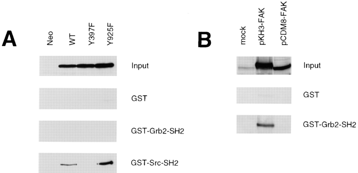
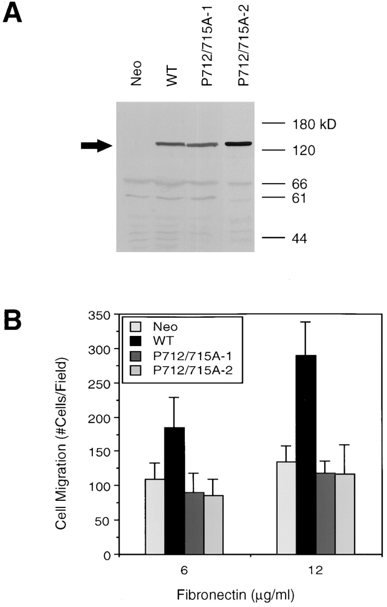
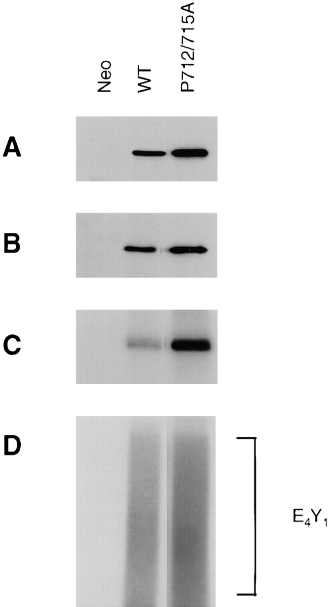
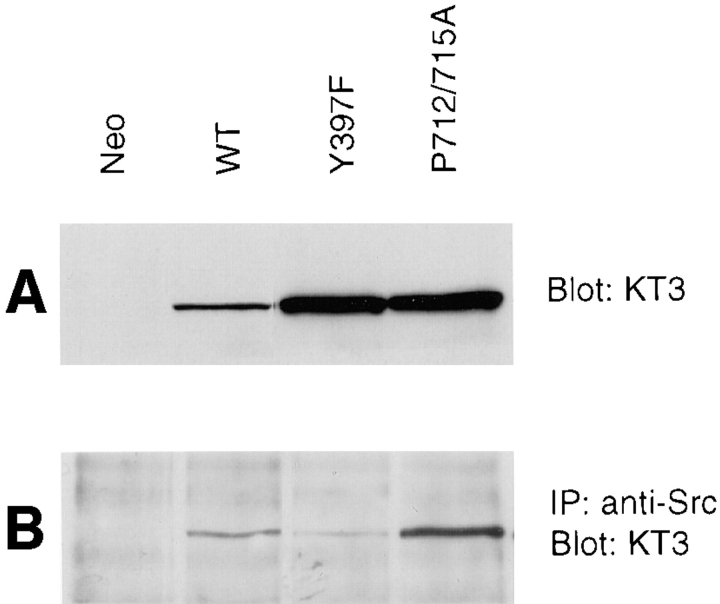
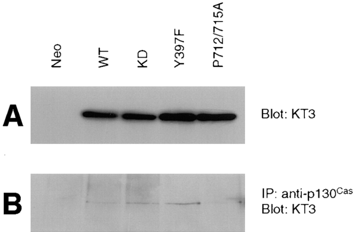
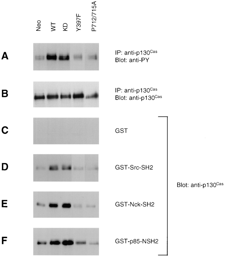
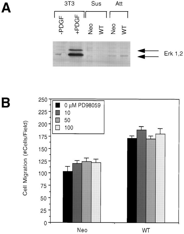
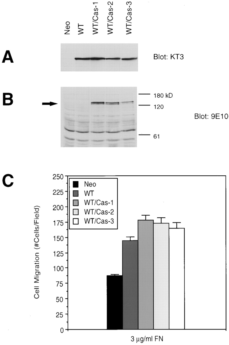
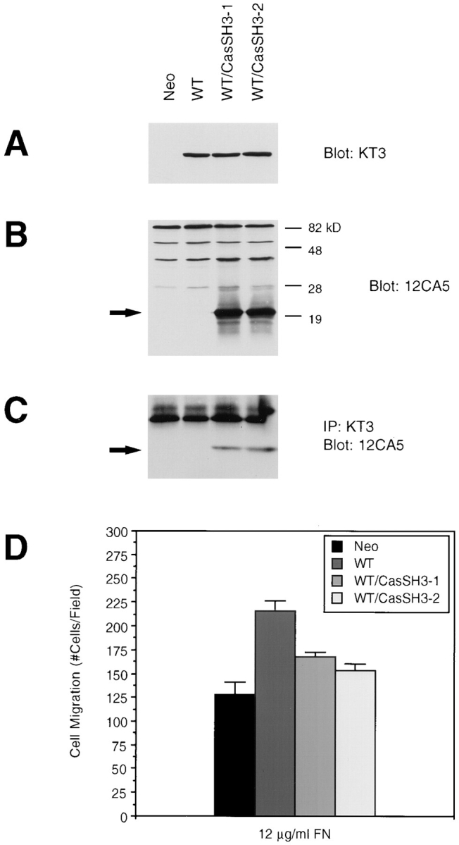
Previously we have demonstrated that focal adhesion kinase (FAK)-promoted migration on fibronectin (FN) by its overexpression in CHO cells is dependent on FAK autophosphorylation at Y397 and subsequent binding of Src to this site. In this report, we have examined the role of FAK association with Grb2 and p130(Cas), two downstream events of the FAK/Src complex that could mediate integrin-stimulated activation of extracellular signal-regulated kinases (Erks). We show that a Y925F FAK mutant was able to promote cell migration as efficiently as FAK and that the transfected FAK demonstrated no detectable association with Grb2 in CHO cells. In contrast, cells expressing a FAK P712/715A mutant demonstrated a level of migration comparable to that of control cells. This mutation did not affect FAK kinase activity, autophosphorylation, or Src association but did significantly reduce p130(Cas) association with FAK. Furthermore, FAK expression in CHO cells increased tyrosine phosphorylation of p130(Cas) and its subsequent binding to several SH2 domains, which depended on both the p130(Cas) binding site and the Src binding site. However, we did not detect increased activation of Erks in cells expressing FAK, and the MEK inhibitor PD98059 did not decrease FAK-promoted cell migration. Finally, we show that coexpression of p130(Cas) further increased cell migration on FN and coexpression of the p130(Cas) SH3 domain alone functioned as a dominant negative mutant and decreased cell migration. Together, these results demonstrate that p130(Cas), but not Grb2, is a mediator of FAK-promoted cell migration and suggest that FAK/ p130(Cas) complex targets downstream pathways other than Erks in mediating FAK-promoted cell migration.


Figures

Figure 1
Figure 1
Grb2 binding to Y925 of FAK does not mediate FAK-promoted cell migration. (A) Cell lysates were generated from two CHO cell clones expressing the Y925F FAK mutant (Y925F-1 and Y925F-2), a clone overexpressing wild-type FAK (WT), and a control clone (Neo), as indicated. Total cell lysate proteins were Western blotted with the mAb KT3, which recognizes the epitope-tagged exogenous FAK, the position of which is indicated by an arrow. Molecular mass positions (kD) are shown on the right. (B) Fibronectin-mediated cell migration of the above clones was analyzed using a NeuroProbe chemotaxis chamber. Cells in the upper chamber were allowed to migrate through a porous membrane toward the lower chamber to which fibronectin had been added at either 6 or 12 μg/ml. Cells that migrated through the membrane were then fixed, stained, and counted using a light microscope. Mean cell counts from at least 10 fields and three experiments are shown. Error bars represent standard deviations. No significant migration was observed in the absence of fibronectin (not shown).
Figure 2
Figure 2
FAK expressed in CHO cells does not bind Grb2 in vitro. (A) Cell lysates were prepared from a CHO control clone (Neo) or clones expressing either wild-type (WT), Y397F, or Y925F mutant FAK, as indicated. Total cell lysate proteins were Western blotted with KT3 to demonstrate levels of FAK expression (Input). The lysates were used for an in vitro binding experiment with the SH2 domains of Grb2 or Src expressed as recombinant GST fusion proteins or with GST alone as a control. The GST fusion proteins were immobilized on glutathione-Sepharose beads with which the above lysates were incubated followed by several washes with lysis buffer. Bound FAK was detected by Western blotting with KT3. (B) Cell lysates were prepared from 293 cells that had been mock transfected or transiently transfected with either pKH3-FAK or pCDM8-FAK, as indicated. Total cell lysate proteins were Western blotted with anti-FAK to demonstrate levels of FAK expression (Input). The lysates were used for an in vitro binding assay with either GST-Grb2-SH2 or GST, as above, and bound FAK was detected by Western blotting with anti-FAK.
Figure 3
Figure 3
The proline-rich p130Cas binding site on FAK is necessary for FAK-promoted cell migration. (A) Cell lysates were generated from a control clone (Neo), a clone expressing wild-type FAK (WT), or two independent clones expressing the P712/715A FAK mutant (P712/715A-1 and P712/715A-2), as indicated. Total cell lysate proteins were Western blotted with KT3 to demonstrate comparable levels of FAK overexpression, the position of which is indicated on the left by an arrow. Molecular mass positions (kD) are shown on the right. (B) Fibronectin-mediated cell migration of the above clones was analyzed using a NeuroProbe chemotaxis chamber as in Fig. 1. Mean cell counts from at least nine fields and two experiments are shown. Error bars represent standard deviations. No significant migration was observed in the absence of FN (not shown).
Figure 4
Figure 4
The P712/715A FAK mutant demonstrates efficient kinase activity and tyrosine phosphorylation. Cell lysates were generated from a control clone (Neo) or clones expressing either wild-type (WT) or P712/715A FAK, as indicated. The exogenous FAK was immunoprecipitated using KT3, and the immune complexes were Western blotted with either KT3 (A) or the antiphosphotyrosine mAb PY20 (B). Alternatively, the immune complexes were subjected to in vitro kinase assays using [γ-32P]ATP in the absence (C) or presence (D) of poly(Glu,Tyr) (E4Y1) as an exogenous substrate, the position of which is indicated on the right.
Figure 5
Figure 5
A mutation of the p130Cas binding site on FAK does not affect its association with Src. Cell lysates were prepared from a control clone (Neo) or from clones expressing either wild-type (WT), Y397F, or P712/715A FAK, as indicated. Total cell lysate proteins were Western blotted with KT3 to demonstrate relative levels of exogenous FAK expression (A). Alternatively, Src was immunoprecipitated from the lysates using the anti-Src mAb 2-17, and the immune complexes were Western blotted with KT3 to demonstrate Src-associated FAK (B).
Figure 6
Figure 6
The P712/715A FAK mutant demonstrates reduced association with p130Cas. Cell lysates from a control clone (Neo), a clone expressing wild-type FAK (WT), or clones expressing the indicated FAK mutants (kinase-defective [KD], Y397F, or P712/715A) were prepared using modified RIPA lysis buffer. Total cell lysate proteins were Western blotted with KT3 to demonstrate relative levels of exogenous FAK expression (A). The lysates were used for immunoprecipitations using anti-p130Cas, followed by Western blotting with KT3 to demonstrate p130Cas-associated FAK (B).
Figure 7
Figure 7
FAK-promoted tyrosine phosphorylation of p130Cas and its subsequent binding to SH2 domain–containing proteins may mediate FAK-promoted cell migration. Cells overexpressing FAK (wild-type [WT] or the mutants kinase-defective [KD], Y397F, or P712/715A) or a control clone (Neo) were treated with sodium vanadate and lysed. Lysates were used for immunoprecipitations with anti-p130Cas, and the immune complexes were Western blotted with PY20 (A) or anti-p130Cas (B). The SH2 domains of Src (D), Nck (E), or the p85 subunit of phosphatidylinositol 3-kinase (F) were expressed as recombinant bacterial GST fusion proteins, or GST alone (C) was expressed as a control. The GST fusion proteins were immobilized on glutathione-Sepharose beads, with which the above cell lysates were incubated followed by several washes with lysis buffer. Bound p130Cas was detected by Western blotting with anti-p130Cas.
Figure 8
Figure 8
The Erk signaling pathway does not mediate FAK-promoted cell migration. (A) Cells overexpressing wild-type FAK (WT) or control cells (Neo) were lysed as either a monolayer of attached cells (Att) or in suspension (Sus). As a control for Erk activation, NIH 3T3 cells were serum-starved and treated with or without PDGF (25 ng/ml for 10 min) followed by lysis. Total cell lysate proteins were Western blotted with anti–phospho-MAPK antibody. Arrows indicate the position of activated Erk1 and Erk2. (B) WT or Neo cells were pretreated for 1 h with the indicated concentrations of the MEK inhibitor PD98059, and their migration on 12 μg/ml FN was then analyzed using a chemotaxis chamber. Mean cell counts from at least five fields and two experiments are shown. Error bars represent standard errors.
Figure 9
Figure 9
Expression of p130Cas enhances FAK-promoted cell migration. Cell lysates were prepared from a clone expressing FAK (WT), three clones expressing both FAK and wild-type p130Cas (WT/Cas-1, 2, and 3), or a control clone (Neo), as indicated. Total cell lysate proteins were Western blotted with mAb KT3 to demonstrate exogenous FAK expression (A) or mAb 9E10 to demonstrate exogenous p130Cas expression, which is indicated by an arrow (B). Molecular mass positions (kD) are shown on the right (B). Migration of the above clones was analyzed using a chemotaxis chamber and 3 μg/ml FN as an attractant (C). Mean cell counts from at least 20 fields and three experiments are shown. Error bars represent standard errors.
Figure 10
Figure 10
Expression of the p130Cas SH3 domain and its association with FAK inhibits FAK-promoted cell migration. Cell lysates were prepared from a clone expressing FAK (WT), two clones expressing both FAK and CasSH3 (WT/CasSH3-1 and 2), or a control clone (Neo), as indicated. Total cell lysate proteins were Western blotted with the mAb KT3 to demonstrate exogenous FAK expression (A) or mAb 12CA5 to demonstrate expression of the SH3 domain of p130Cas (B). Lysates were immunoprecipitated with KT3 and Western blotted with 12CA5 to show association of exogenous FAK with CasSH3 (C). The position of CasSH3 is indicated by arrows (B and C). Molecular mass positions (kD) are shown on the right (B). Migration of the above clones was analyzed using a chemotaxis chamber and 12 μg/ml FN as an attractant (D). Mean cell counts from at least 21 fields and three experiments are shown. Error bars represent standard errors.

References

    1. Alessi DR, Cuenda A, Cohen P, Dudley DT, Saltiel AR. PD 098059 is a specific inhibitor of the activation of mitogen-activated protein kinase kinase in vitro and in vivo. J Biol Chem. 1995;270:27489–27494. - PubMed
    1. Cary L, Chang J, Guan J-L. Stimulation of cell migration by overexpression of focal adhesion kinase and its association with Src and Fyn. J Cell Sci. 1996;109:1787–1794. - PubMed
    1. Chan P-Y, Kanner S B, Whitney G, Aruffo A. A transmembrane-anchored chimeric focal adhesion kinase is constitutively activated and phosphorylated at tyrosine residues identical to pp125FAK . J Biol Chem. 1994;269:20567–20574. - PubMed
    1. Chen H-C, Guan J-L. Association of focal adhesion kinase with its potential substrate phosphatidylinositol 3-kinase. Proc Natl Acad Sci USA. 1994;91:10148–10152. - PMC - PubMed
    1. Chen H-C, Appeddu PA, Parsons JT, Hildebrand JD, Schaller MD, Guan J-L. Interaction of focal adhesion kinase with cytoskeletal protein talin. J Biol Chem. 1995;270:16995–16999. - PubMed

Publication types

MeSH terms