. 1997 Dec 18;241(2):297-304.
doi: 10.1006/bbrc.1997.7705.

The A3 adenosine receptor mediates cell spreading, reorganization of actin cytoskeleton, and distribution of Bcl-XL: studies in human astroglioma cells

Affiliations

The A3 adenosine receptor mediates cell spreading, reorganization of actin cytoskeleton, and distribution of Bcl-XL: studies in human astroglioma cells

M P Abbracchio et al. Biochem Biophys Res Commun. .

Abstract

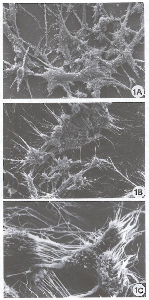
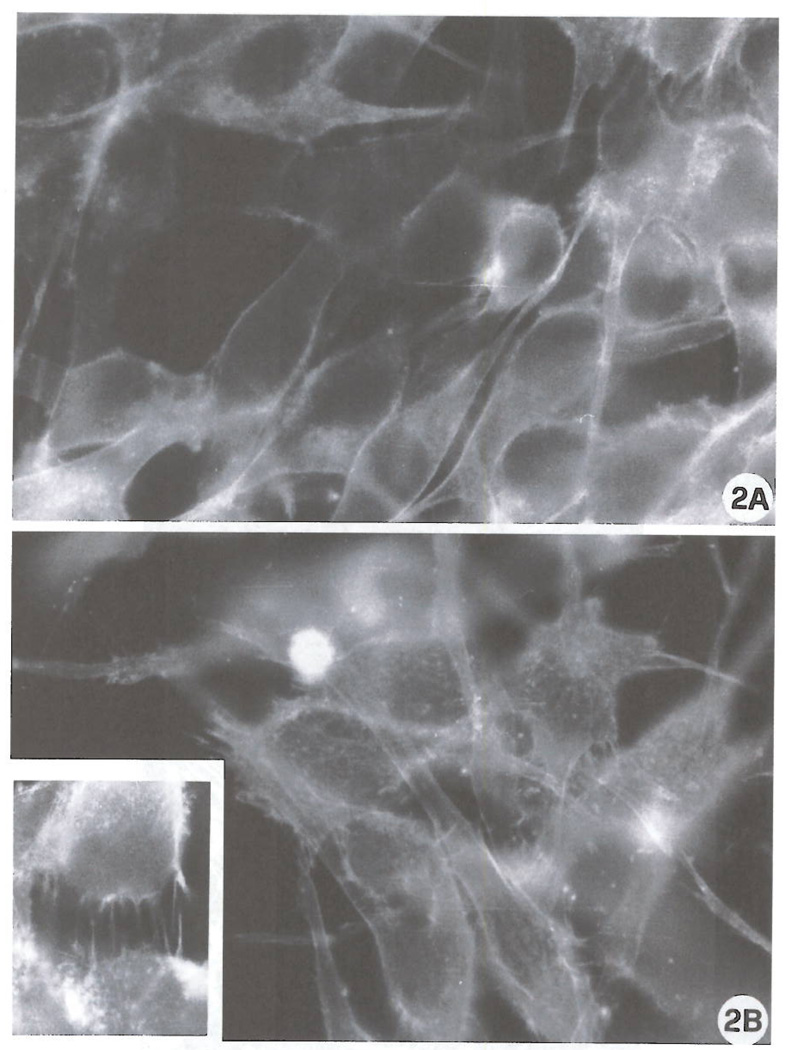
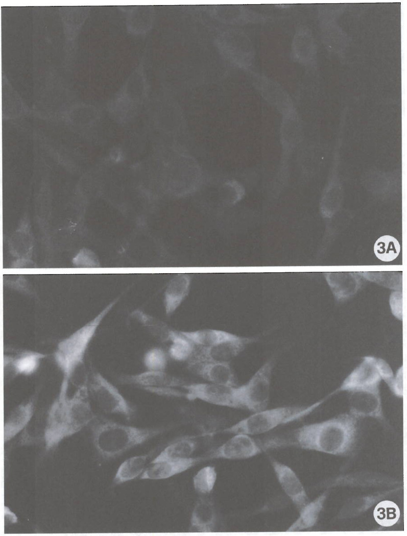
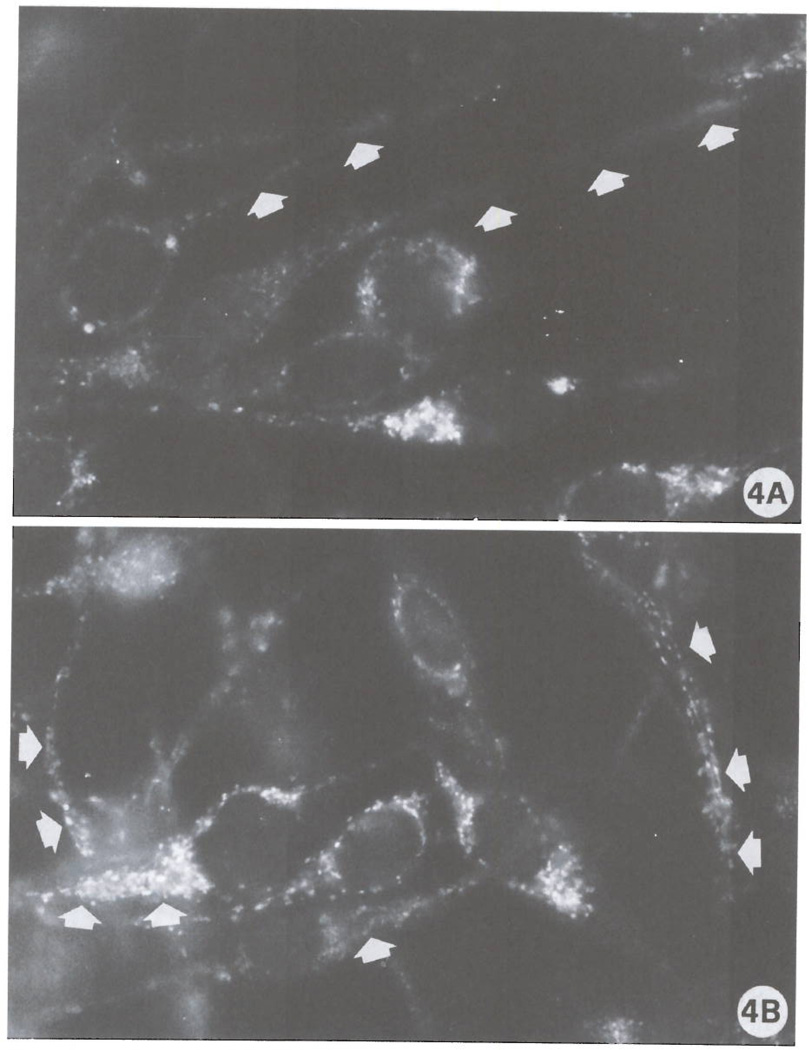
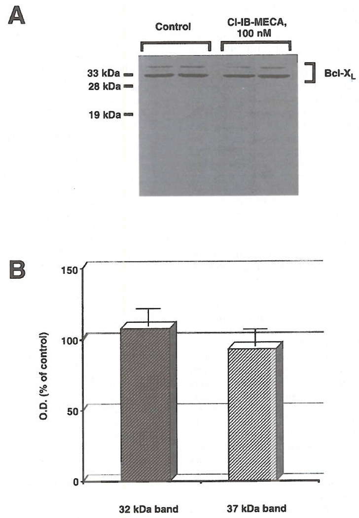
The pathophysiological role of the adenosine A3 receptor in the central nervous system is largely unknown. We have investigated the effects of the selective A3 receptor agonist 2-chloro-N6-(3-iodobenzyl)-adenosine, Cl-IB-MECA, in cells of the astroglial lineage (human astrocytoma ADF cells). A marked reorganization of the cytoskeleton, with appearance of stress fibers and numerous cell protrusions, was found following exposure of cells to low (nM) concentrations of Cl-IB-MECA. These "trophic" effects were accompanied by induction of the expression of Rho, a small GTP-binding protein, which was virtually absent in control cells, and by changes of the intracellular distribution of the antiapoptotic protein Bcl-XL, that, in agonist-exposed cells, became specifically associated to cell protrusions. This is the first demonstration that the intracellular organization of Bcl-XL can be modulated by the activation of a G-protein-coupled membrane receptor, such as the A3 adenosine receptor. Moreover, modulation of the astrocytic cytoskeleton by adenosine may have intriguing implications in both nervous system development and in the response of the brain to trauma and ischemia.


Figures

FIG 1
FIG 1
Scanning election micrographs of ADF control cells (A) and ADF cells treated with 100 nM Cl-IB-MECA for 72 h (B, C). Control cells show a typical bipolar shape with a relatively low number of cell protrusions (A). Exposure to Cl-IB-MECA induces marked morphological changes represented by a dramatic increase of both the number and length of cellular processes (B, C). (Original magnification: A, B 1000X; C 3300X).
FIG 2
FIG 2
F-actin analysis. Stress fibers, which were hardly detectable in control cultures (Fig. 2A), are markedly increased after exposure to 100 nM Cl-IBMECA for 72 h (Fig. 2B). Note elongated processes (Fig. 2B, inset), (Original magnification: A, B, inset 3000X).
FIG 3
FIG 3
The expression of Rho in control cells (A) and in cultures exposed to 100 nM Cl-IB-MECA for 72 h (B). Note the marked positivity in the cell body and cell protrusions in agonist-exposed cells. (Original magnification: A, B 1500X).
FIG 4
FIG 4
Intracellular distribution of Bcl-xL in ADF control cells (A) and in ADF cells exposed to Cl-IB-MECA for 72 h (B). Arrows in (A) indicate cell protrusions expressing low levels of Bcl-xL. By contrast, in treated cultures, positivity to the anti-Bcl-xL antibody is mainly found as very bright dots particularly abundant in cell protrusions (arrows). (Original magnifications: A, B, 3000X).
FIG 5
FIG 5
Immunoblot analysis of Bcl-x in control ADF cells and in ceils exposed for 48 h to 100 nM Cl-IB-MECA. Bcl-xL is detected as a doublet of proteins with an apparent molecular mass of 32 and 37 kDa. In B, immunoblot analysis from 4 independent experiments run in triplicate and subjected to densitometric analysis of autoradiograms; data (optical density, O.D., in arbitrary units) are expressed as % of control.

References

    1. Fredholm BB, Abbracchio MP, Burnstock G, Daly JW, Harden TK, Jacobson KA, Leff P, Williams M. Pharmacol. Rev. 1994;46:143–156. - PMC - PubMed
    1. Rudolphi KA, Schubert P. In: Novel Therapies for CNS Injuries: Rationales and Results. Peterson PL, Phillis JW, editors. 1996. pp. 327–346.
    1. Jacobson KA, Kim HO, Siddiqi SM, Olah ME, Stiles GL, von Lubitz DKJE. Drugs of the Future. 1995;20:689–699. - PMC - PubMed
    1. Gallo-Rodriguez C, Ji X-D, Melman N, Siegman BD, Sanders LH, Orlina J, Pu Q, Olah ME, van Galen PJM, Stiles GL, Jacobson KA. J. Med. Chem. 1994;37:636–646. - PMC - PubMed
    1. Kim HO, Ji KD, Siddiqi SM, Olah ME, Stiles GL, Jacobson KA. J. Med. Chem. 1994;37:3614–3621. - PMC - PubMed

Publication types