. 1998 Jan 19;187(2):197-204.
doi: 10.1084/jem.187.2.197.

Circulating activated platelets reconstitute lymphocyte homing and immunity in L-selectin-deficient mice

Affiliations

Circulating activated platelets reconstitute lymphocyte homing and immunity in L-selectin-deficient mice

T G Diacovo et al. J Exp Med. .

Abstract

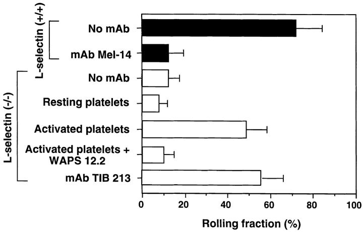
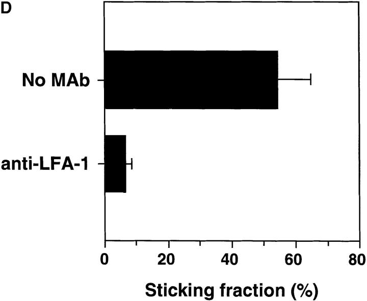
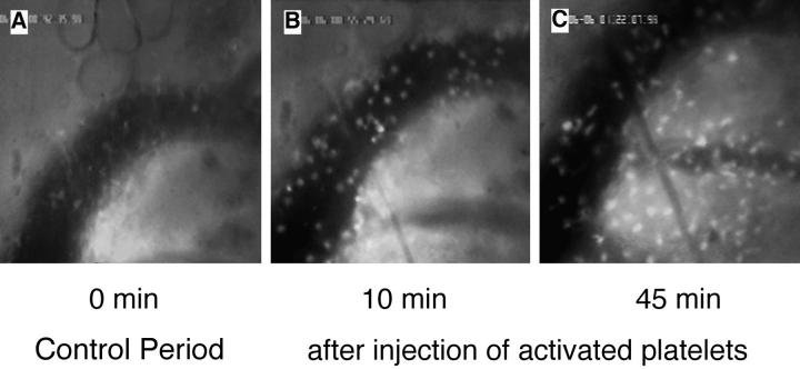
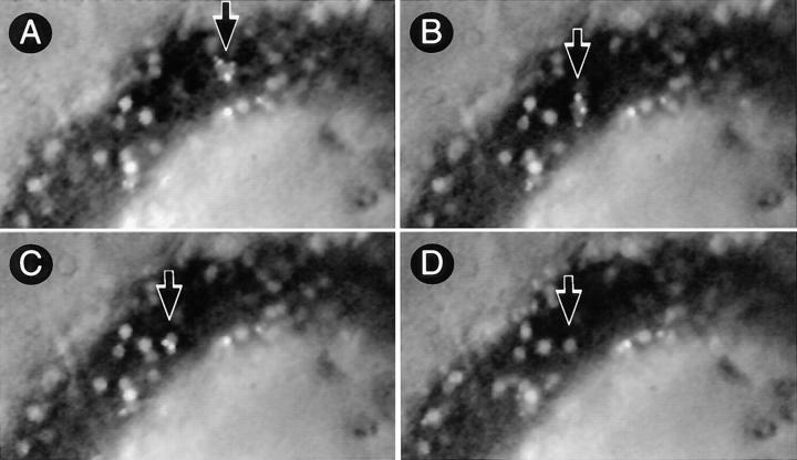
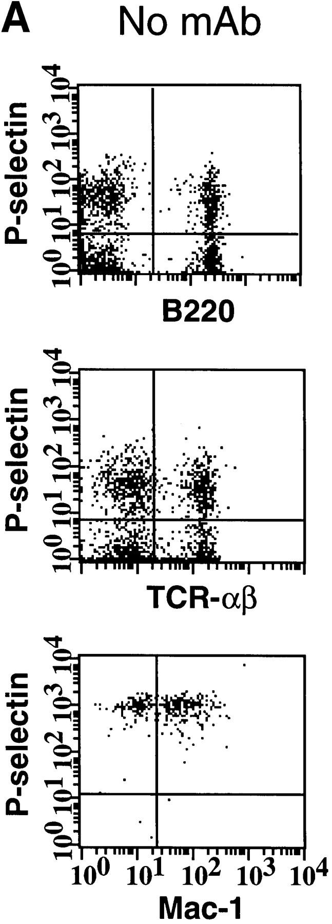
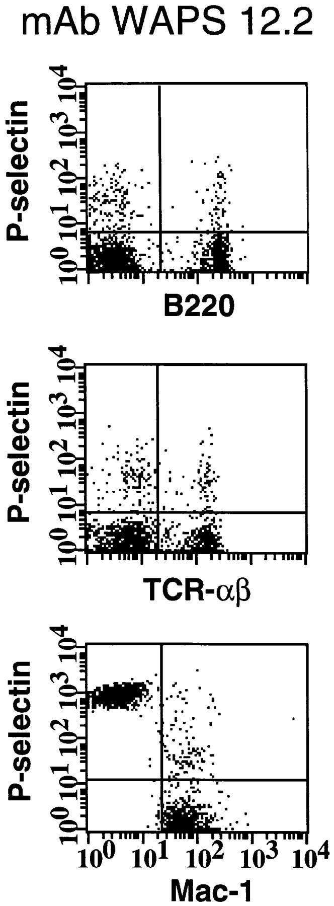
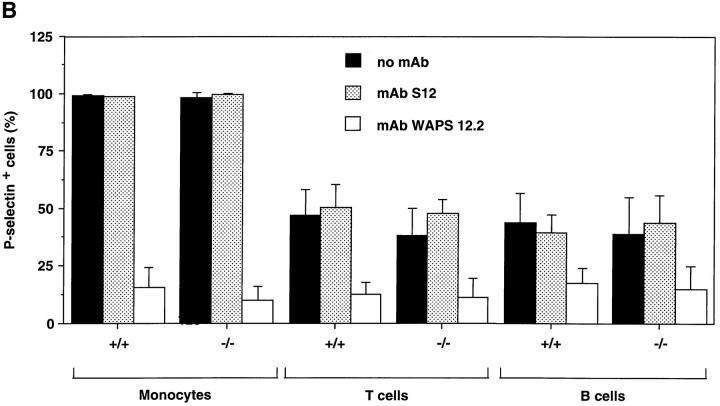
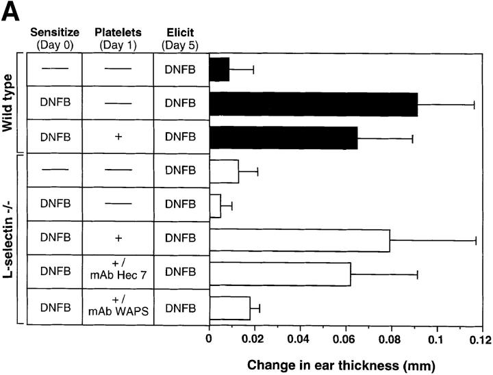
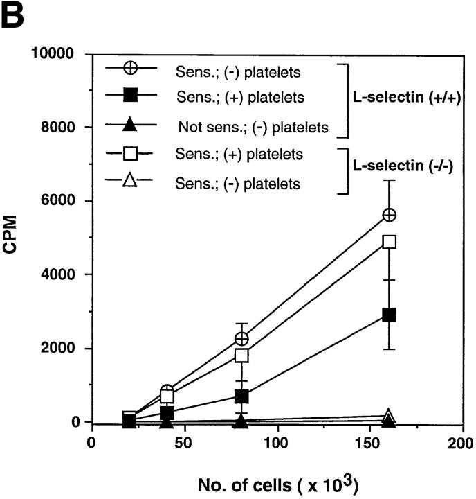
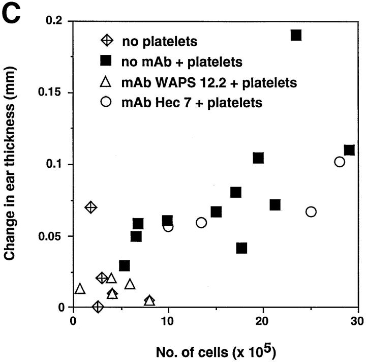
Peripheral lymph nodes (PLN) are critical for immunologic memory formation in response to antigens that penetrate the skin. Blood-borne lymphocytes first encounter such antigens after they home to PLN through a multi-step adhesion process that is normally initiated by L-selectin (CD62L) in high endothelial venules (HEV). Since naive T cells can not enter PLN normally in L-selectin-deficient mice, a delayed type hypersensitivity response to cutaneously applied antigen cannot be mounted. In this study, we report that the administration of activated platelets into the systemic circulation of L-selectin knockout mice restores lymphocyte trafficking to PLN, and reconstitutes T cell-mediated immunity in response to a cutaneous antigen. These effects required platelet-expressed P-selectin that allows activated platelets to transiently form a bridge between lymphocytes and HEV, thereby enabling lymphocytes to undergo subsequent beta2 integrin-dependent firm adhesion. These profound effects of platelet-mediated cell-cell interactions on lymphocyte trafficking and formation of immunologic memory may impact on a variety of autoimmune and inflammatory conditions.


Figures

Figure 1
Figure 1
Effect of circulating, activated platelets on WBCs rolling in PLN–HEV of mice deficient in L-selectin. WBCs (mononuclear cells and granulocytes) were fluorescently labeled in situ with rhodamine 6G, visualized in HEVs of PLN of wild-type (closed bars) and L-selectin–deficient (open bars) mice by fluorescence intravital microscopy, and rolling fractions quantitated as previously described (10). The effect of mAbs and platelet injections on the WBC rolling fraction was determined in identical fields of view. The L-selectin blocking mAb Mel-14 was tested in 10 venules of 4 wild-type animals. The ability of platelets to reconstitute rolling in mutant animals was examined by injecting TRAP-activated or resting, human platelets (12 venules in 5 animals). The ability of the human P-selectin mAb WAPS 12.2 and murine LFA-1 mAb TIB 213 to inhibit WBC rolling was also evaluated (four venules in three animals each). Data are shown as mean ± SD.
Figure 2
Figure 2
Activated human platelets support WBCs homing to subiliac PLN in L-selectin–deficient mice. WBCs were fluorescently labeled in situ using rhodamine 6G and recorded during their passage through single HEV. (A) Digitized intravital fluorescence micrograph of a typical HEV in an L-selectin–deficient mouse. Relatively few blood-borne cells (arrows) interacted with the vascular wall over a 45-min control period (0 min). (B) Intraarterial injection of TRAP-stimulated human platelets over a 10-min period resulted in rolling and accumulation of sticking endogenous WBCs (10 min). (C) A significant number of stuck cells subsequently transmigrated into the node (45 min). (D) LFA-1 (CD11a) receptor blockade abrogates firm adhesion of L-selectin–deficient WBCs delivered to HEV by platelets. The percentage of rolling cells that became firmly adherent for a mininum of 30 s (sticking fraction) was determined in the presence or absence of LFA-1 mAb TIB 213. The data shown represent the mean ± SD of seven venules in three animals.
Figure 2
Figure 2
Activated human platelets support WBCs homing to subiliac PLN in L-selectin–deficient mice. WBCs were fluorescently labeled in situ using rhodamine 6G and recorded during their passage through single HEV. (A) Digitized intravital fluorescence micrograph of a typical HEV in an L-selectin–deficient mouse. Relatively few blood-borne cells (arrows) interacted with the vascular wall over a 45-min control period (0 min). (B) Intraarterial injection of TRAP-stimulated human platelets over a 10-min period resulted in rolling and accumulation of sticking endogenous WBCs (10 min). (C) A significant number of stuck cells subsequently transmigrated into the node (45 min). (D) LFA-1 (CD11a) receptor blockade abrogates firm adhesion of L-selectin–deficient WBCs delivered to HEV by platelets. The percentage of rolling cells that became firmly adherent for a mininum of 30 s (sticking fraction) was determined in the presence or absence of LFA-1 mAb TIB 213. The data shown represent the mean ± SD of seven venules in three animals.
Figure 3
Figure 3
Lymphocyte delivery to PLN of L-selectin–deficient mice does not lead to platelet accumulation in HEV. The micrographs show a segment of the same HEV as in Fig. 2 (blood flow from right to left) 10 min after injection of activated human platelets. (A) A rolling aggregate of bright 2′7′-bis-(2-carboxyethyl)-5(and 6) carboxyfluorescein– labeled platelets with a fainter rhodamine 6G labeled WBC in their midst can be seen (arrow). (B) 2 s later, the platelet–WBC aggregate has arrested firmly. Labeled platelets are initially distributed randomly on the surface of the stuck cell. (C) Within 5 s after the WBC had become stuck, platelets roll slowly downstream across the body of the adherent cell and eventually detach. (D) 30 s later, all platelets have detached and returned to the blood stream, whereas the WBC remains stuck at the HEV surface.
Figure 4
Figure 4
Activated human platelets bind to murine lymphocytes in suspension. Freshly isolated murine PBMCs were stained with FITC-conjugated antibodies to TCR-α/β, B220, and Mac-1 to identify T cells, B cells, and monocytes, respectively. Activated human platelets were stained with PE-conjugated nonblocking antibody to P-selectin and then mixed with labeled PBMCs. (A) Identification of PBMC subsets from L-selectin–deficient mice capable of aggregating with platelets as determined by two-color flow cytometry. The quadrant markers were drawn based on appropriate negative controls after gating separately for lymphocytes and monocytes. Double positive cells (upper right quadrant) represent the members of each subset capable of platelet binding. Events in the monocyte gate that show positive staining for P-selectin, but not Mac-1, are platelet aggregates (upper left quadrant). P-selectin–dependent adhesion was evaluated by treating platelets with mAb WAPS 12.2 before and during incubation with PBMCs. Similar results were obtained in wild-type mice. (B) Percentage of monocytes, T cells, and B cells of wild-type (+/+) and L-selectin-deficient (−/−) donor mice that interacted with activated human platelets. The effect of P-selectin blocking mAb WAPS 12.2 (open bar) and nonblocking mAb S12 (dotted bars) was evaluated as described in Materials and Methods. Bars reflect mean ± SD of four independent experiments.
Figure 4
Figure 4
Activated human platelets bind to murine lymphocytes in suspension. Freshly isolated murine PBMCs were stained with FITC-conjugated antibodies to TCR-α/β, B220, and Mac-1 to identify T cells, B cells, and monocytes, respectively. Activated human platelets were stained with PE-conjugated nonblocking antibody to P-selectin and then mixed with labeled PBMCs. (A) Identification of PBMC subsets from L-selectin–deficient mice capable of aggregating with platelets as determined by two-color flow cytometry. The quadrant markers were drawn based on appropriate negative controls after gating separately for lymphocytes and monocytes. Double positive cells (upper right quadrant) represent the members of each subset capable of platelet binding. Events in the monocyte gate that show positive staining for P-selectin, but not Mac-1, are platelet aggregates (upper left quadrant). P-selectin–dependent adhesion was evaluated by treating platelets with mAb WAPS 12.2 before and during incubation with PBMCs. Similar results were obtained in wild-type mice. (B) Percentage of monocytes, T cells, and B cells of wild-type (+/+) and L-selectin-deficient (−/−) donor mice that interacted with activated human platelets. The effect of P-selectin blocking mAb WAPS 12.2 (open bar) and nonblocking mAb S12 (dotted bars) was evaluated as described in Materials and Methods. Bars reflect mean ± SD of four independent experiments.
Figure 5
Figure 5
Circulating, activated platelets reconstitute CHS response in L-selectin–deficient mice. (A) Mice were sensitized to DNFB on day 0. CHS was elicited at day 5, and the subsequent ear swelling response was measured 24 h later. 18 h after sensitization, some animals were injected intravenously with TRAP-activated human platelets (3 × 10⁸ in 3 ml over 6 h). Some L-selectin−/− animals were also treated with either P-selectin mAb WAPS 12.2 or CD31 mAb Hec 7. There was no significant difference between the response of L-selectin–deficient mice and wild-type mice which were elicited without earlier sensitization (P >0.05). Ear swelling in sensitized wild-type animals with or without platelet infusion was highly significant (P <0.001 versus elicit only). In sensitized L-selectin−/− mice, elicitation alone had no effect, whereas mutant mice that also received activated platelets displayed a dramatic swelling response (P <0.001) that was not different from that in sensitized wild-type animals with or without platelet treatment (P >0.05 versus both). MAb Hec 7 had no significant effect (P >0.05 versus sensitized L-selectin−/− receiving activated platelets only), whereas coinjection of platelets and MAb WAPS12.2 abolished the delayed type hypersensitivity effect (P >0.05 versus both L-selectin−/− groups without platelet infusion; P <0.001 versus sensitized, platelet-treated L-selectin−/− animals; P <0.01 versus sensitized, platelet plus MAb Hec 7–treated L-selectin−/− animals). Data shown are mean ± SD from 4 to 11 mice in each group (5 measurements per ear before and after elicitation). (B) Antigen–specific T cells are present in PLN of L-selectin–deficient mice only after treatment with activated platelets. Cells from mice sensitized 5 d earlier with DNFB were cultured for 36 h in the presence of DNBS after which the cultures were pulsed with [3H]thymidine for 18 h. Each point represents the mean ± SD of three separate mouse groups, with each group consisting of pooled PLN from two mice. Each group was done in triplicate. Background proliferation was subtracted as previously described (7). (C) Magnitude of CHS response in L-selectin–deficient mice receiving activated platelets correlates with the number of resident lymphocytes in PLN. After eliciting a CHS response, six PLN per mouse (subiliac, axillary, and cervical) were harvested and the number of resident cells was counted and correlated to the extent of the ear swelling. PLN were obtained from mice which had received no platelets (n = 5), activated platelets (n = 11), activated platelets pretreated with control mAb Hec 7 (n = 4), or activated platelets pretreated with mAb WAPS 12.2 (n = 5).
Figure 5
Figure 5
Circulating, activated platelets reconstitute CHS response in L-selectin–deficient mice. (A) Mice were sensitized to DNFB on day 0. CHS was elicited at day 5, and the subsequent ear swelling response was measured 24 h later. 18 h after sensitization, some animals were injected intravenously with TRAP-activated human platelets (3 × 10⁸ in 3 ml over 6 h). Some L-selectin−/− animals were also treated with either P-selectin mAb WAPS 12.2 or CD31 mAb Hec 7. There was no significant difference between the response of L-selectin–deficient mice and wild-type mice which were elicited without earlier sensitization (P >0.05). Ear swelling in sensitized wild-type animals with or without platelet infusion was highly significant (P <0.001 versus elicit only). In sensitized L-selectin−/− mice, elicitation alone had no effect, whereas mutant mice that also received activated platelets displayed a dramatic swelling response (P <0.001) that was not different from that in sensitized wild-type animals with or without platelet treatment (P >0.05 versus both). MAb Hec 7 had no significant effect (P >0.05 versus sensitized L-selectin−/− receiving activated platelets only), whereas coinjection of platelets and MAb WAPS12.2 abolished the delayed type hypersensitivity effect (P >0.05 versus both L-selectin−/− groups without platelet infusion; P <0.001 versus sensitized, platelet-treated L-selectin−/− animals; P <0.01 versus sensitized, platelet plus MAb Hec 7–treated L-selectin−/− animals). Data shown are mean ± SD from 4 to 11 mice in each group (5 measurements per ear before and after elicitation). (B) Antigen–specific T cells are present in PLN of L-selectin–deficient mice only after treatment with activated platelets. Cells from mice sensitized 5 d earlier with DNFB were cultured for 36 h in the presence of DNBS after which the cultures were pulsed with [3H]thymidine for 18 h. Each point represents the mean ± SD of three separate mouse groups, with each group consisting of pooled PLN from two mice. Each group was done in triplicate. Background proliferation was subtracted as previously described (7). (C) Magnitude of CHS response in L-selectin–deficient mice receiving activated platelets correlates with the number of resident lymphocytes in PLN. After eliciting a CHS response, six PLN per mouse (subiliac, axillary, and cervical) were harvested and the number of resident cells was counted and correlated to the extent of the ear swelling. PLN were obtained from mice which had received no platelets (n = 5), activated platelets (n = 11), activated platelets pretreated with control mAb Hec 7 (n = 4), or activated platelets pretreated with mAb WAPS 12.2 (n = 5).

Similar articles

Cited by

References

    1. Butcher EC, Picker LJ. Lymphocyte homing and homeostasis. Science. 1996;272:60–66. - PubMed
    1. Gallatin WM, Weissman IL, Butcher EC. A cell-surface molecule involved in organ-specific homing of lymphocytes. Nature. 1983;304:30–34. - PubMed
    1. Berg EL, Robinson MK, Warnock RA, Butcher EC. The human peripheral lymph node vascular addressin is a ligand for LECAM-1, the peripheral lymph node homing receptor. J Cell Biol. 1991;114:343–349. - PMC - PubMed
    1. Imai Y, Singer MS, Fennie C, Lasky LA, Rosen SD. Identification of a carbohydrate based endothelial ligand for a lymphocyte homing receptor. J Cell Biol. 1991;113:1213–1221. - PMC - PubMed
    1. von Andrian UH. Intravital microscopy of the peripheral lymph node microcirculation in mice. Microcirculation. 1996;3:287–300. - PubMed

Publication types

MeSH terms