. 1998 Jan 19;187(2):205-16.
doi: 10.1084/jem.187.2.205.

Molecular mechanisms of lymphocyte homing to peripheral lymph nodes

Affiliations

Molecular mechanisms of lymphocyte homing to peripheral lymph nodes

R A Warnock et al. J Exp Med. .

Abstract

To characterize the adhesion cascade that directs lymphocyte homing to peripheral lymph nodes (PLNs), we investigated the molecular mechanisms of lymphocyte interactions with the microvasculature of subiliac lymph nodes. We found that endogenous white blood cells and adoptively transferred lymph node lymphocytes (LNCs) tethered and rolled in postcapillary high endothelial venules (HEVs) and to a lesser extent in collecting venules. Similarly, firm arrest occurred nearly exclusively in the paracortical HEVs. Endogenous polymorphonuclear (PMNs) and mononuclear leukocytes (MNLs) attached and rolled in HEVs at similar frequencies, but only MNLs arrested suggesting that the events downstream of primary rolling interactions critically determine the specificity of lymphocyte recruitment. Antibody inhibition studies revealed that L-selectin was responsible for attachment and rolling of LNCs, and that LFA-1 was essential for sticking. LFA-1-dependent arrest was also abolished by pertussis toxin, implicating a requirement for G alpha i--protein-linked signaling. alpha 4 integrins, which play a critical role in lymphocyte homing to Peyer's Patches, made no significant contribution to attachment, rolling, or sticking in resting PLNs. Velocity analysis of interacting LNCs revealed no detectable contribution by LFA-1 to rolling. Taken together, our results suggest that lymphocyte- HEV interactions within PLNs are almost exclusively initiated by L-selectin followed by a G protein-coupled lymphocyte-specific activation event and activation-induced engagement of LFA-1. These events constitute a unique adhesion cascade that dictates the specificity of lymphocyte homing to PLNs.


Figures

Figure 1
Figure 1
Both PMNs and MNLs roll, but only MNLs stick in PLN-HEVs. Endogenous circulating WBCs were labeled in situ with the nuclear dye rhodamine 6G. Cells were recorded by fluorescence microscopy through an ×100 objective during their passage through HEVs. The micrographs show typical scenes in a segment of an order III venule (50-μm diameter, blood flow from top to bottom). Lines were drawn to demarcate lateral borders of the lumenal compartment. WBCs in the plane of focus show two distinct nuclear staining patterns allowing the identification of PMNs (irregular shape) and MNLs (homogenous round or oval staining pattern). (A) Two rolling PMNs (arrows) and several sticking MNLs (stars) can be seen. (B) 3.7 s later, one of the PMNs (A, lower left) has rolled out of the field of view, whereas the other PMN (arrowhead) has continued to roll slowly through the vascular segment. Sticking MNLs did not change their position. In the meantime, several additional rolling PMNs and MNLs entered the field of view.
Figure 2
Figure 2
Frequency of rolling and sticking MNLs and PMNs in noninflamed PLN-HEVs. Nuclei of circulating WBCs were fluorescently labeled by intravenous injection of rhodamine 6G. Differential counts of PMNs and MNLs in systemic blood were determined in parallel to video recordings of WBC behavior in PLN-HEVs. The frequency of PMNs and MNLs in the total flux of identifiable rolling cells (16–83 consecutive rolling cells in 7 HEVs of 3 separate preparations) was determined. 10 HEVs in 3 preparations were scanned to determine the frequency of sticking cell subsets (i.e., cells that did not move during a 30 s time period). *P <0.01; **P <0.001 versus MNL (Student's t test).
Figure 3
Figure 3
Lymphocyte interactions in the nodal venular tree are spatially restricted. (A) Micrographs show a typical subiliac LN preparation at low (×4 objective, left) and high (×20 objective, right) magnification. The panel on the left shows the LN embedded in fatty tissue and partially covered by the superficial epigastric artery (SEA) and vein (SEV). Venous blood from the LN drains into an extralymphoid side branch of the SEV via a large order I venule in the nodal medulla (I in left panel). An area of the PLN (rectangle on left) containing some of the tributaries (orders II–V) to this collecting venule is shown on the right 30 min after injection of BCECF-labeled LNCs (some cells appear larger because of bleeding of the bright fluorescence signal and/or because they are out of focus). Interacting LNCs are preferentially found in high-order HEV, and rarely stick in order I or II venules. (B) Rolling fractions and (C) sticking fractions of BCECF-labeled LNCs in PLN venules were correlated with venular order. LNCs were retrogradely injected into the right femoral artery, and their rolling and sticking behavior during their initial passage through the LN was analyzed as described in Materials and Methods. Data shown are means ± SEM of 9 experiments (4–10 venules each). (D) LNC accumulation (10 min after injection of 107 cells) in a representative subiliac LN preparation. Bars depict mean ± SD of accumulated LNCs in a total of 67 venules.
Figure 3
Figure 3
Lymphocyte interactions in the nodal venular tree are spatially restricted. (A) Micrographs show a typical subiliac LN preparation at low (×4 objective, left) and high (×20 objective, right) magnification. The panel on the left shows the LN embedded in fatty tissue and partially covered by the superficial epigastric artery (SEA) and vein (SEV). Venous blood from the LN drains into an extralymphoid side branch of the SEV via a large order I venule in the nodal medulla (I in left panel). An area of the PLN (rectangle on left) containing some of the tributaries (orders II–V) to this collecting venule is shown on the right 30 min after injection of BCECF-labeled LNCs (some cells appear larger because of bleeding of the bright fluorescence signal and/or because they are out of focus). Interacting LNCs are preferentially found in high-order HEV, and rarely stick in order I or II venules. (B) Rolling fractions and (C) sticking fractions of BCECF-labeled LNCs in PLN venules were correlated with venular order. LNCs were retrogradely injected into the right femoral artery, and their rolling and sticking behavior during their initial passage through the LN was analyzed as described in Materials and Methods. Data shown are means ± SEM of 9 experiments (4–10 venules each). (D) LNC accumulation (10 min after injection of 107 cells) in a representative subiliac LN preparation. Bars depict mean ± SD of accumulated LNCs in a total of 67 venules.
Figure 4
Figure 4
Lymphocyte rolling in PLNs is L-selectin–dependent. LNCs rolling fractions were analyzed before (control) and after treatment of cells and animals with blocking mAbs (α4, mAb PS/2; LFA-1, mAb Tib 213; L-selectin, mAb Mel-14). Bars depict rolling fractions as percentage of control cell rolling in the same venule; data shown are mean ± SEM of rolling fractions from 3–5 experiments of 4–10 vessels each. **P <0.001 versus all groups (Kruskal Wallis test with Bonferoni correction).
Figure 5
Figure 5
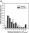
The strength of LNC rolling in HEVs is not augmented by α4 or LFA-1 integrins, and is not PTX sensitive. Frequency histograms were generated by measuring rolling velocities of individual LNCs in order III venules before (control) and after treatment with (A) anti–LFA-1, (B) anti-α4, or (C) PTX or MTX. Vroll, Vfast, and Vblood were determined in the same vessels before and after mAb, MTX, or PTX treatment as described in Materials and Methods (results of hemodynamic measurements are given in Table 1). To account for hemodynamic differences between control and treatment period, Vrel was calculated as percentage of Vblood. Frequency distributions were calculated after cells were assigned to velocity classes with Vrel >0% to <1%, 1% to <2%, and so on. Statistical comparison of population means and distributions revealed no significant differences between rolling velocities (P >0.05).
Figure 5
Figure 5
The strength of LNC rolling in HEVs is not augmented by α4 or LFA-1 integrins, and is not PTX sensitive. Frequency histograms were generated by measuring rolling velocities of individual LNCs in order III venules before (control) and after treatment with (A) anti–LFA-1, (B) anti-α4, or (C) PTX or MTX. Vroll, Vfast, and Vblood were determined in the same vessels before and after mAb, MTX, or PTX treatment as described in Materials and Methods (results of hemodynamic measurements are given in Table 1). To account for hemodynamic differences between control and treatment period, Vrel was calculated as percentage of Vblood. Frequency distributions were calculated after cells were assigned to velocity classes with Vrel >0% to <1%, 1% to <2%, and so on. Statistical comparison of population means and distributions revealed no significant differences between rolling velocities (P >0.05).
Figure 6
Figure 6
Lymphocyte sticking in PLN-HEVs is mediated by LFA-1. The fraction of rolling LNCs that became stuck for ⩾30 s was determined before (control) and after treatment of cells and animals with blocking mAbs (α4, mAb PS/2; LFA-1, mAb Tib 213; L-selectin, mAb Mel-14). Bars depict sticking fractions as percentage of control cell sticking in the same venule; data shown are mean ± SEM of sticking fractions from 3–5 experiments (4–10 venules each). **P <0.001 versus α4 and control group (Kruskal Wallis test with Bonferoni correction).
Figure 7
Figure 7
Sticking of LNCs in PLN-HEV, but not rolling, is blocked by PTX. To test the role of Gαi-linked proteins, LNCs were treated with PTX or the inactive mutant form, MTX, as a control. Cells were labeled with BCECF and injected into the feeding artery of a subiliac PLN. (A) Rolling and (B) sticking fractions of treated LNCs in subiliac LN venules were determined. Data are shown as mean ± SEM of 3 experiments (5–10 venules each). **P <0.001 (Student's t test).
Figure 7
Figure 7
Sticking of LNCs in PLN-HEV, but not rolling, is blocked by PTX. To test the role of Gαi-linked proteins, LNCs were treated with PTX or the inactive mutant form, MTX, as a control. Cells were labeled with BCECF and injected into the feeding artery of a subiliac PLN. (A) Rolling and (B) sticking fractions of treated LNCs in subiliac LN venules were determined. Data are shown as mean ± SEM of 3 experiments (5–10 venules each). **P <0.001 (Student's t test).
Figure 8
Figure 8
Homing to PLN is mediated by a unique cascade of molecular adhesive and signaling events. At least three distinct steps must occur for lymphocyte homing via HEVs. Only cells that express L-selectin at sufficient density can tether and roll via binding to PNAd (Step 1). Most lymphocytes (such as subsets of activated/memory cells) that express little or no L-selectin pass HEVs without interacting. Subsequently, rolling cells must encounter a Gαi-linked activating stimulus (Step 2) which triggers functional upregulation of LFA-1 (Step 3). Rolling PMNs cannot respond to this lymphocyte-specific chemoattractant signal. The unique combination of these consecutive steps results in the highly efficient and selective recruitment of preferentially naive lymphocytes.

Similar articles

Cited by

References

    1. Gowans JL, Knight EJ. The route of re-circulation of lymphocytes in the rat. Proc R Soc Lond B Biol. 1964;159:257–282. - PubMed
    1. Butcher EC, Scollay RG, Weissman IL. Organ specificity of lymphocyte migration: mediation by highly selective lymphocyte interaction with organ-specific determinants on high endothelial venules. Eur J Immunol. 1980;10:556–561. - PubMed
    1. Butcher EC, Picker LJ. Lymphocyte homing and homeostasis. Science. 1996;272:60–66. - PubMed
    1. Butcher EC. Leukocyte-endothelial cell recognition: three (or more) steps to specificity and diversity. Cell. 1991;67:1033–1036. - PubMed
    1. Shimizu Y, Newman W, Tanaka Y, Shaw S. Lymphocyte interactions with endothelial cells. Immunol Today. 1992;13:106–112. - PubMed

Publication types

MeSH terms

Substances