. 1998 Jan 20;95(2):525-30.
doi: 10.1073/pnas.95.2.525.

DNA-dependent protein kinase: DNA binding and activation in the absence of Ku

Affiliations

DNA-dependent protein kinase: DNA binding and activation in the absence of Ku

O Hammarsten et al. Proc Natl Acad Sci U S A. .

Abstract

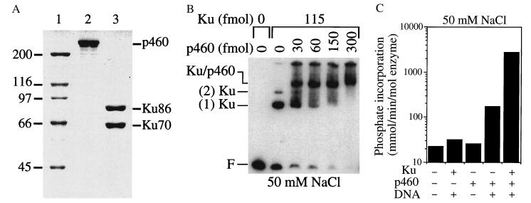
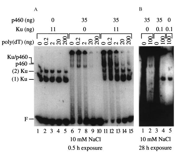
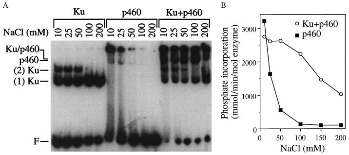
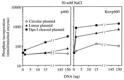
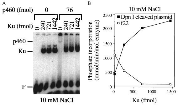
In mammalian cells, double-strand break repair and V(D)J recombination require DNA-dependent protein kinase (DNA-PK), a serine/threonine kinase that is activated by DNA. DNA-PK consists of a 460-kDa subunit (p460) that contains a putative kinase domain and a heterodimeric subunit (Ku) that binds to double-stranded DNA ends. Previous reports suggested that the activation of DNA-PK requires the binding of Ku to DNA. To investigate this further, p460 and Ku were purified separately to homogeneity. Surprisingly, p460 was capable of binding to DNA in the absence of Ku. The binding of p460 to double-stranded DNA ends was salt-labile and could be disrupted by single-stranded or supercoiled DNA, properties distinct from the binding of Ku to DNA. Under low salt conditions, which permitted the binding of p460 to DNA ends, the kinase was activated. Under higher salt conditions, which inhibited the binding of p460, activation of the kinase required the addition of Ku. Significantly, when the length of DNA decreased to 22 bp, Ku competed with p460 for DNA binding and inhibited kinase activity. These data demonstrate that p460 is a self-contained kinase that is activated by direct interaction with double-stranded DNA and that the role of Ku is to stabilize the binding of p460 to DNA ends.


Figures

Figure 1
Figure 1
Analysis of purified p460 and Ku. (A) SDS/PAGE. Purified p460 (lane 2, 350 ng) and Ku (lane 3, 350 ng) were resolved by SDS/PAGE in 8% gels and stained with Coomassie blue. Size markers (lane 1) are defined by their molecular masses in kilodaltons. (B) EMSA. Labeled f32 DNA (0.2 ng) was incubated with Ku (18 ng) and increasing amounts of p460 in 5 μl of buffer B containing 50 mM NaCl and analyzed by EMSA. F denotes the position of free probe. (C) Kinase activity. Peptide substrate (5 μg) was incubated with various combinations of Ku (11 ng), p460 (35 ng), and linear plasmid DNA (300 ng) in 5 μl of buffer C containing 50 mM NaCl, and kinase activity was determined.
Figure 2
Figure 2
DNA binding of Ku, p460, and the Ku–p460 complex. (A) DNA binds to Ku, p460, and Ku–p460 complexes in distinct complexes that are affected differently by poly(dT). Labeled f32 DNA (0.2 ng) and various amounts of poly(dT) were incubated with Ku (11 ng), p460 (35 ng), or an equimolar mixture of both in 5 μl of buffer B containing 10 mM NaCl and analyzed by EMSA. Positions of individual complexes are indicated to the left of the gel. (B) Absence of Ku binding activity in the p460 preparation. Labeled f32 DNA (0.2 ng) with or without poly(dT) was incubated with Ku, p460, or a mixture of Ku and p460 (in a molar ratio of 1:100) in 5 μl of buffer B containing 10 mM NaCl and analyzed by EMSA. Gels were exposed to x-ray film for the indicated times.
Figure 3
Figure 3
Effect of salt on DNA binding and kinase activities of p460 and Ku–p460 complexes. (A) Increasing salt disrupts DNA binding of p460 but not of Ku. Labeled f32 DNA (0.2 ng) was incubated with Ku (11 ng), p460 (35 ng), or an equimolar mixture of both in 5 μl of buffer B containing various concentrations of NaCl and analyzed by EMSA. (B) Increasing salt inhibits kinase activity strongly for p460 and weakly for Ku–p460 complexes. Kinase activities of p460 or the Ku–p460 complex were measured after stimulation by f32 DNA (0.2 ng) in buffer C containing various concentrations of NaCl by using the same protein concentrations as in A.
Figure 4
Figure 4
p460 and the Ku–p460 complex are both specifically activated by DNA ends. Kinase activities of p460 or of Ku–p460 complexes were measured using different forms of plasmid DNA as substrate. Plasmid DNA was linearized with BamHI, digested to 15 fragments with DpnI, or left as supercoiled DNA and incubated with p460 (35 ng) with or without Ku (11 ng) in buffer C containing 50 mM NaCl.
Figure 5
Figure 5
DNA binding and kinase activation of p460 by f22 DNA are inhibited by the addition of Ku. (A) DNA binding. Labeled f22 DNA (1.4 ng, 100 fmol) was incubated with p460 (35 ng, 76 fmol) plus different amounts of Ku in 5 μl of buffer B containing 10 mM NaCl and then analyzed by EMSA. (B) Kinase activation. Kinase activity was measured for p460 (76 fmol) with different amounts of Ku in 5 μl of buffer C containing 10 mM NaCl, after stimulation by f22 DNA (100 fmol) or DpnI-cleaved plasmid DNA (13 ng, corresponding to 100 fmol of DNA fragments).
Figure 6
Figure 6
Model for the assembly and activation of DNA-PK in vivo. Ku binds to the double-stranded DNA end, translocates in an ATP-independent manner to uncover the DNA end, and stabilizes the binding of p460 to the DNA end. Cooperative interactions among Ku, p460, and the DNA lead to efficient activation of the kinase. Our data indicate that both Ku and p460 contact the DNA, but their relative positions at the DNA end remain undetermined.

References

    1. Keeney S, Giroux C N, Kleckner N. Cell. 1997;88:375–384. - PubMed
    1. Smider V, Chu G. Sem Immun. 1997;9:189–197. - PubMed
    1. Chu G. J Biol Chem. 1997;272:24097–24100. - PubMed
    1. Smider V, Rathmell W K, Lieber M, Chu G. Science. 1994;266:288–291. - PubMed
    1. Taccioli G, Gottlieb T, Blunt T, Priestly A, Demengeot J, Mizuta R, Lehmann A, Alt F, Jackson S, Jeggo P. Science. 1994;265:1442–1445. - PubMed

Publication types

MeSH terms

Substances