. 1998 Jan 20;95(2):582-7.
doi: 10.1073/pnas.95.2.582.

Cloning and characterization of hSRP1 gamma, a tissue-specific nuclear transport factor

Affiliations

Cloning and characterization of hSRP1 gamma, a tissue-specific nuclear transport factor

M V Nachury et al. Proc Natl Acad Sci U S A. .

Abstract

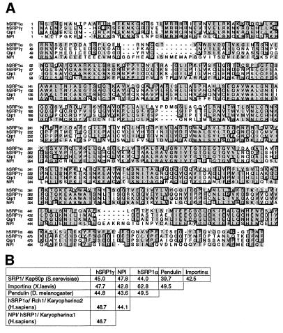
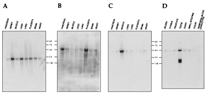
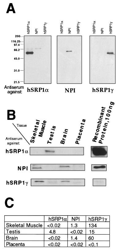
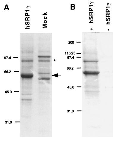
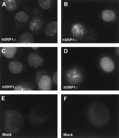
Nuclear import of proteins containing a nuclear localization signal (NLS) is dependent on the presence of a cytoplasmic NLS receptor, the GTPase Ran, and p10/ NTF2. The NLS receptor is a heterodimeric proteins consisting of subunits of approximately 60 and 97 kDa, which have been termed importin alpha/beta, karyopherin alpha/beta, or PTAC 58/ 97. Members of the 60-kDa/importin alpha subunit family directly bind to the NLS motif and have been shown to function as adaptors that tether NLS-containing proteins to the p97/ importin beta subunit and to the downstream transport machinery. Herein we report the identification and characterization of hSRP1 gamma, a human importin alpha homologue. The hSRP1 gamma protein is around 45% identical to the previously identified human importin alpha homologues hSRP1 alpha/Rch1 and NPI/ hSRP1. hSRP1 gamma can form a complex with importin beta and is able to mediate import of a BSA-NLS substrate in an in vitro nuclear import system. Interestingly, hSRP1 gamma shows a very selective expression pattern and is most abundantly expressed in skeletal muscle, representing more than 1% of the total protein in this tissue. A potential role for hSRP1 gamma in tissue-specific transport events is discussed.


Figures

Figure 1
Figure 1
(A) Multiple sequence alignment of different human importin α homologues. The amino acid sequence of hSRP1γ, hSRP1α (GenBank U09559), Qip1 (GenBank AB002533), and NPI/hSRP1 (GenBank S75295) were aligned with clustal w (53) and the alignment was displayed with seqvu using the ges homology matrix. Boxes indicate that at least three out of four amino acids are identical at that position and shading indicates a homology of more than 85%. (B) Identity matrix of hSRP1γ (this study), SRP1 (S. cerevisiae, GenBank M75849), Importin α (Xenopus laevis, GenBank G623602), Pendulin (Drosophila melanogaster, GenBank S57866), hSRP1α (Homo sapiens), and NPI (Homo sapiens). Alignment was performed with fasta (54), and the percentage of identity between the indicated amino is displayed.
Figure 2
Figure 2
Northern blots showing the expression pattern of hSRP1α (A), NPI/hSRP1 (B), and hSRP1γ (C and D) and in different human tissues. Two multitissue Northern blots containing poly(A)+ RNA isolated from the indicated human tissues were analyzed with probes derived from the cDNAs of hSRP1α (A), NPI (B), or hSRP1γ (C and D). Molecular sizes are in kilobases.
Figure 3
Figure 3
Protein expression pattern of hSRP1γ, hSRP1α, and NPI/hSRP1 in different human tissues. (A) Specificity of the antisera used in this study. About 500 ng of the indicated recombinant proteins were separated by SDS/PAGE, transferred to nitrocellulose, and subsequently probed with the antiserum against hSRP1α, NPI, or hSRP1γ. (B) Multitissue Western blots probed with the antiserum against hSRP1α, NPI, or hSRP1γ. Five micrograms (hSRP1α, NPI) or 1 μg (hSRP1γ) of tissue protein extracts (CLONTECH) and 50 μg (hSRP1α, NPI) or 10 μg (hSRP1γ) were loaded in the first four lanes. The indicated amount of recombinant protein loaded in the last lane was determined by densitometry scanning in a Coomassie-stained gel. (C) Result of quantitation of protein expression. The values correspond to the amount (in ng) of each importin-α-like factor per 10 μg of total protein.
Figure 4
Figure 4
(A) Expression and purification of recombinant His6-hSRP1γ. Histidine-tagged hSRP1γ was expressed in HeLa cells with the vaccinia virus expression system and purified over Ni-NTA agarose, and its purity was analyzed by Coomassie staining after separation on 10% SDS/PAGE gels (lane 1, hSRP1γ). A similar purification was performed from HeLa cells that had been infected with the wild-type vaccinia virus (lane 2, mock). The arrow indicates hSRP1γ. The asterisk shows a contaminating protein of ∼90 kDa that also was purified from the wild-type-infected extract. Western blotting demonstrated that this band is not importin β (data not shown). Molecular sizes are in kilodaltons. (B) Coselection of importin β by hSRP1γ. Untagged recombinant importin β was incubated with Ni-NTA agarose beads either in the presence (lane 1) or absence (lane 2) of His6-tagged hSRP1 γ. After selection the proteins were separated on a 10% SDS/PAGE gel and analyzed by Coomassie staining.
Figure 5
Figure 5
In vitro nuclear protein import assays. Import of fluorescein-labeled BSA-NLS conjugate into digitonin-permeabilized HeLa cells was assayed in the presence of hSRP1α and importin β (A); hSRP1α, importin β, Ran, and p10/NTF2 (B); hSRP1γ and importin β (C); hSRP1γ, importin β, Ran, and p10/NTF2 (D); mock-purified extract and importin β (E); or mock-purified extract, importin β, Ran, and p10/NTF2 (F). The localization of the BSA-NLS substrate was analyzed by epifluorescence. Images were recorded by using a charge-coupled device camera. Quantitation of the uptake of BSA-NLS in B and D shows that hSRP1γ has only ∼30% of the import activity of hSRP1α. The protein amounts of hSRP1γ and α used in the assay were normalized with respect to their binding activity to importin β. Coomassie blue staining showed that roughly equal amounts of the two proteins were present in the transport assay (data not shown).

Similar articles

Cited by

References

    1. Davis L I. Annu Rev Biochem. 1995;64:865–896. - PubMed
    1. Doye V, Hurt E. Curr Opin Cell Biol. 1997;9:401–411. - PubMed
    1. Pante N, Aebi U. J Cell Sci (Suppl) 1995;19:1–11. - PubMed
    1. Goldberg M W, Allen T D. Curr Biol. 1995;7:301–309. - PubMed
    1. Dingwall C, Laskey R A. Trends Biochem Sci. 1991;16:478–481. - PubMed

Publication types

Associated data