. 1998 Jan 20;95(2):735-40.
doi: 10.1073/pnas.95.2.735.

5-Hydroxytryptamine2A serotonin receptors in the primate cerebral cortex: possible site of action of hallucinogenic and antipsychotic drugs in pyramidal cell apical dendrites

Affiliations

5-Hydroxytryptamine2A serotonin receptors in the primate cerebral cortex: possible site of action of hallucinogenic and antipsychotic drugs in pyramidal cell apical dendrites

R L Jakab et al. Proc Natl Acad Sci U S A. .

Abstract

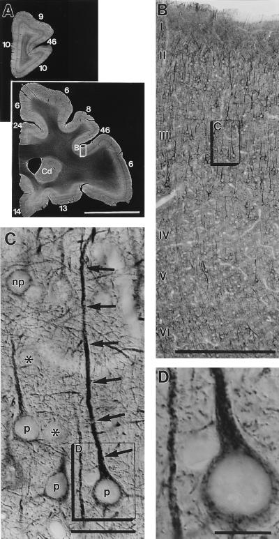
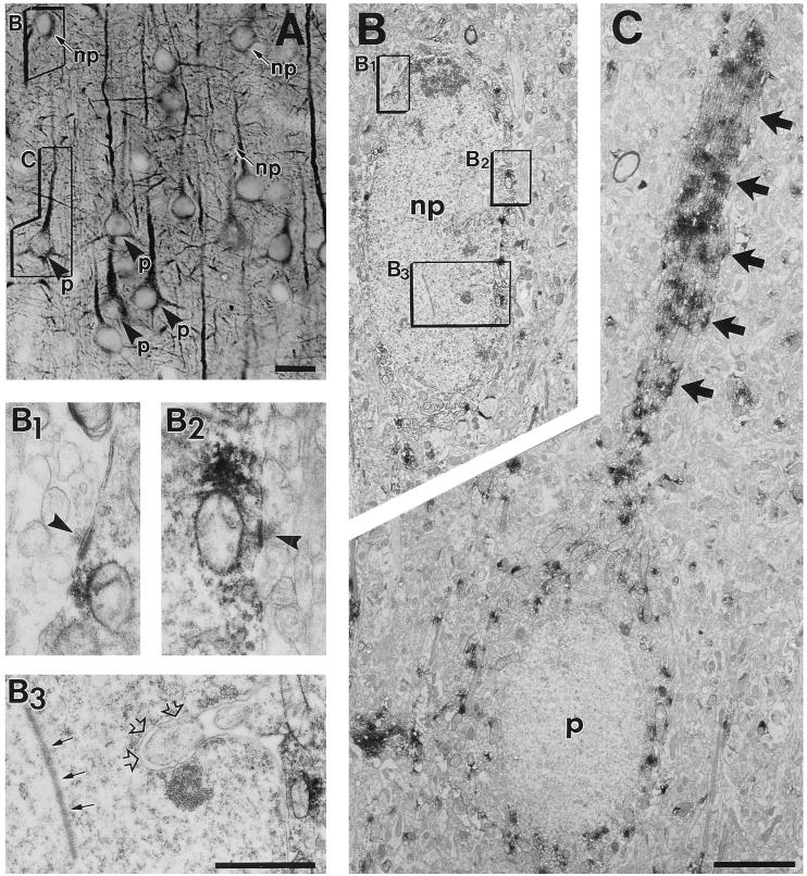
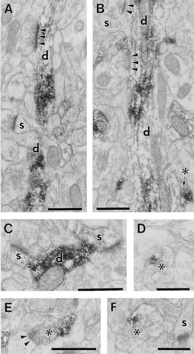
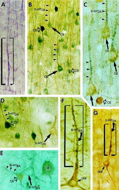
To identify the cortical sites where 5-hydroxytryptamine2A (5-HT2A) serotonin receptors respond to the action of hallucinogens and atypical antipsychotic drugs, we have examined the cellular and subcellular distribution of these receptors in the cerebral cortex of macaque monkeys (with a focus on prefrontal areas) by using light and electron microscopic immunocytochemical techniques. 5-HT2A receptor immunoreactivity was detected in all cortical layers, among which layers II and III and layers V and VI were intensely stained, and layer IV was weakly labeled. The majority of the receptor-labeled cells were pyramidal neurons and the most intense immunolabeling was consistently confined to their parallelly aligned proximal apical dendrites that formed two intensely stained bands above and below layer IV. In double-label experiments, 5-HT2A label was found in calbindin D28k-positive, nonphosphorylated-neurofilament-positive, and immuno-negative pyramidal cells, suggesting that probably all pyramidal cells express 5-HT2A receptors. 5-HT2A label was also found in large- and medium-size interneurons, some of which were immuno-positive for calbindin. 5-HT2A receptor label was also associated with axon terminals. These findings reconcile the data on the receptor's cortical physiology and localization by (i) establishing that 5-HT2A receptors are located postsynaptically and presynaptically, (ii) demonstrating that pyramidal neurons constitute the major 5-HT2A-receptor-expressing cells in the cortex, and (iii) supporting the view that the apical dendritic field proximal to the pyramidal cell soma is the "hot spot" for 5-HT2A-receptor-mediated physiological actions relevant to normal and "psychotic" functional states of the cerebral cortex.


Figures

Figure 1
Figure 1
(A) Dark-field photographs show the distribution of 5-HT2A receptor immunoreactivity in two representative sections of the prefrontal cortex of an adult macaque monkey. Cortical areas are numbered according to Walker’s nomenclature. Cd, caudate nucleus. Note that receptor labeling is weak in a stripe denoting layer IV throughout the cortical sheet. The framed cortical region, enlarged in B, exemplifies the receptor immunoreactivity in area 46 of the prefrontal cortex. 5-HT2A labeling is found in most (if not all) pyramidal neurons throughout cortical layers II and III and layers V and VI, including their dendritic branches in layer I. Receptor labeling is weak in layer IV because only moderately labeled interneurons and some en passant apical dendrites of layer V pyramids are present in this layer. The boxed area in layer III (enlarged in C and D) demonstrates receptor-labeled pyramidal cells (p), unlabeled (asterisks) and labeled nonpyramidal (np) cells, and receptor-positive fine processes (their ultrastructure is shown in Fig. 3). Note that the immunoreaction is strongest in the apical dendrites (arrows) of pyramidal cells and relatively weak in perikarya. [Bars = 1 cm (A), 0.5 mm (B), 50 μm (C), and 20 μm (D).]
Figure 2
Figure 2
Cellular (A) and subcellular (B and C) localization of 5-HT2A receptor immunoreactivity in pyramidal (p) and nonpyramidal (np) neurons, detected by correlated light and electron microscopy in layer III of prefrontal area 46. (A) The framed nonpyramidal and pyramidal cells were selected for electron microscopic examination and enlargements are shown in B and C. (B) Labeled medium-size nonpyramidal neuron (np) exhibiting intranuclear rod (B3, small arrows), nuclear infolding (B3, open arrows), and axosomatic asymmetric synapses (B1 and B2, arrowheads), three typical features of cortical interneurons. (C) The receptor label concentration is high in the pyramidal cell apical dendrite (arrows) and low in the perikaryal cytoplasm. [Bars = 25 μm (A), 5 μm (B and C), and 0.5 μm (B1B3).]
Figure 3
Figure 3
Postsynaptic (A–C) and presynaptic (B, D–F) localization of 5-HT2A receptor immunoreactivity in area 46 of the prefrontal cortex. (A and B) 5-HT2A label is localized to dendritic shafts (d), but dendritic spines (s) are generally immunonegative. Unlabeled axons form asymmetric synapses (arrowheads) on these dendrites. (C) A rare example of two weakly labeled dendritic spines (s). (B, D–F) Immunolabeled axon terminals (asterisks) forming asymmetric synapse (E, arrowheads) or containing dense core vesicle (F). 5-HT2A label is restricted to a portion of the axoplasm. (Bars = 0.5 μm.)
Figure 4
Figure 4
Color light micrographs demonstrate the accumulation of 5-HT2A receptor immunoreactivity in the proximal dendrites of pyramidal and nonpyramidal neurons in prefrontal area 46. The neurons were neurochemically identified by using 5-HT2A/CB (B–E) or 5-HT2A/SMI-32 (F–G) double immunocytochemistry. Arrowheads point to dendrites, arrows point to pyramidal cell somata, and open arrows point to nonpyramidal cell bodies. (A) 5-HT2A-positive pyramidal cell in layer III (stained purple with the Vector VIP staining kit). (B and C) Two 5-HT2A/CB double-stained sections from cortical layer III demonstrate that 5-HT2A immunoreactivity (arrowheads) is colocalized with CB immunolabel (arrows) in pyramidal neurons. (B) 5-HT2A label is light brown and CB label is bluish gray. (C) Double labeling is “reverse,” 5-HT2A label is bluish gray, and CB label is light brown. (D and E) Two micrographs from layer V demonstrate that large- (asterisk) and medium-size interneurons (open arrows) colocalize 5-HT2A receptor and CB and show that 5-HT2A receptor is also present in CB-negative pyramidal cells (arrows), typical of the infragranular layers. (D) 5-HT2A, light brown; CB, bluish gray. (E) 5-HT2A, bluish gray; CB, light brown. (F and G) 5-HT2A/SMI-32 double-labeled pyramidal cells from layer III. Note that the gray color of 5-HT2A label dominates only in the proximal apical dendrites (within the frame), whereas thinner dendritic branches and the cell body are overshadowed by the brown-colored SMI-32 staining.

References

    1. Aghajanian G K. In: Psychopharmacology: The Fourth Generation of Progress. Bloom F E, Kupfer D J, editors. New York: Raven; 1995. pp. 451–460.
    1. Buhot M-C. Curr Opin Neurobiol. 1997;7:243–254. - PubMed
    1. Deutch A Y, Moghaddam B, Innis R B, Krystal J H, Aghajanian G K, Bunney B S, Charney D S. Schizophr Res. 1991;4:121–156. - PubMed
    1. Meltzer H Y, Nash J F. Pharmacol Rev. 1991;43:587–604. - PubMed
    1. Schmidt C J, Sorensen S M, Kehne J H, Carr A A, Palfreyman M G. Life Sci. 1995;56:2209–2222. - PubMed

Publication types

MeSH terms

LinkOut - more resources