

Overcoming cellular senescence in human cancer pathogenesis
- PMID: 9436977
- PMCID: PMC316442
- DOI: 10.1101/gad.12.2.163
Overcoming cellular senescence in human cancer pathogenesis
Abstract
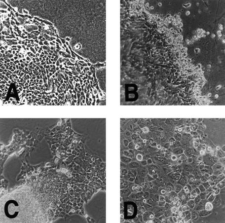
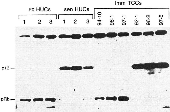
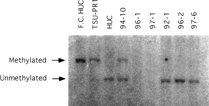
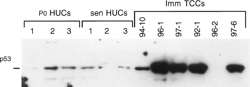
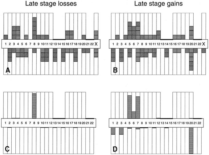
Elevation of p16, the CDKN2/p16 tumor suppressor gene (TSG) product, occurs at senescence in normal human uroepithelial cells (HUC). Immortal HUCs and bladder cancer cell lines show either alteration of p16 or pRb, the product of the retinoblastoma (RB) TSG. In addition, many human cancers show p16 or pRb alteration along with other genetic alterations that we associated with immortalization, including +20q and -3p. These observations led us to hypothesize that p16 elevation plays a critical role in senescence cell cycle arrest and that overcoming this block is an important step in tumorigenesis in vivo, as well as immortalization in vitro. Using a novel approach, we tested these hypotheses in the present study by examining p16 and pRb status in primary culture (P0) and after passage in vitro of transitional cell carcinoma (TCC) biopsies that represented both superficial bladder tumors and invasive bladder cancers. We demonstrated that all superficial TCCs showed elevated p16 after limited passage in vitro and then senesced, like normal HUCs. In contrast, all muscle invasive TCCs contained either a p16 or a pRb alteration at P0 and all spontaneously bypassed senescence (P = 0.001). Comparative genomic hybridization (CGH) was used to identify regions of chromosome loss or gain in all TCC samples. The application of a statistical model to the CGH data showed a high probability of elevated alteration rates of +20q11-q12 (0.99) and +8p22-pter (0.94) in the immortal muscle invasive TCCs, and of -9q (0.99) in the superficial TCCs. Three myoinvasive TCCs lost 3p13-p14. In this study, four of six myoinvasive TCCs also showed TP53 mutation that associated well with genome instability (P = 0.001), as previously hypothesized. Notably, TP53 mutation, which has been used as a marker of tumor progression in many human cancers, was less significant in associating with progression in this study (P = 0.04) than was p16 or pRb alteration (P = 0.001). Thus, these data support a new model in which overcoming senescence plays a critical role in human cancer pathogenesis and requires at least two genetic changes that occur in several combinations that can include either p16 or pRb loss and at least one additional alteration, such as +20q11-q12, -3p13-p14, or -8p21-pter.
Figures


Similar articles
-
Different combinations of genetic/epigenetic alterations inactivate the p53 and pRb pathways in invasive human bladder cancers.Cancer Res. 2000 Jul 15;60(14):3862-71. Cancer Res. 2000. PMID: 10919661
-
p16/pRb pathway alterations are required for bypassing senescence in human prostate epithelial cells.Cancer Res. 1999 Jun 15;59(12):2957-64. Cancer Res. 1999. PMID: 10383161
-
Increased p16 levels correlate with pRb alterations in human urothelial cells.Cancer Res. 1995 Feb 1;55(3):493-7. Cancer Res. 1995. PMID: 7834615
-
A molecular genetic model of human bladder cancer pathogenesis.Semin Oncol. 1996 Oct;23(5):571-84. Semin Oncol. 1996. PMID: 8893868 Review.
-
Molecular and kinetic features of transitional cell carcinomas of the bladder: biological and clinical implications.Virchows Arch. 2001 Mar;438(3):289-97. doi: 10.1007/s004280000289. Virchows Arch. 2001. PMID: 11315626 Review.
Cited by
-
Caveolin-1 regulates the antagonistic pleiotropic properties of cellular senescence through a novel Mdm2/p53-mediated pathway.Cancer Res. 2009 Apr 1;69(7):2878-86. doi: 10.1158/0008-5472.CAN-08-2857. Epub 2009 Mar 24. Cancer Res. 2009. PMID: 19318577 Free PMC article.
-
Cellular senescence, an unpopular yet trustworthy tumor suppressor mechanism.Cancer Sci. 2003 Nov;94(11):944-7. doi: 10.1111/j.1349-7006.2003.tb01382.x. Cancer Sci. 2003. PMID: 14611669 Free PMC article. Review.
-
Identification of prefoldin amplification (1q23.3-q24.1) in bladder cancer using comparative genomic hybridization (CGH) arrays of urinary DNA.J Transl Med. 2013 Aug 1;11:182. doi: 10.1186/1479-5876-11-182. J Transl Med. 2013. PMID: 23914742 Free PMC article.
-
Genetic and epigenetic changes in human epithelial cells immortalized by telomerase.Am J Pathol. 2000 May;156(5):1537-47. doi: 10.1016/S0002-9440(10)65025-0. Am J Pathol. 2000. PMID: 10793065 Free PMC article.
-
Measurement of relative copy number of CDKN2A/ARF and CDKN2B in bladder cancer by real-time quantitative PCR and multiplex ligation-dependent probe amplification.J Mol Diagn. 2004 Nov;6(4):356-65. doi: 10.1016/S1525-1578(10)60532-6. J Mol Diagn. 2004. PMID: 15507675 Free PMC article.
References
-
- American Cancer Society. Cancer facts and figures—1996, 96-300M–No. 5008.96. Bethesda, MD: American Cancer Society; 1997.
-
- Balazs M, Carroll P, Kerschmann R, Sauter S, Waldman FM. Frequent homozygous deletion of cyclin-dependent kinase inhibitor 2 (MTS1, p16) in superficial bladder cancer detected by fluorescence in situ hybridization. Genes, Chrom Cancer. 1997;19:84–89. - PubMed
-
- Bane BL, Rao JY, Hemstreet GP. Pathology and staging of bladder cancer. Semin Oncol. 1996;23:546–570. - PubMed
-
- Cairns P, Mao L, Merlo A, Lee DJ, Schwab D, Eby Y, Tokino K, van der Reit P, Blaugrund JE, Sidransky D. Rates of p16 (MTS1) mutations in primary tumors with 9p loss. Science. 1994;265:415–416. - PubMed
MeSH terms
Substances
LinkOut - more resources
Full Text Sources
Other Literature Sources
Medical
Research Materials
Miscellaneous