. 1998 Jan 15;12(2):246-60.
doi: 10.1101/gad.12.2.246.

Regulation of POU genes by castor and hunchback establishes layered compartments in the Drosophila CNS

Affiliations

Regulation of POU genes by castor and hunchback establishes layered compartments in the Drosophila CNS

R Kambadur et al. Genes Dev. .

Abstract

POU transcription factors participate in cell-identity decisions during nervous system development, yet little is known about the regulatory networks controlling their expression. We report all known Drosophila POU genes require castor (cas) for correct CNS expression. drifter and I-POU depend on cas for full expression, whereas pdm-1 and pdm-2 are negatively regulated. cas encodes a zinc finger protein that shares DNA-binding specificity with another pdm repressor: the gap segmentation gene regulator Hunchback (Hb). Our studies reveal that the embryonic CNS contains sequentially generated neuroblast sublineages that can be distinguished by their expression of either Hb, Pdm-1, or Cas. Hb and Cas may directly silence pdm expression in early and late developing sublineages, given that pdm-1 cis-regulatory DNA contains >=32 Hb/Cas-binding sites and its enhancer(s) are ectopically activated in cas- neuroblasts. In addition, the targeted misexpression of Cas in all neuroblast lineages reduces Pdm-1 expression without altering Hb expression. By ensuring correct POU gene expression boundaries, hb and cas maintain temporal subdivisions in the cell-identity circuitry controlling CNS development.


Figures

Figure 1
Figure 1
Cas and Hb share consensus DNA-binding sites. Cas DNA-binding sites were identified from an unbiased degenerate population of dsDNA fragments by use of the cyclic amplification of selected target DNA-binding site procedure (see Materials and Methods). After six cycles of selection and PCR amplification, which included four rounds of immunoprecipitation followed by two rounds of gel shifts, bound fragments were subcloned and sequenced. (A) Alignment of common DNA sequences (upper strand shown) present in Cas–DNA-bound fragments. Note, clone 4 contains two recognition sequences (4a,b) separated by 8 bp. (B) Gel mobility-shift assays with radiolabeled fragments containing either one binding site (clone 1 and 2), two Cas recognition sites (clone 4a+b), or a randomly-selected fragment with no core-recognition homology (clone 3). Antibody supershifting of fragment  1 DNA–Cas complexes with anti-Cas antibodies establishes Cas involvement in the protein–DNA complexes. Note, the sequence specificity of Cas DNA-binding was confirmed by competition assays and base-pair substitutions (see Fig. 7H). Also note, no Cas protein was added to the samples run in lanes marked (−). (C) Alignment of Cas and Hb consensus DNA-binding sites. Hb consensus DNA recognition sequences were obtained from Stanojevic et al. 1989 (Hb1) and Treisman and Desplan 1989 (Hb2).
Figure 2
Figure 2
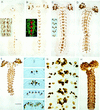
Cas expression during embryonic CNS development. Dissected fillets or transverse sections (6 μm thick) of whole-mount immunostained embryos reveal Cas expression dynamics during embryonic CNS development (embryo staging according to Hartenstein and Campos-Ortega 1984; anterior up in A–E, G, and H; ventral up in F). The same magnification was used in A–E; scale bar in E represents 80 μm. In F, panels 1–5, the same scale bar represents 25 μm, in G it equals 20 μm, and in H it is equivalent to 35 μm. (A) Stage 9 embryos stained with either Cas (main panel) or Hb (inset) antisera. Cas protein is detected first in segmental clusters of ventral midline mesectodermal cells (arrow) and in cells that line the anterior midgut primordium (arrowhead). At this time, no Cas expression is detected in either cephalic lobe or ventral cord NBs (see also panel F, 1). The initial reduced expression observed in the thoracic and first abdominal midline cells reflects the delayed onset of cas mRNA expression in these segments (data not shown). (Inset) By late stage 9, NB delaminations from the lateral ventral cord neurogenic regions have produced three rows of Hb-positive NBs flanking the midline in all gnathal, thoracic, and abdominal segments (see also Fig. 3). Note, NBs were identified on the basis of their large cell body diameters and on their position underlining the ectoderm. (B) During stage 10, bilaterally symmetrical subsets of cephalic lobe and ventral cord NBs initiate Cas expression. The first ventral cord NBs to express Cas are the late delaminating NB6-1s located on the posterior edge of the gnathal, thoracic, and abdominal a1–8 segments (also see F, panel 2). At this time, some of the NB6-1s have produced Cas-positive GMCs (arrows). (Inset) Confocal colocalization views of Hb (green) and Cas (red) reveal no coexpression in NB6-1s or in midline precursors. (C) By early stage 11, the number of cephalic lobe Cas-positive NBs has nearly doubled and the number of Cas-expressing ventral cord NBs has increased to 5–7 per hemisegment (see also F, panel 3). (Inset) Following activation of Cas expression in NB6-1s, the next NBs to express Cas are the earlier delaminating (S1) NB5-2s (arrow). At this time, NB5-2s have terminated Hb expression and their Hb positive sublineages take up dorsal positions relative to ventrally located Cas-positive NBs (see Fig. 3). (D) During late stage 11, Cas expressing NBs in ventral cord neuromeres have now increased to 9–10 and 7–8 per thoracic and abdominal hemisegments, respectively. (E) Stage 12, germ-band contraction; by late stage 11/early stage 12, most CNS neuroblasts express Cas. Cas-expressing NB sublineages are positioned on the outer surface of the cephalic lobes and on the ventral/ventral–lateral surfaces of the developing subesophageal ganglion (arrow) and ventral cord neuromeres (see also F, panels 4 and 5; and Fig. 4). Also note, no Cas expression is detected outside the CNS. (F) Transverse sections (6 μm thick) through abdominal segments of stage 9–13 embryos (1–5, respectively) reveal that Cas-expressing NBs, and their sublineages take up ventral/ventral–lateral positions in the developing neuromeres. (G) Cas is concentrated in GMCs during NB cytokinesis. Shown is a ventral view of a stage 11 ventral cord showing Cas-positive NBs and GMCs in the second and third thoracic and first three abdominal segments. Note, the more intense immunostained GMCs (arrows) relative to their weaker stained NBs. Also note, nuclear-located Cas in nondividing NBs (arrowhead). In situ mRNA localizations show that cas is not expressed in GMCs (data not shown; Mellerick et al. 1992). (H) By stage 15, Cas immunopositive sublineages are evenly distributed on the outer cephalic lobe surfaces and on the ventral/ventral-lateral regions of the subesophageal and ventral cord ganglia.
Figure 3
Figure 3
CNS neuroblasts produce layered sublineages distinguished by their expression of Hb, Pdm, or Cas. The dynamics of Hb (A,C,E,G) and Pdm-1 (B,D,F,H) expression during ventral cord development are shown in transverse (6 μm thick) sections through abdominal segments of stage 9 (A,B), stage 10 (C,D), stage 11 (E,F), and stage 12 (G,H) embryos immunostained in whole-mount with either Hb or Pdm-1 antibodies. (A,B) Sections through stage 9 embryos reveal that most, if not all, fully delaminated NBs express Hb (see also Fig. 2A, inset) whereas Pdm-1 expression is restricted to neuroectoderm cells. (C,D) By stage 10, many NBs and GMCs are immunostained with Hb antibodies, however, not all NBs contain detectable levels of Hb (arrow). In addition to its high levels of neuroectoderm expression, low levels of Pdm-1 immunostaining are now detected in a small subset of NBs and GMCs. (E,F) Starting at late stage 10, and through stage 11, there is a progressive reduction in the number of NBs containing detectable levels of Hb, whereas many GMCs and/or their progeny maintain high levels of Hb immunostaining. Loss of Hb expression in NBs parallels the activation of Pdm-1 NB expression. The number of GMCs containing detectable levels of Pdm-1 also rises during stage 11 and follows that observed in NBs. Given their close apposition to Pdm-1-positive NBs, many of these GMCs were most likely products of Pdm-1-expressing NBs. (G,H) Stage 12; during this period, Pdm-1 NB expression is reduced such that only low levels of Pdm-1 immunostaining are found in NBs. Hb and Pdm-1-positive sublineages are now positioned dorsal to late dividing NBs. Scale bar in H is equivalent to 28 μm and also applies to A–G. (I) By stage 13, Hb and Pdm-1 immunopositive sublineages form layered subpopulations of cells in all CNS neuromeres. In the ventral cord, Pdm-1 positive sublineages are juxtaposed to the more dorsal/internal Hb sublineages. Shown are lateral, confocal fluorescent views from a double-labeled, Hb (green) and Pdm-1 (red), stage 13 embryo (anterior is left and ventral up). (J) Confocal immunofluorescent views of stage 12 and older embryos reveal that Hb (green) and Cas (red) positive sublineages do not overlap and occupy the dorsal and ventral neuromere surfaces, respectively. Shown is a stage 13 ventral cord (lateral view; anterior is left and ventral up). Note the more ventral position of the Cas-expressing sublineages compared with the internal Pdm-1 positive cells in I. (K) Triple-labeling [Hb (blue), Pdm-1 (green), and Cas (red)] reveals that by stage 13 most, if not all, sublineages express one of these transcription factors. For confocal ventral views of adjacent Pdm-1- and Cas-expressing sublineages, see Fig. 4. Note, the arrow in K indicates a NB that coexpresses Pdm-1 and Cas. Scale bar in J is 30 μm and also applies to I and K.
Figure 4
Figure 4
By mid-CNS development, Cas and Pdm-1 colocalizations reveal little overlap in their expression. Confocal immunofluorescent views of embryos doubled-labeled for Cas (red) and Pdm-1 (green) reveal that the activation of Cas expression in CNS NBs coincides with the loss of Pdm-1 NB expression. (A–C) Shown are ventral to dorsal (left to right) optical section views of a late stage 12 ventral cord (anterior, up). Note the more ventral/ventral–lateral position of the Cas-positive cells relative to the deeper/internal Pdm-1 immunostained sublineages. (D–F) Cephalic lobe expression of Cas and Pdm-1; shown are dorsal to ventral (left to right), serial views of a late stage 12 brain. Arrow in E points to a NB immunostained for both Cas and Pdm-1. Note, the red staining between and below the cephalic lobes is caused by the nonspecific binding of the rhodamine-tagged secondary antibody to yolk cells. Same magnification in all panels. Scale bar in A, 50 μm.
Figure 5
Figure 5
Loss of hb disrupts axon fasciculation and triggers ectopic Pdm-1 expression in early developing neuroblast sublineages. (A,B) Correct formation/organization of both longitudinal and commissural axon fascicle tracks requires hb function. Shown are dorsal views of dissected wild-type (A) and hb4 null mutant (B) stage 13 ventral cords immunostained in whole-mount with an axon marker, the BP102 mAb (anterior is up). Note the lack of longitudinal connectives and missing or disorganized commissures in the hb ventral cord. (C–F) In hb mutant embryos, early delaminating CNS NBs (S1 and S2 waves) fail to terminate Pdm-1 expression. Comparisons between wild-type (C) and the hb4 null allele (D) late stage 12 embryos reveal increased numbers of NB sublineages expressing Pdm-1 in all CNS ganglia of the hb mutant. Shown are dissected flattened fillets of whole-mount immunostained embryos (anterior is up). Transverse sections through abdominal segments of late stage 9 (E) and stage 12 (F) hb4 embryos reveal ectopic Pdm-1 expression in early delaminating NBs and in their sublineages. Note the increased numbers and thicker dorsal/ventral field of Pdm-1-positive cells in the stage 12 mutant ventral cord (F) when compared with the Pdm-1 immunoreactive cells present in a similar transverse section from a wild-type embryo shown in Fig. 3H. (G,H) Shown are a transverse section through the second abdominal segment (G) and a dissected fillet (H) both from late stage 12 hb4 embryos immunostained with Cas antibodies. In hb4 mutants, late developing NB sublineages express Cas. When compared with wild-type Cas expression patterns, however, its onset is delayed in certain sublineages (data not shown) and the overall position of Cas-expressing cells is altered in hb disrupted neuromeres. In hb embryos, Cas-positive late sublineages are still found on the outer ventral surfaces of the ventral cord neuromeres.
Figure 6
Figure 6
Loss of castor results in ectopic Pdm-1 and Pdm-2 expression in late developing neuroblast sublineages. (A,B) Comparisons of Pdm-1 immunostaining in wild-type (A) and cas null (B) stage 13 ventral cords reveals ectopic pdm expression in late-stage cas NBs and in their progeny. Pdm-1 misexpression is particularly evident in the ventral–lateral regions of the cas ventral cord. In wild-type ventral cords, these ventral–lateral areas are predominantly made up of Cas-expressing sublineages (see Fig. 4C). Shown are the third thoracic through third abdominal neuromeres of early stage 13 embryos (anterior is up). (C,D) Ectopic Pdm-1 expression is also evident in late developing cephalic lobe NB sublineages. Shown are dorsal views of wild-type (C) and cas (D) stage 13 Pdm-1 immunostained embryos (anterior is up). (E,F) Ni-enhanced, Pdm-2 immunostaining of wild-type (E) and cas (F) embryos also reveals ectopic Pdm-2 expression in late-stage cas cephalic lobe NBs. Note, focal planes are similar to C and D.
Figure 7
Figure 7
Cas silences pdm-1 neuroblast enhancer(s). (A,B) Whole-mount in-situ mRNA localizations in transformant embryos shows that a 5.3-kb fragment of pdm-1 regulatory DNA can activate the divergently transcribed reporter genes, mini-white (A) and GAL4 (B), in a manner similar to endogenous cellular blastoderm pdm-1 expression (anterior, left; see Fig. 7G and Materials and Methods for P-element vector details). Note, pdm cellular blastoderm expression boundaries are set by Hb repression (Lloyd and Sakonju 1991; Cockerill et al. 1993). (C–F) In situ hybridizations performed on wild-type (C,E) and cas (D,F) transformant embryos reveal that the mini-white (C,D) and Gal4 (E,F) reporter genes, are activated in cas CNS NBs. Shown are dorsal views of stage 13 cephalic lobes (anterior, up; focal plane passes through the dorsal surface of the cephalic lobes). (G) The pdm-1 regulatory DNA/reporter gene P-element construct showing locations (solid ovals) of 32 potential Cas/Hb DNA-binding sites. These sites share 8 of 10 bp with the consensus Hb-binding site and all contain the core A/T-rich sequence (see Fig. 1C for consensus DNA-binding sequences). Core recognition sequences (upper strand) for sites 6 and 12 are shown plus a mutated site (m12) used to test Cas-DNA binding sequence-specificity (see H). Both sites were selected because they match known Hb recognition sites (Stanojevic et al. 1989; Treisman and Desplan 1989). Dashes in m12 represent same bases as in the wild-type site. The pdm-1 embryonic transcriptional start site is shown as a rightward-facing arrow. (H) Electrophoretic mobility-shift assays show that Cas binds specifically to sites in pdm-1 regulatory DNA. Shown are gel-shift assays with 30-bp dsDNA fragments matching either Hb recognition site 6 or 12. Increasing concentrations (5- and 50-fold) of cold competitor DNA (6 or 12) reduce labeled DNA–Cas complexes, whereas the mutated fragment (m12) fails to bind Cas or compete (5- and 50-fold) with wild-type 12 DNA–Cas binding. Note, no recombinant Cas was added to the samples run in lanes marked −.
Figure 8
Figure 8
Targeted misexpression of Cas in all neuroblast lineages reduces Pdm-1 expression. (A) Activation of a UAS.cas transgene by the prospero.GAL4 driver results in Cas misexpression in most, if not all, NB lineages in both the CNS and in the PNS (arrows). Shown is a transverse section through an abdominal segment of a stage 13 prospero.GAL4/UAS.cas transformant embryo immunostained in whole-mount with Cas antibodies (ventral is up). Scale bar equals 25 μm. (C,D) Comparisons of ventral cord fillets from Pdm-1 immunostained wild-type (C) and prospero.GAL4/UAS.cas (D) stage 13 embryos, processed under identical conditions, reveal that ectopic Cas reduces, but does not eliminate, Pdm-1 expression. Shown are the second and third thoracic neuromeres plus abdominal neuromeres 1–5. The focal plane for both C and D passes through the layers containing the highest density of Pdm-1-positive cells.
Figure 9
Figure 9
Both Drf and I-POU require cas for full-expression. (A,B) Confocal views of Cas (red) and Drf (green) simultaneous localizations in whole-mount immunostained stage 13 wild-type (A) and cas (B) embryos shows: (1) many late developing NB sublineages coexpress Drf and Cas (yellow cells in A); and (2) loss of cas results in reduced numbers of Drf expressing NB sublineages (B). Shown are lateral views with focal planes passing through the right cephalic lobes (dorsal is to the right, anterior is up). Note, Drf expression in the epidermis of cas embryos is unaltered. Also note, the absence of Cas immunostaining in cas embryos confirms the specificity of the Cas antisera. (C,D) In situ mRNA localization of I-POU expression in wild-type (C) and cas (D) embryos show that cas function is necessary for full I-POU expression. Shown are ventral focal plane views of dissected stage 13 ventral cords (third thoracic and first three abdominal segments; anterior, up). Note, I-POU expression in the medial–lateral cluster (arrowhead in C) is significantly diminished in the cas ventral cord.

Similar articles

Cited by

References

    1. Anderson MG, Perkins GL, Chittick P, Shrigley RJ, Johnson WA. drifter, a Drosophila POU-domain transcription factor, is required for correct differentiation and migration of tracheal cells and midline glia. Genes & Dev. 1995;9:123–137. - PubMed
    1. Artavanis-Tsakonas SA, Matsuno K, Fortini ME. Notch Signaling. Science. 1995;268:225–232. - PubMed
    1. Ashburner M. Drosophila: A laboratory manual. Cold Sping Harbor, NY: Cold Spring Harbor Laboratory Press; 1989.
    1. Bellefroid EJ, Poncelet DA, Lecocq PJ, Revelant O, Martial JA. The evolutionarily conserved Kruppel-associated box domain defines a subfamily of eukaryotic multifingered proteins. Proc Natl Acad Sci. 1991;88:3608–3612. - PMC - PubMed
    1. Bhat K, Schedl P. The Drosophila miti-mere gene, a member of the POU family, is required for the specification of the RP2/sibling lineage during neurogenesis. Development. 1994;120:1483–1501. - PubMed

MeSH terms

Associated data