. 1998 Jan 15;12(2):261-73.
doi: 10.1101/gad.12.2.261.

Functional and regulatory interactions between Hox and extradenticle genes

Affiliations

Functional and regulatory interactions between Hox and extradenticle genes

N Azpiazu et al. Genes Dev. .

Abstract

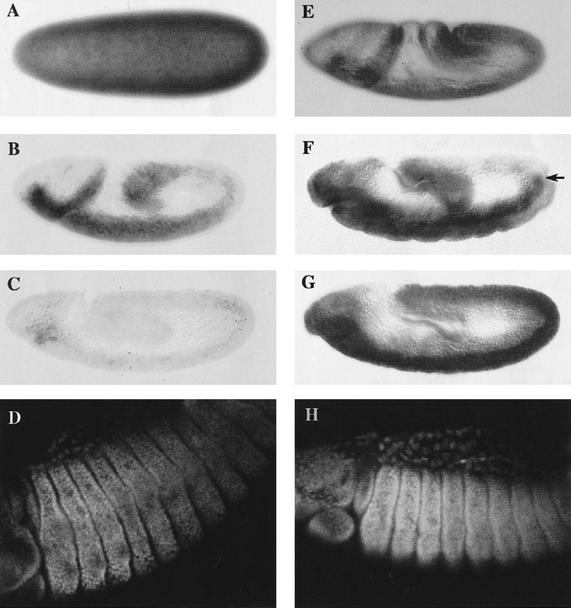
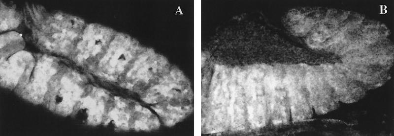
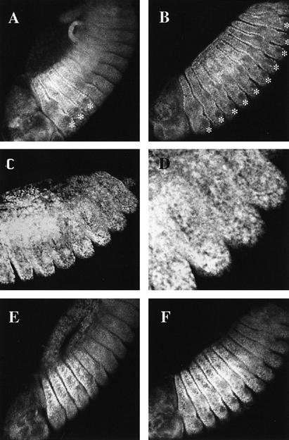
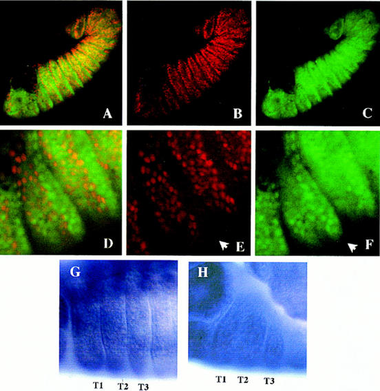
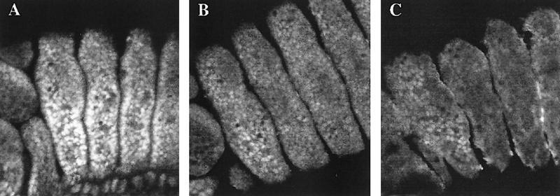
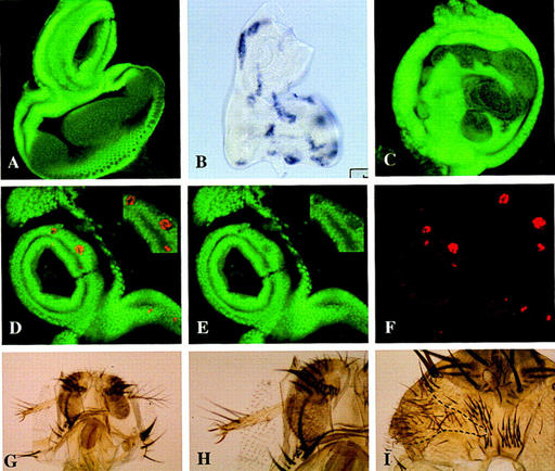
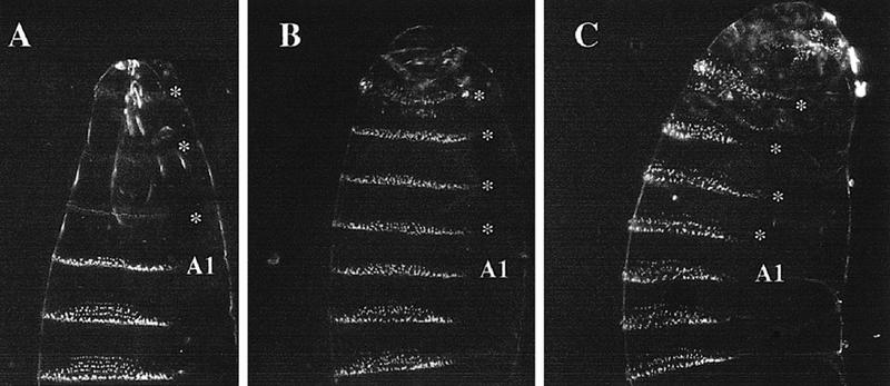
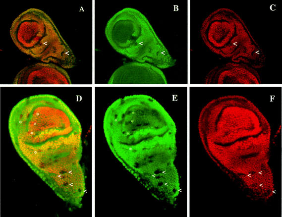
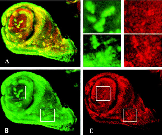
The homeobox gene extradenticle (exd) acts as a cofactor of Hox function both in Drosophila and vertebrates. It has been shown that the distribution of the Exd protein is developmentally regulated at the post-translational level; in the regions where exd is not functional Exd is present only in the cell cytoplasm, whereas it accumulates in the nuclei of cells requiring exd function. We show that the subcellular localization of Exd is regulated by the BX-C genes and that each BX-C gene can prevent or reduce nuclear translocation of Exd to different extents. In spite of this negative regulation, two BX-C genes, Ultrabithorax and abdominal-A, require exd activity for their maintenance and function. We propose that mutual interactions between Exd and BX-C proteins ensure the correct amounts of interacting molecules. As the Hoxd10 gene has the same properties as Drosophila BX-C genes, we suggest that the control mechanism of subcellular distribution of Exd found in Drosophila probably operates in other organisms as well.


Figures

Figure 1
Figure 1
Distribution of exd RNA and protein in maternal and zygotic exd mutants. All panels show embryos with anterior to the left and dorsal up. (A–D) Embryos of exdY012 genotype and therefore, have Exd product exclusively from maternal origin (mat+ zyg). (E–H) Embryos containing only Exd product of zygotic origin (mat zyg+). (A) mat+ zyg embryo at the blastoderm stage stained for exd RNA. The maternal product is ubiquitously distributed throughout the embryo. (B) mat+ zyg embryo at stage 8–9 of embryogenesis. Maternal exd RNA is still present, but at lower levels. (C) Stage 10 embryo of the same genotype in which the maternal RNA is almost undetectable. (D) mat+ zyg embryo after germ-band retraction stained for Exd protein. Although no maternal RNA is detectable in the embryo from stage 11 onwards, the protein is still present in later stages of embryogenesis. Note the nuclear localization of Exd in the cells of thoracic segments. (E) Stage 8 mat zyg+ embryo lacking the maternal contribution stained for exd mRNA. The zygotic product is present throughout the body. (F) Stage 11 embryo showing a modulation in the levels of zygotic RNA along the A/P axis. In the ectoderm there is very little expression in the more posterior segments, but note that there is mesodermal exd expression (arrow) that is not in register with the ectoderm. (G) Embryo of approximately the same age as in F stained with the anti-Exd antibody. The Exd zygotic protein is still present in the posterior segments of the embryo, where the mRNA is not detected anymore. (H) Stage 14 mat zyg+ embryo stained with anti-Exd antibody. The zygotic protein shows high levels of nuclear distribution in the thoracic segments and lower levels in the abdominal segments. The distribution of the zygotic protein is the same as the maternal one, but the levels of the maternal protein are lower (cf. D and H).
Figure 2
Figure 2
Ectopic expression of exd. (A) Stage 11 embryo with induced exd expression in the ptc domain stained for Exd protein. The levels of ectopic Exd are high in every anterior compartment. (B) Embryo with the same genotype as A shortly before completion of germ band retraction, close to stage 12, stained as in A. The high levels of Exd are not observed in the posterior end of the embryo where Exd is clearly cytoplasmic.
Figure 3
Figure 3
Effect of BX-C mutations on the subcellular localization of Exd. (A) Wild-type distribution (see text for details). Note the predominant nuclear localization in the three thoracic segments, except in the precursors of the Keilin’s organs (*). (B) Exd distribution in homozygous Df(3R)P9 embryos lacking the BX-C genes. Exd is nuclear in all the thoracic and abdominal segments. Because of the homeotic transformation, all thoracic and abdominal segments develop Keilin’s organs whose precursor cells (*) have cytoplasmic Exd. (C) Exd localization in a mat+ zyg embryo also homozygous for Df(3R)P9. The product is nuclear and evenly localized in thoracic and abdominal segments. (D) Magnification of the posterior segments of the embryo in C. (E) Embryo homozygous for the UbxPlacZ mutation. The region of high nuclear expression of Exd extends to the A1 segment (cf. A). (F) Embryo homozygous for the UbxMX9 abd-AMI chromosome and, therefore, lacking the Ubx and abd-A functions. The region of high nuclear Exd expression now extends to segment A4 (cf. with A and E).
Figure 4
Figure 4
Ectopic expression of Ubx in embryos. (A–F) Exd antigen is in green, and Ubx is in red. (A) Embryo with Ubx ectopic expression in the ptc domain doubly stained for Exd and Ubx proteins. The domains of high nuclear expression of both proteins do not overlap. (B) Same embryo as A, showing ectopic Ubx protein in the anterior compartments. (C) exd expression in the same embryo as A. Nuclear Exd localization in the thoracic segments is restricted to the posterior compartment, where no ectopic Ubx is induced. (D) Higher magnification of the thoracic segments of the embryo in A, showing that Ubx and Exd are mutually exclusive. (E) Ubx expression induced in the thorax by the ptc–Gal4 driver. The posterior compartments do not show ectopic Ubx (arrow). (F) Expression of exd in the same region as in E. Nuclear protein is mostly confined to the posterior compartments (arrow). (G, H) exd RNA expression in the thoracic segments (T1, T2, and T3) of wild-type (G) and in ptc–Gal4/UAS–Ubx embryo (H), showing no significant differences.
Figure 5
Figure 5
Ectopic expression of Ubx and Abd-B in embryos shows their different ability in preventing the nuclear translocation of Exd. (A) Stage 13 wild-type embryo showing the nuclear distribution of Exd in the three thoracic segments, aligned left to right. (B) Stage 13 embryo of genotype Antp–Gal4/UAS–Ubx reared at 17°C stained for Exd protein. The Antp domain occupies the posterior part of the first thoracic to the end of the third thoracic segment (see text). The moderate excess of Ubx protein in the second and third thoracic segments reduce, but do not eliminate, the nuclear transport of Exd. Note higher levels of nuclear Exd in the anterior region of the first thoracic segments, outside the Antp domain. (B) Stage 13 embryo of the genotype Gal4–Antp/UAS–Abd-B, also reared at 17°C. The presence of the Abd–Bm protein abolishes Exd nuclear expression, except in the anterior first thoracic segment.
Figure 6
Figure 6
Ectopic expression of Hoxd10 or Ubx prevents the nuclear translocation of Exd in the antennal cells. (A) Confocal image with the expression of Exd in a wild-type eye–antennal disc. Exd is nuclear in the proximal and cytoplasmic in the more distal regions. (B) Eye–antennal disc dissected from a larvae of FLP; Arm>y>Gal4/UAS–Hoxd10 genotype heat shocked at 37°C and stained for Hoxd10 mRNA. Clones of Hoxd10-expressing cells can be detected all over the disc. (C) Confocal image of an eye–antennal disc from a larva of the same genotype and treated as in B, stained with anti-exd antibody. The patches of cytoplasmic Exd are likely to be clones of Hoxd10-expressing cells in which the nuclear translocation of Exd is prevented. (D–F) Clones of Ubx-expressing cells (red) affecting Exd (green) localization. (D) Confocal image of an eye–antennal disc with several clones containing ectopic Ubx activity stained for Exd and Ubx. No overlapping of the nuclear expression of both proteins is observed. (E) Confocal image of the same disc as D, stained for Exd and showing cytoplasmic expression. (F) The same portion of the disc as in E showing the clones containing Ubx expression. (G) Adult head with a clone of Hoxd10-expressing cells marked with y and showing a leg transformation. (E) Higher magnification of the clone shown in G. Note the presence of two claws. (F) Clone of Hoxd10+ cells (encircled) in the head capsule exhibiting a transformation toward thorax.
Figure 7
Figure 7
Different amounts of Ubx protein cause different transformations. (A) Ventral aspect of a wild-type larva showing head, thorax, and the first three abdominal segments. The first abdominal segment (A1) has a characteristic thin belt of denticles, whereas those of more posterior abdominal segments are wider and with a trapezoidal shape. (B) Larva of genotype Gal4-444; UAS–Ubx, grown at 17°C, in which Ubx has been overexpressed during embryogenesis (see text). At this temperature the three thoracic and one head segments (*) are transformed toward A1, and the A1 segment remains unaltered. (C) Larva of the same genotype as B grown at 28°C. The greater amount of Ubx protein produces a transformation of A1, the three thoracic segments, and one head segment (*) into an A3–A5 segment type.
Figure 8
Figure 8
exd function is necessary to maintain Ubx expression in the haltere imaginal disc. (A–F) Exd protein is in green; Ubx is in red. Overlapping regions of expression appear yellow. (A–C) Confocal image of a haltere disc with exd clones induced at 48–72 hr, doubly stained for Exd and Ubx. exd clones are detected by the lack of Exd protein. The upper pointer (<) indicates an exd clone crossing the border of nucleo/cytoplasmic exd expression. The portion of the clone in the appendage part contains Ubx protein, but in the proximal part of the clone Ubx protein is not detected. Other exd clones showing loss of Ubx antigen are also visible in the thoracic part of the haltere (<). (B, C) The same clones in the green (Exd) and red (Ubx) channels. (D–F) Confocal image of another haltere disc with several small exd clones induced at 72–96 hr of development and double-stained as in A–C. exd clones in the haltere pouch (*) have no effect on Ubx expression, whereas many of those in the metanotum (<) reduces Ubx expression.
Figure 9
Figure 9
Effect of increasing levels of Exd on Ubx expression in the haltere imaginal disc. Confocal images of a third instar disc containing clones of excess of exd expression double-stained with anti-Exd (green) and anti-Ubx (red) antibody. (A) Double staining of the disc, where cells with both proteins appear yellow. Clones can be seen in the proximal and distal part of the disc and are detected by higher levels of the two proteins. (B) The same disc only showing Exd protein. (C) Ubx protein in the same disc to show the general correspondence between the increase of Exd and Ubx proteins. Insets in B and C are amplified in the upper right panel for a better assessment of the protein levels in the clones. Note that the excess of Exd protein in the clones in the pouch gives rise to nuclear Exd localization and that the increase of nuclear Exd produces an increase of Ubx protein, although the correspondence is sometimes not strictly cell by cell.

Similar articles

Cited by

References

    1. Aspland SE, White RAH. Nucleocytoplsmic localisation of extradenticle protein is spatially regulated throughout development in Drosophila. Development. 1997;124:741–747. - PubMed
    1. Bachiller D, Macias A, Duboule D, Morata G. Conservation of a functional hierarchy between mammalian and insect Hox/HOM genes. EMBO J. 1994;13:1930–1941. - PMC - PubMed
    1. Brand AH, Perrimon N. Targeted gene expression as a means of altering cell fates and generating dominant phenotypes. Development. 1993;118:401–415. - PubMed
    1. Casanova J, Sanchez-Herrero E, Morata G. Identification and characterisation of a parasegment specific regulatory element of the Abdominal-B gene of Drosophila. Cell. 1986;47:627–636. - PubMed
    1. Casares F, Calleja M, Sanchez-Herrero E. Functional similarity in appendage specification by the Ultrabithorax and abdominal-A Drosophila Hox genes. EMBOJ. 1996;15:3934–3942. - PMC - PubMed

Publication types

LinkOut - more resources