. 1998 Jan 15;12(2):274-84.
doi: 10.1101/gad.12.2.274.

DRhoGEF2 encodes a member of the Dbl family of oncogenes and controls cell shape changes during gastrulation in Drosophila

Affiliations

DRhoGEF2 encodes a member of the Dbl family of oncogenes and controls cell shape changes during gastrulation in Drosophila

U Häcker et al. Genes Dev. .

Abstract

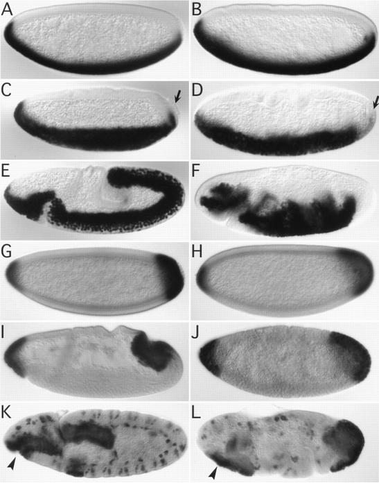
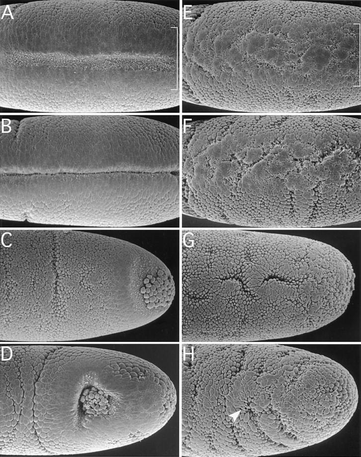
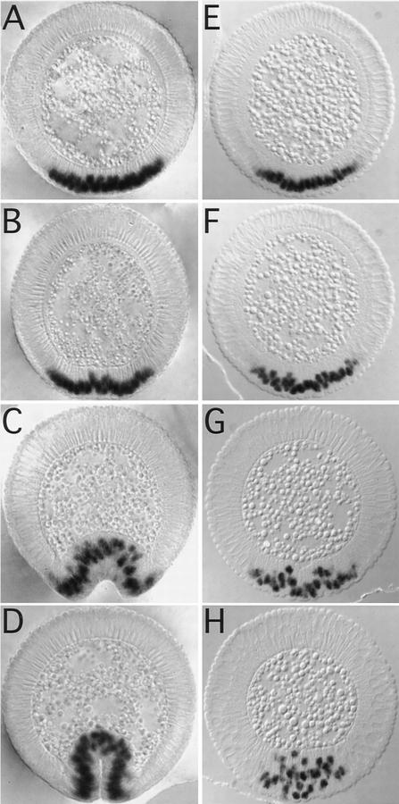
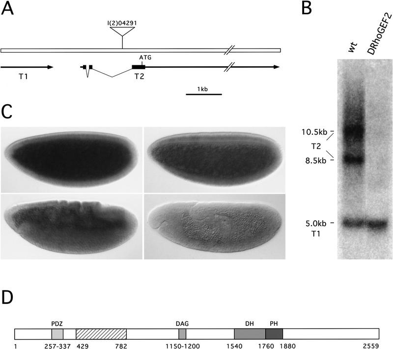
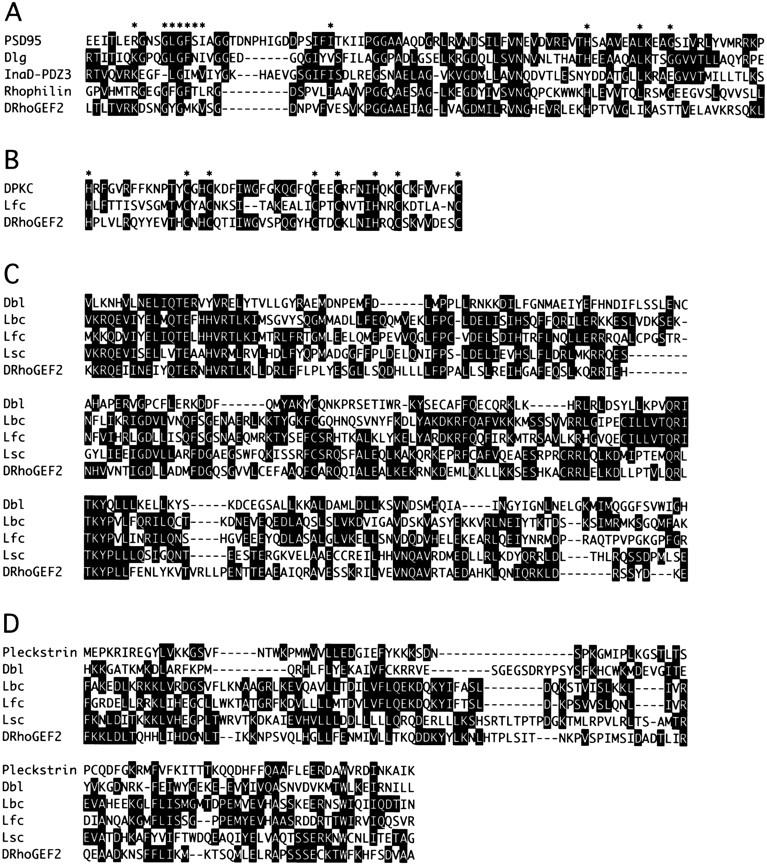
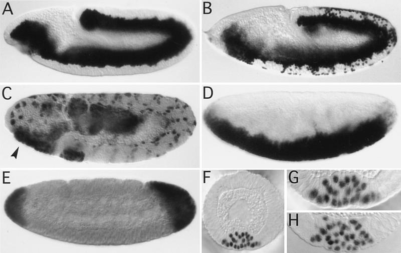
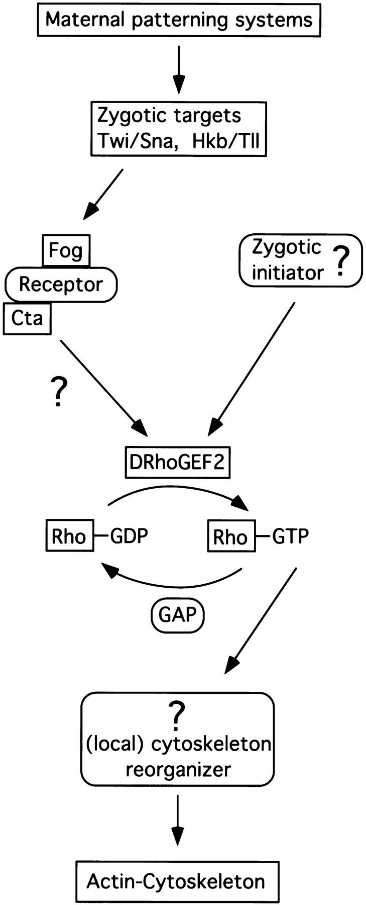
We have identified a gene, DRhoGEF2, which encodes a putative guanine nucleotide exchange factor belonging to the Dbl family of oncogenes. DRhoGEF2 function is essential for the coordination of cell shape changes during gastrulation. In the absence of maternal DRhoGEF2 gene activity, mesodermal and endodermal primordia fail to invaginate. The phenotype seen in DRhoGEF2 mutants is more severe than the defects associated with mutations in two previously identified gastrulation genes, folded gastrulation and concertina, suggesting that DRhoGEF2 acts in a signaling pathway independent of these genes. Expression of dominant-negative DRhoA during gastrulation results in phenocopies of the DRhoGEF2 mutant, suggesting that a signaling cascade involving DRhoGEF2 and the small GTPase DRhoA is responsible for the regulation of cell shape changes during early Drosophila morphogenesis.


Figures

Figure 1
Figure 1
Invagination of mesodermal and endodermal primordia in wild-type and DRhoGEF2 mutant embryos. All embryos are stained immunohistochemically for the transcription factors Twi (A–F) or Fkh (G–L). Expression of Twi is initiated in the wild-type (A) and DRhoGEF2l(2)04291 (B) embryos during syncytial blastoderm. At stage 6 the ventral furrow has invaginated in the wild type. The PMG primordium has flattened and moved to a dorsally shifted position (C). The pole cells are attached to the PMG primordium (arrow in C). In DRhoGEF2l(2)04291 embryos no ventral furrow is formed (embryo shown is at a slightly later stage than in C). The mesoderm is not extending to the posterior pole as in the wild type. The PMG primordium is not moving dorsally and the pole cells are deeply embedded into the epithelium (arrow in D). At stage 9 the mesoderm, with the exception of the most lateral cells of the primordium, has completely invaginated in the wild type. The germ band is extending around the posterior pole to the dorsal side of the embryo (E). In DRhoGEF2l(2)04291 embryos at the same stage the mesoderm remains on the surface of the embryo. The germ band is not moving around the posterior pole (F). Expression of Fkh is established in the PMG primordium at the late syncytial blastoderm stage in wild type (G) and DRhoGEF2l(2)04291 embryos (H). At stage seven the posterior midgut has started to invaginate in the wild type (I). In DRhoGEF2l(2)04291 embryos no invagination is formed at this stage. Note the folds forming in the dorsal epithelium indicating that the germ band has started to elongate (J). At stage 10 the PMG and the AMG (arrowhead) are fully internalized and have started to elongate in the wild type (K). In DRhoGEF2l(2)04291 embryos PMG and AMG remain uninvaginated (arrowhead). The germ band forms deep transverse folds (L).
Figure 2
Figure 2
Cell shape changes in wild type (A–D) and DRhoGEF2 mutants (E–H) analyzed by SEM. Following cellularization, cells in an ∼20-cell-wide stripe on the ventral surface of the embryo (indicated by brackets in A and E) flatten their surfaces and move into close contact with each other. Subsequently apical constriction of cells near the midline (notice the membrane blebs indicative of this process) forces the epithelium to bend inward and form a shallow groove (A). As more lateral cells constrict, the furrow deepens and closes over to invaginate the mesoderm (B). In DRhoGEF2l(2)04291 embryos the initial flattening of the cell surfaces appears to occur. However, only very few cells undergo apical constriction in a spatially disorganized fashion (E). As the germ band starts to elongate (indicated by the formation of transverse folds laterally) temporary clefts form at random in the ventral mesoderm (F). No ventral furrow is formed. The PMG primordium is invaginated by a very similar mechanism as the mesoderm. After the initial flattening, cells located dorsal to the pole cells constrict first, as seen by the occurrence of membrane blebs in this region (C). Subsequently a cup shaped invagination is formed harboring the pole cells (D). In DRhoGEF2l(2)04291 embryos, cells in the dorsal polar region do not undergo any specific shape changes and no invagination is formed. The pole cells are deeply embedded into the epithelium. On the dorsal surface of the embryo, groups of cells undergo apical flattening or apical constriction in random regional patterns (G). As the germ band starts to elongate some dorsal cells form cytoplasmic protrusions (arrowhead in H). Altogether cell shape changes appear randomized (H). A, B, E, and F are ventral views; C, D, G, and H are dorsal views. All embryos are oriented with anterior to the left.
Figure 3
Figure 3
Analysis of cell shape changes during ventral furrow formation in wild-type (A–D) and in DRhoGEF2 (E–H) mutants in cross sections immunohistochemically stained for Twi. Shortly after cellularization is completed on the ventral side of the embryo (A), nuclei of cells at the ventral midline start to move basally (B). Apical constriction of midventral cells forces the epithelial sheet to bend inward (B,C). Shortening of cells along their apical–basal axis supports this process and results in internalization of the mesoderm (D). DRhoGEF2l(2)04291 embryos appear normal during cellularization. Cell nuclei are lined up along the apical end of cells (E). At the onset of gastrulation, nuclei are seen to move basally in a random manner (F). Very few cells undergo apical constriction (G). Cells do not shorten along their apical basal axes; they lose their epithelial character and pile up in several layers. No ventral furrow is formed (H).
Figure 4
Figure 4
Molecular cloning, Northern analysis, and expression of the DRhoGEF2 gene. (A) Genomic map of the region surrounding the P-element insertion l(2)04291. Two transcription units, T1 and T2, were identified near the P-element insertion point. The P element was found to be inserted into an intron in the untranslated leader region of the T2 transcript. The 3′ end of T2 was mapped ∼20 kb from the P-element insertion. Solid boxes indicate exons; arrows denote transcribed regions. (B) Northern blot analysis of T1 and T2 on poly(A)+ RNA derived from 0- to 12-hr wild type (left lane) and DRhoGEF2l(2)04291 (right lane) embryos. A single T1 transcript, 5.0 kb in length, is detected by the T1 cDNA in RNA prepared from wild-type and DRhoGEF2l(2)04291 embryos, suggesting that the P insertion is not interfering with the expression of this gene. In wild-type RNA cDNAs of T2 detect two transcripts, 8.5 and 10.5 kb in length. Both transcripts are absent in RNA derived from DRhoGEF2l(2)04291 embryos indicating that expression of T2 is abolished by the P element insertion. (C) In situ hybridization of a genomic DNA fragment, encompassing part of the T2 transcription unit, to wild-type embryos. DRhoGEF2 transcripts are abun dant at the syncytial blastoderm stage (top left). DRhoGEF2 mRNAs are distributed evenly throughout the embryo during gastrulation (top right and bottom left). DRhoGEF2 transcripts are no longer detectable after extension of the germ band (bottom right). (D) Schematic representation of the DRhoGEF2 protein. The DRhoGEF2 cDNA contains an ORF for a protein of 2559 amino acids with several regions of homology to sequences present in the databases. A PDZ domain is located near the amino terminus (PDZ). In the central region DRhoGEF2 contains a cysteine-rich diacylglycerol-binding motif (DAG). A DH and a PH domain are located in tandem in the carboxy-terminal half of the protein and identify DRhoGEF2 as a putative GEF for small GTPases of the Rho family. The hatched box in the amino-terminal third of the protein represents the region not encoded in the shorter version of the cDNA (see Materials and Methods). Numbers represent amino acid positions.
Figure 5
Figure 5
Conserved sequence motifs found in DRhoGEF2. (A) Sequence alignment of the PDZ domains of PSD95, Discs large (Dlg), the third PDZ domain of InaD, Rhophilin, and DRhoGEF2. Stars above the sequence indicate residues implicated in substrate binding. (B) Sequence alignment of the DAG-binding motifs of PKC (Drosophila eye isoform), the mouse Dbl family oncogene Lfc, and DRhoGEF2. Stars indicate conserved histidine and cysteine residues. (C) Sequence alignment of the DH domains of the human oncoproteins Dbl and Lbc, the mouse oncoproteins Lfc, and Lsc, and DRhoGEF2. (D) Sequence alignment of the PH domains of Pleckstrin and the Dbl-family genes Dbl, Lbc, Lfc, Lsc, and DRhoGEF2. Sequences where aligned by the Clustal method using DNASTAR software. Solid boxes indicate residues identical to a consensus sequence.
Figure 6
Figure 6
Expression of dominant-negative forms of Rho family GTPases during gastrulation. Embryos are stained immunohistochemically for either Twi (A,B,D,F–H) or Fkh (C,E). In wild-type embryos all mesodermal cells have been internalized at stage 10 (A). In twiGAL4/UASDRhoAN19 embryos at the same stage patches of mesodermal cells along the germ band have failed to invaginate (B). Invagination of the AMG is blocked (arrowhead in C, cf. Fig. 1L). (D–G) matα4-GAL–VP16/UASDRhoAN19 embryos: The phenotype caused by the maternal expression of dominant-negative DRhoA strongly resembles that seen in DRhoGEF2l(2)04291 embryos. No ventral furrow is formed (D; cf. Fig. 1D) and no PMG invagination is seen in the posterior dorsal region (E, cf. Fig. 1J). Cells fail to undergo shape changes during ventral furrow formation (F,G) in a way very similar to DRhoGEF2l(2)04291 embryos (H).
Figure 7
Figure 7
Model of the signaling events regulating cell shape changes during gastrulation. Boxed components have been identified in Drosophila. Boxes with rounded corners represent postulated components. (see text for details).

Similar articles

Cited by

References

    1. Ahmed S, Kozma R, Lee J, Monfries C, Harden N, Lim L. The cysteine-rich domain of human proteins, neuronal chimaerin, protein kinase C and diacylglycerol kinase binds zinc. Evidence for the involvement of a zinc-dependent structure in phorbol ester binding. Biochem J. 1991;280:233–241. - PMC - PubMed
    1. Bell RM, Burns DJ. Lipid activation of protein kinase C. J Biol Chem. 1991;266:4661–4664. - PubMed
    1. Brand AH, Perrimon N. Targeted gene expression as a means of altering cell fates and generating dominant phenotypes. Development. 1993;118:401–415. - PubMed
    1. Brönner G, Chu-LaGraff Q, Doe CQ, Cohen B, Weigel D, Taubert H, Jäckle H. Sp1/egr-like zinc-finger protein required for endoderm specification and germ-layer formation in Drosophila. Nature. 1994;369:664–668. - PubMed
    1. Brown NH, Kafatos FC. Functional cDNA libraries from Drosophila embryos. J Mol Biol. 1988;203:425–437. - PubMed

Publication types

Substances

Associated data

LinkOut - more resources