. 1998 Jan;9(1):47-61.
doi: 10.1091/mbc.9.1.47.

Developmental expression of spectrins in rat skeletal muscle

Affiliations
Free PMC article

Developmental expression of spectrins in rat skeletal muscle

D Zhou et al. Mol Biol Cell. 1998 Jan.
Free PMC article

Abstract

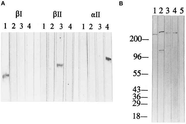
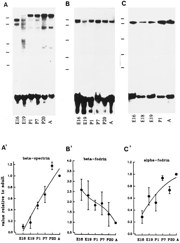
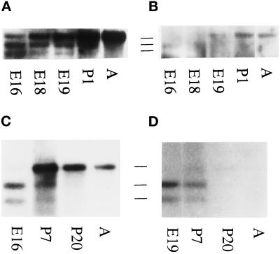
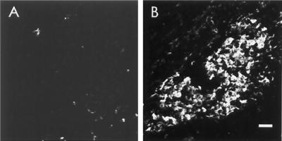
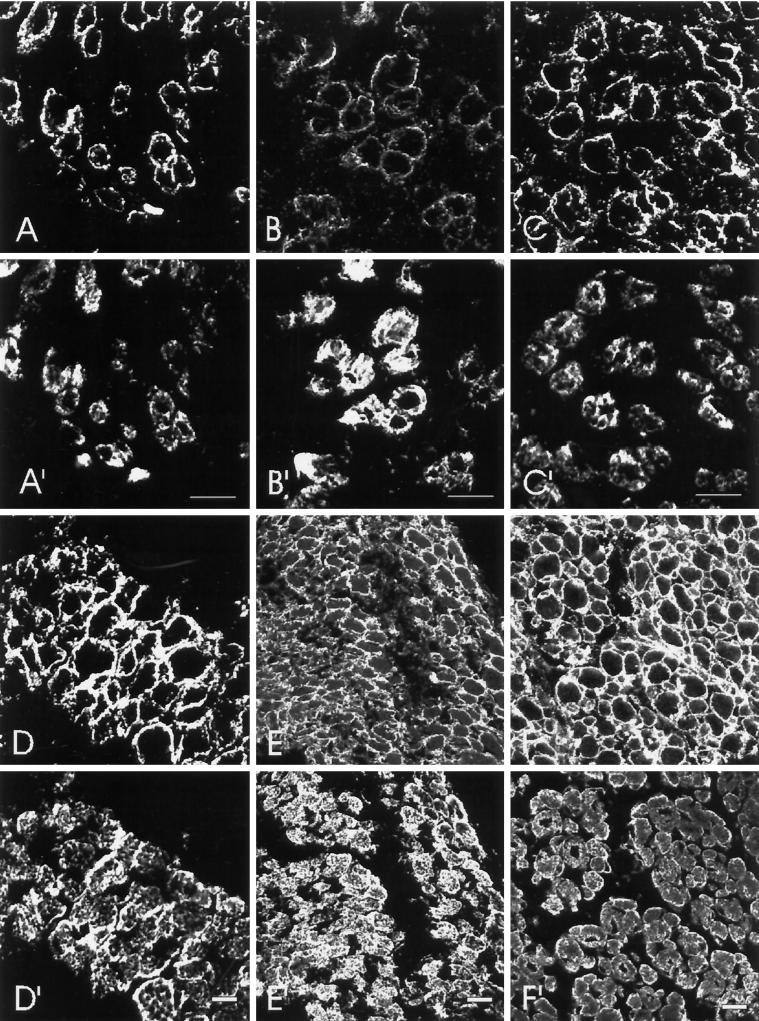
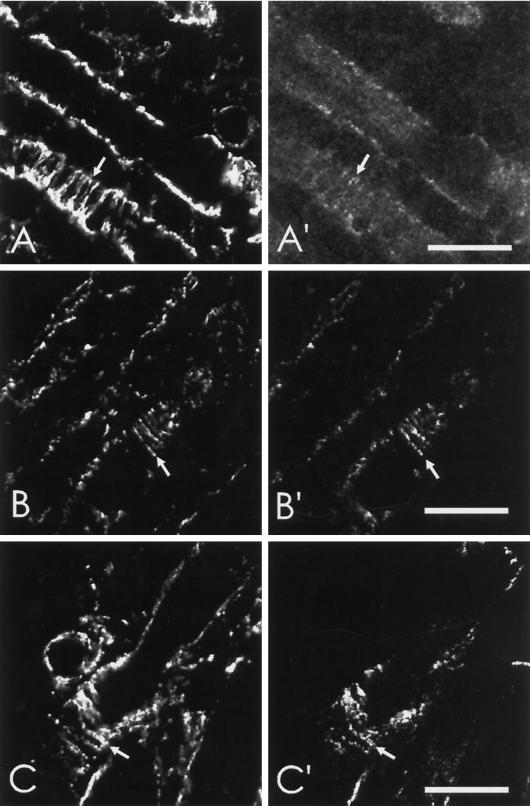
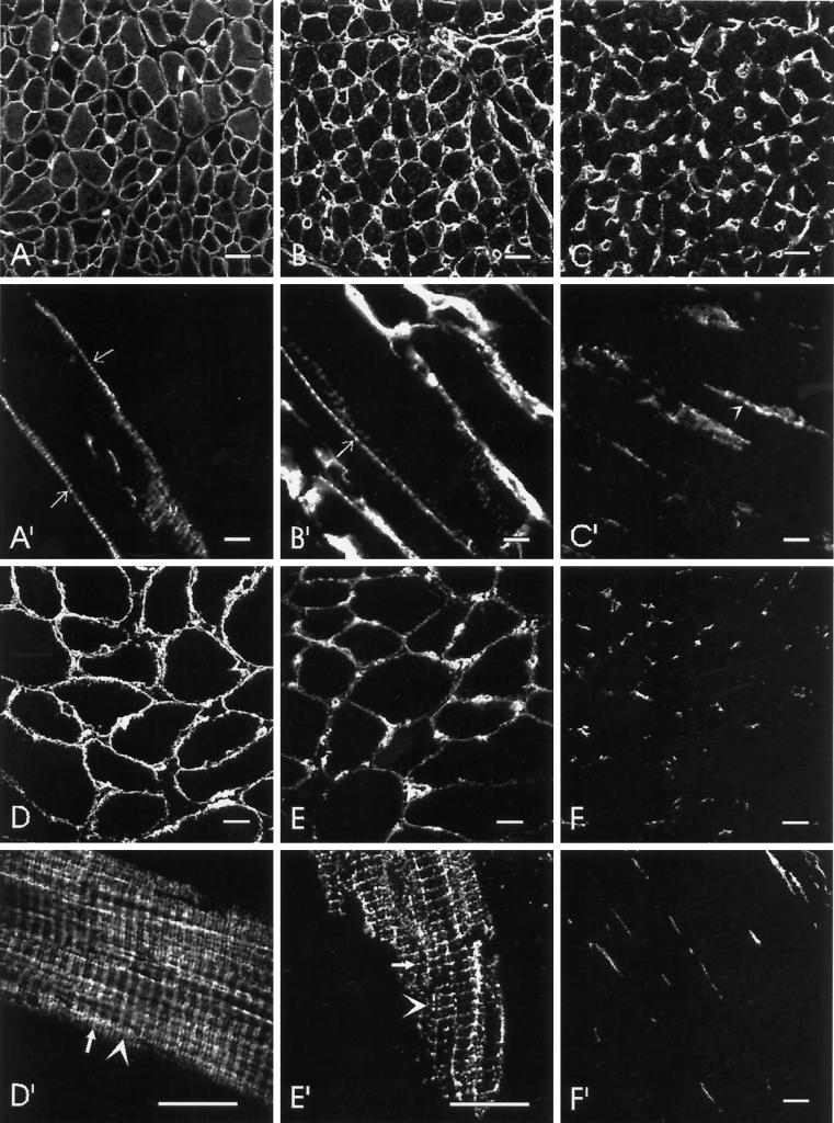
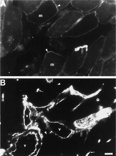
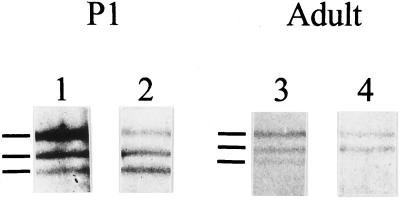
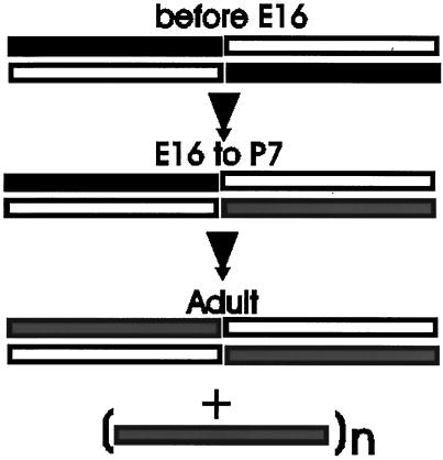
Skeletal muscle contains spectrin (or spectrin I) and fodrin (or spectrin II), members of the spectrin supergene family. We used isoform-specific antibodies and cDNA probes to investigate the molecular forms, developmental expression, and subcellular localization of the spectrins in skeletal muscle of the rat. We report that beta-spectrin (betaI) replaces beta-fodrin (betaII) at the sarcolemma as skeletal muscle fibers develop. As a result, adult muscle fibers contain only alpha-fodrin (alphaII) and the muscle isoform of beta-spectrin (betaISigma2). By contrast, other types of cells present in skeletal muscle tissue, including blood vessels and nerves, contain only alpha- and beta-fodrin. During late embryogenesis and early postnatal development, skeletal muscle fibers contain a previously unknown form of spectrin complex, consisting of alpha-fodrin, beta-fodrin, and the muscle isoform of beta-spectrin. These complexes associate with the sarcolemma to form linear membrane skeletal structures that otherwise resemble the structures found in the adult. Our results suggest that the spectrin-based membrane skeleton of muscle fibers can exist in three distinct states during development.


Figures

Figure 1
Figure 1
Specificity of anti-spectrin antibodies determined by immunoblotting. Purified proteins (A) or tissue extracts (B) were boiled in sample buffer, subjected to SDS-PAGE in gels containing 5% acrylamide (A) or 5–15% acrylamide gradients (B), transferred to nitrocellulose, and subjected to immunoblotting with antibodies generated against β-spectrin (βIΣ*), β-fodrin (βIIΣ*), and α-fodrin (αIIΣ*) that were affinity purified and cross-adsorbed (see MATERIALS AND METHODS). (A) Lanes 1–4 contain purified β-spectrin, α-spectrin, β-fodrin, and α-fodrin, respectively. (B) Immunoblots of rat muscle or hippocampal (lane 4 only) extracts were incubated with antibodies to β-spectrin (lane 1), α-fodrin (lane 2), β-fodrin (lanes 3 and 4), or to a control rabbit IgG (lane 5). Anti-β-spectrin recognizes a single band or a closely spaced doublet at ∼265 kDa. Anti-α-fodrin recognizes a single band at ∼280 kDa and a well-characterized proteolytic fragment (Harris et al., 1988) at ∼150 kDa. Anti-β-fodrin reacts faintly with bands at ∼270 and ∼170 kDa in extracts of skeletal muscle (lane 3) but reacts strongly with a single band at ∼270 kDa in hippocampal extracts (lane 4). The results indicate that each of the antibodies reacts specifically with its antigen and not with other proteins in tissue extracts or with the other spectrins assayed.
Figure 2
Figure 2
Developmental expression of β-spectrin, α-fodrin, and β-fodrin in rat skeletal muscle analyzed by Northern blot. RNAs were isolated from rat hindlimb muscle at different ages, fractionated on 1% agarose gels, and transferred to nylon membranes. Three different membranes were hybridized to cDNA probes specific for β-spectrin (A), β-fodrin (B), and α-fodrin (C). The membranes were then stripped and hybridized with probes to 18S rRNA to indicate the relative amounts of RNA loaded (shown at the bottom of each blot). After normalization against the rRNA signal, the level of expression of each message at each age was plotted relative to its corresponding adult value: A′, β-spectrin (only the 11-kb transcript is shown); B′, β-fodrin; C′, α-fodrin. The values were averages of at least three independent experiments; values for the mean ± SEM molecular size standards, indicated to the left of each blot, are (from top to bottom) 9.5, 7.5, 4.4, 2.4, and 1.4 kb. The developmental stages tested were E16, E18, and E19 (embyronic days 16, 18, and 19), P1 (postnatal day 1), and adult (A).
Figure 3
Figure 3
Isoforms of β-spectrin encoded by the 7.5-, 9.0-, and 11-kb transcripts. Two RNA blots were hybridized to a probe shared by both erythroid and skeletal muscle β-spectrin cDNA (panels A and C) and show three bands at 11, 9.0, and 7.5 kb. The membranes were then stripped and rehybridized with probes for muscle-specific (βIΣ2, panel B) and erythrocyte-specific (βIΣ1, panel D) 3′ sequences. The results indicate that the predominant 11-kb transcript in postnatal muscle is muscle specific, while the 7.5- and 9.0-kb transcripts encode the alternatively spliced form found in erythrocytes.
Figure 4
Figure 4
Absence of erythrocyte β-spectrin in E16 muscle fibers. Cryosections of E16 rat hindlimb were double-labeled with polyclonal antibodies to the extreme C-terminal sequence of erythroid β-spectrin (βIΣ1, panel A) and monoclonal antibodies to desmin (panel B), to identify the muscle cells, followed by rhodamine-conjugated anti-rabbit and FITC-conjugated anti-mouse secondary antibodies. The result shows that the desmin-positive muscle cells seen in E16 hindlimb do not contain the erythrocyte isoform of β-spectrin. Bar, 20 μm.
Figure 5
Figure 5
Presence of β-spectrin, α-fodrin, and β-fodrin at the sarcolemma of embryonic rat skeletal muscle. Cross-sections of E16 (A–C, A′-C′) and E19 (D–F, D′–F′) rat diaphragm were labeled with isoform-specific polyclonal antibodies to β-spectrin (A and D), α-fodrin (B and E), and β-fodrin (C and F) along with monoclonal antibodies to desmin (A′–F′). The results show that all three subunits are expressed in embryonic rat skeletal muscle cells, where they assemble at or near the sarcolemma. Bar, 20 μm.
Figure 6
Figure 6
β-Fodrin and α-fodrin codistribute with β-spectrin at the sarcolemma of embryonic skeletal muscle. Longitudinal sections of E16 (A and A′) or E19 (B, B′, C, and C′) diaphragm were double-labeled with polyclonal antibodies to α-fodrin (A and B) or β-fodrin (C) and 4C3 monoclonal antibodies to β-spectrin (A′–C′). Each pair of labeled subunits codistributes in strands at the sarcolemma of embryonic muscle fibers (arrows). Bar, 20 μm.
Figure 7
Figure 7
Spectrins in P7 and adult rat skeletal muscle. Cross-sections (A–F) and longitudinal sections (A′–F′) of 7-d-old (A–C, A′–C′) and adult (D–F, D′–F′) rat diaphragms were labeled with polyclonal antibodies to β-spectrin (A, A′, D, and D′), α-fodrin (B, B′, E, and E′), and β-fodrin (C, C′, F, and F′). The labeling of β-fodrin seen in C and C′ is mostly in capillaries, which are also labeled by anti-α-fodrin (B and B′), but not by anti-β-spectrin. Labeling of the sarcolemma by anti-β-fodrin at P7 is barely detectable. However, some residual punctate labeling could still be seen in longitudinal sections (e.g., arrowheads in C′). By contrast, β-spectrin and α-fodrin are readily apparent at the sarcolemma of P7 muscle, where they are present at punctate structures typical of costameres (e.g., arrows in A′ and B′). β-Spectrin is seen over both the Z lines and M lines in adult muscle (D′, Z line indicated by arrow; M line indicated by arrowhead), with the labeling pattern split into two lines over the Z line. α-Fodrin also localizes over the Z line, but does not show a split there; it is present at M lines to a much lesser extent (E′). Note also that labeling by anti-β-spectrin but not by anti-α-fodrin is present in longitudinally oriented structures, as previously reported (Porter et al., 1997). Bars: A–F, D′–F′, 20 μm; A′–C′, 10 μm.
Figure 8
Figure 8
β-Fodrin and β-spectrin in adult rat skeletal muscle. Cross-sections of adult rat diaphragm were double-labeled with monoclonal antibodies to β-spectrin (A) and polyclonal antibodies to β-fodrin (B). Although anti-β-fodrin antibodies label the blood vessels (v) and nerves (n) brightly, β-fodrin was not detected at the sarcolemma of skeletal myofibers (white arrowheads). β-Spectrin was found at the sarcolemma (A, white arrowheads) and was particularly concentrated at the neuromuscular junction (B, double black arrowhead). Capillaries are indicated with small white arrows; β-fodrin at the membrane of a cell in the blood vessel wall is indicated with a small black arrow. Nerve fibers are indicated with single black arrowheads. m, muscle fiber; v, blood vessel. Bar, 20 μm.
Figure 9
Figure 9
Immunoprecipitation of spectrin and fodrin from skeletal muscle using anti-fodrin antibodies. Spectrin and fodrin were immunoprecipitated from homogenates of P1 (lanes 1 and 2) and adult skeletal muscle (lanes 3 and 4) using anti-α-fodrin (lanes 1 and 3) or anti-β-fodrin (lanes 2 and 4) antibodies. The immunoprecipitated products were electrophoresed on 5% acrylamide gels and visualized by silver staining. The immunoprecipitated proteins are marked by lines to the left of each set. From top to bottom, they are: α-fodrin, β-fodrin, and muscle β-spectrin.
Figure 10
Figure 10
Proposed model of changes in spectrin during skeletal muscle development. Open bar, α-fodrin; solid bar, β-fodrin; shaded bar, muscle β-spectrin. Our results can most easily be explained by a progressive change in the composition of spectrin tetramers as muscle fibers develop. Specifically, we propose that embryonic myoblasts and early myotubes contain tetramers of α- and β-fodrin (Weed, 1996), but that mixed tetramers containing these two subunits together with muscle β-spectrin begin to form with the onset of myogenesis and persist through early postnatal life. Adult muscle fibers contain heteromers composed of only α-fodrin and β-spectrin as well as β-spectrin that assembles at the sarcolemma without α-fodrin (Porter et al., 1997); the state of oligomerization of the latter population of β-spectrin is still unknown. This model does not include the proteins that bind to spectrin (e.g., actin, adducin, ankyrin, band 4.1) or other members of the spectrin superfamily that have not yet been identified, and so it may need to be revised when the associations of these proteins with spectrin are studied in mature and developing muscle fibers.

Similar articles

Cited by

References

    1. Appleyard ST, Dunn MJ, Dubowitz V, Scott ML, Pittman SJ, Shotton DM. Monoclonal antibodies detect a spectrin-like protein in normal and dystrophic human skeletal muscle. Proc Natl Acad Sci USA. 1984;81:776–780. - PMC - PubMed
    1. Beck KA, Nelson WJ. The spectrin-based membrane skeleton as a membrane protein-sorting machine. Am J Physiol. 1996;270:C1263–1270. - PubMed
    1. Bennett V. Protein involved in membrane-cytoskeleton association in human erythrocytes: spectrin, ankyrin, and band 3. Methods Enzymol. 1983;96:313–324. - PubMed
    1. Bennett V, Baines AJ, Davis J. Purification of brain analogs of red blood cell membrane skeletal proteins: ankyrin, protein 4.1 (synapsin), spectrin, and spectrin subunits. Methods Enzymol. 1986;134:55–69. - PubMed
    1. Bennett V, Gilligan DM. The spectrin-based membrane skeleton and micron-scale organization of the plasma membrane. Annu Rev Cell Biol. 1993;9:27–66. - PubMed

Publication types