. 1998 Jan;9(1):89-101.
doi: 10.1091/mbc.9.1.89.

A fungal kinesin required for organelle motility, hyphal growth, and morphogenesis

Affiliations
Free PMC article

A fungal kinesin required for organelle motility, hyphal growth, and morphogenesis

Q Wu et al. Mol Biol Cell. 1998 Jan.
Free PMC article

Abstract

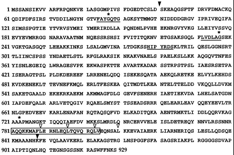
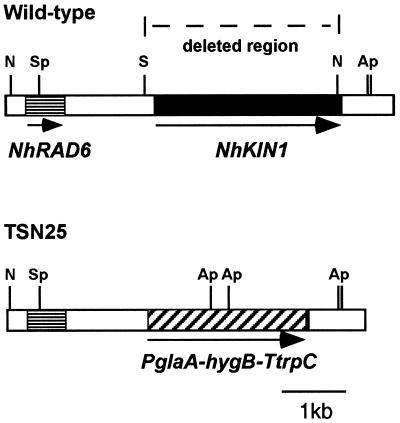
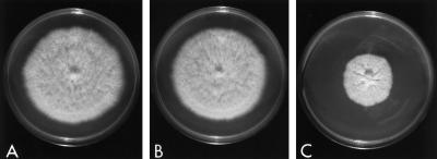
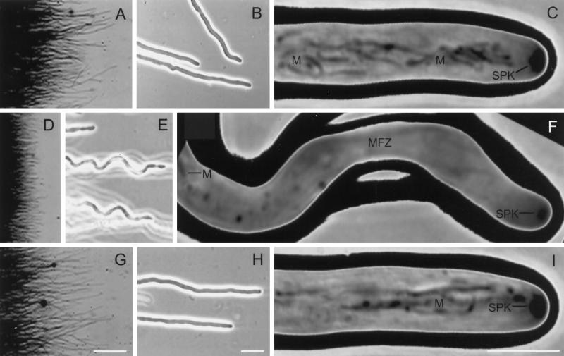
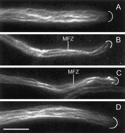
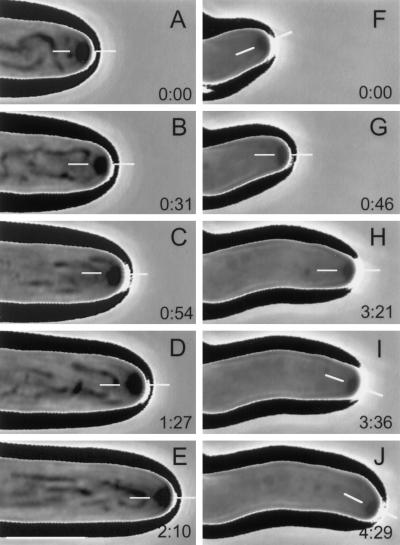
A gene (NhKIN1) encoding a kinesin was cloned from Nectria haematococca genomic DNA by polymerase chain reaction amplification, using primers corresponding to conserved regions of known kinesin-encoding genes. Sequence analysis showed that NhKIN1 belongs to the subfamily of conventional kinesins and is distinct from any of the currently designated kinesin-related protein subfamilies. Deletion of NhKIN1 by transformation-mediated homologous recombination caused several dramatic phenotypes: a 50% reduction in colony growth rate, helical or wavy hyphae with reduced diameter, and subcellular abnormalities including withdrawal of mitochondria from the growing hyphal apex and reduction in the size of the Spitzenkörper, an apical aggregate of secretory vesicles. The effects on mitochondria and Spitzenkörper were not due to altered microtubule distribution, as microtubules were abundant throughout the length of hyphal tip cells of the mutant. The rate of spindle elongation during anaphase B of mitosis was reduced 11%, but the rate was not significantly different from that of wild type. This lack of a substantial mitotic phenotype is consistent with the primary role of the conventional kinesins in organelle motility rather than mitosis. Our results provide further evidence that the microtubule-based motility mechanism has a direct role in apical transport of secretory vesicles and the first evidence for its role in apical transport of mitochondria in a filamentous fungus. They also include a unique demonstration that a microtubule-based motor protein is essential for normal positioning of the Spitzenkörper, thus providing a new insight into the cellular basis for the aberrant hyphal morphology.


Figures

Figure 1
Figure 1
Predicted amino acid sequence of NhKIN1. Filled arrowheads denote positions of introns. Splice junctions were verified by sequencing of NhKIN1 cDNA. Conserved amino acids from which degenerate primers were made are underlined. Asterisks indicate the first amino acid in each of three potential nucleotide-binding motifs, identified by the program PROSITE (Bairock, 1992). Boxed amino acids designate a highly conserved region shared among conventional kinesins, as noted for Nkin (Steinberg and Schliwa, 1995). Open arrowheads depict positions of leucines in the leucine zipper motif.
Figure 2
Figure 2
Deletion of NhKIN1 from the wild-type genome. The wild-type chromosome shows NhKIN1, the deleted region, and RAD6, an apparent homologue of genes encoding ubiquitin-conjugating proteins. The SphI-KpnI fragment of pTMS5 was used to delete most of NhKIN1. A double cross-over event resulted in the recombinant chromosome, which is deleted for a region starting 182 bp 5′ of the translational start site and stopping 26 bp 5′ of the translation stop site of NhKIN1. The deleted sequence was replaced with hygB. Certain restriction enzyme sites mentioned in the text are noted: N, NheI; Ap, ApaLI; K, KpnI; S, SacII; and Sp, SphI.
Figure 3
Figure 3
Growth of wild-type isolate T213 (A), NhKIN1+ transformant TSN20 (B), and Nhkin1 mutant TSN25, which lacks KIN1 (C), on CMX at 30°C for 6 d.
Figure 4
Figure 4
Videomicrographs depicting the development of distinctive mycelial, hyphal, and cellular characteristics of Nhkin1 deletion mutant TSN25 relative to those of NhKIN1+ transformant TSN20 and WT. (A), (D), and (G), Reverse dark-field images of colony margins of WT (A), TSN25 (D), and TSN20 (G), illustrating the relatively larger number of hyphal tip cells arranged more uniformly in the mutant (D) than in the other two isolates. (B), (E), and (H) Phase-contrast images. At low magnification, the mutant tip cells (E) exhibit a regular undulation that describes a spiral in three dimensions, whereas the WT (B) and TSN20 (H) exhibit relatively straight growth. (C), (F), and (I) Phase-contrast images. High magnification of apical portions of living hyphal tip cells, to illustrate some of the effects of NhKIN1 deletion on organelles. In the mutant cell (F), mitochondria (M) do not occupy the apical region (MFZ, mitochondria-free zone) as they do in the WT (C) and TSN20 (I) cells. Also, the Spitzenkörper (SPK) in the mutant is smaller than in either the WT or TSN20 cells. Bars: (A), (D), and (G), 0.5 mm; (B), (E), and (H), 20 μm; (C), (F), and (I), 5 μm.
Figure 5
Figure 5
Fluorescence videomicrographs showing immunocytochemical localization of MTs in hyphal tip cells of the wild type (A), the NhKIN1 deletion mutant TSN25 (B and C), and the NhKIN1+ transformant TSN20 (D). The MT distribution in the mutant cells appears normal, with MTs present throughout the mitochondrion-free zone (MFZ). White arcs mark the positions of the hyphal apices. Bar, 5 μm.
Figure 6
Figure 6
Phase-contrast, time-lapse videomicrographs of growing hyphal tip cells illustrating the effects of kinesin deletion on positioning of the Spitzenkörper and, consequently, on hyphal morphogenesis. Elapsed time (min:sec) is shown in the lower right corner of each panel, and thin, white bars in each panel indicate the position of the Spitzenkörper relative to the hyphal apex. (A–E) A representative tip cell of the ectopic transformant-control. The central position of the Spitzenkörper was maintained throughout, resulting in a straight hypha. (F–J) A representative tip cell of the kinesin mutant. Central positioning of the Spitzenkörper produced a short, straight segment of hyphae (F). Then the position of the Spitzenkörper was shifted (G) and maintained long enough to produce another straight segment of hypha (H) with a different growth orientation. Finally, another shift of the Spitzenkörper (I) produced another short segment of hypha (J) with yet another growth orientation. Bar (in the lower left corner of panel E), 5 μm.

Similar articles

Cited by

References

    1. Aist JR, Bayles CJ. Video motion analysis of mitotic events in living cells of the fungus Fusarium solani. Cell Motil Cytoskel. 1988;9:325–336.
    1. Aist JR, Bayles CJ. Detection of spindle pushing forces in vivo during anaphase B in the fungus Nectria haematococca. Cell Motil Cytoskel. 1991;19:18–24.
    1. Aist JR, Bayles CJ, Tao W, Berns MW. Direct experimental evidence for the existence, structural basis and function of astral forces during anaphase B in vivo. J Cell Sci. 1991;100:279–288. - PubMed
    1. Aist JR, Wu Q, Wirsel SG, Turgeon BG, Yoder OC. Disruption of a fungal kinesin gene affects both morphogenesis and mitosis. Mol Biol Cell. 1996;7:408a. - PMC - PubMed
    1. Allan V. Membrane traffic motors. FEBS Lett. 1995;369:101–106. - PubMed

Publication types

Associated data