. 1998 Feb 1;18(3):914-22.
doi: 10.1523/JNEUROSCI.18-03-00914.1998.

Interaction of presenilins with the filamin family of actin-binding proteins

Affiliations

Interaction of presenilins with the filamin family of actin-binding proteins

W Zhang et al. J Neurosci. .

Abstract

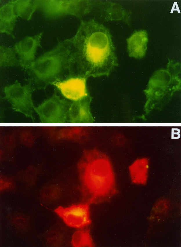
Mutations in presenilin genes PS1 and PS2 account for approximately 50% of early-onset familial Alzheimer's disease (FAD). The PS1 and PS2 genes encode highly homologous transmembrane proteins related to the Caenorhabditis elegans sel-12 and spe-4 gene products. A hydrophilic loop region facing the cytoplasmic compartment is likely to be functionally important because at least 14 mutations in FAD patients have been identified in this region. We report here that the loop regions of PS1 and PS2 interact with nonmuscle filamin (actin-binding protein 280, ABP280) and a structurally related protein (filamin homolog 1, Fh1). Overexpression of PS1 appears to modify the distribution of ABP280 and Fh1 proteins in cultured cells. A monoclonal antibody recognizing ABP280 and Fh1 binds to blood vessels, astrocytes, neurofibrillary tangles, neuropil threads, and dystrophic neurites in the AD brain. Detection of ABP280/Fh1 proteins in these structures suggests that these presenilin-interacting proteins may be involved in the development of AD and that interactions between presenilins and ABP280/Fh1 may be functionally significant. The ABP280 gene is located on the human X chromosome, whereas the newly identified Fh1 gene maps to human chromosome 3. These results provide a new basis for understanding the function of presenilin proteins and further implicate cytoskeletal elements in AD pathogenesis.


Figures

Fig. 1.
Fig. 1.
Alignment of ABP280 and Fh1 peptide sequences in the C-terminal region. Two clones of ABP280 cDNA and four clones of Fh1 cDNA were identified from screening a human brain cDNA yeast two-hybrid library, using the PS1 loop region as bait. The ABP280 cDNAs encode the C-terminal 387 residues of the protein, whereas the Fh1 cDNAs encode the C-terminal 291 amino acids. The alignment of the C-terminal 291 residues of the two proteins as predicted from cDNA sequences is shown. The amino acid residues identical between the two proteins are inbold and underlined, with the similar residues underlined. Over this region of 291 residues, identity is 69% and similarity is 80% without the introduction of any gaps.
Fig. 2.
Fig. 2.
Fh1 maps to human chromosome 3. A sequenced tagged site (STS) for the Fh1 gene was developed, and PCR was used to determine the Fh1 chromosomal location. The PCR products from individual monochromosome somatic cell hybrids and control cell lines, as well as DNA molecular size markers, were separated on a 2% agarose gel and visualized under UV transillumination with ethidium bromide. In total human DNA, the STS generated two PCR fragments, one of the expected size (230 bp) and a second band of 800 bp. Direct sequencing of the PCR products revealed that the 230 bp band was Fh1 and that the 800 bp band was a nonspecific PCR product. The 220 bp Fh1 band maps to chromosome 3.
Fig. 3.
Fig. 3.
ABP280 and Fh1 interact with PS1/2 in coimmunoprecipitation experiments. The individual proteins were labeled by in vitro translation in the presence of [35S]methionine. The monoclonal antibody NCL-FIL precipitated both ABP280 (labeled as ABP orA; lanes 1 and 6) and Fh1 (labeled as Fh1 or F;lanes 4 and 9) from in vitrotranslation products. The antibody did not cross-react with in vitro-translated PS1 or PS2 loop region (lanes 3and 8). After coincubation with either ABP280 or Fh1 proteins, PS1 (lanes 2 and 5) or PS2 (lanes 7 and 10) was coimmunoprecipitated by the antibody. The immunoprecipitated products were separated on SDS-PAGE. An autoradiograph of the gel is shown.
Fig. 4.
Fig. 4.
PS1 overexpression may modify intracellular distribution of ABP280/Fh1 proteins in transfected cells. After Cos-1 cells were transfected with a plasmid expressing the full-length PS1 protein, cells were fixed and double-stained, using polyclonal rabbit anti-PS1 antisera (B) and the monoclonal antibody NCL-FIL, which recognizes both ABP280 and Fh1 (A). Fluorescein-conjugated anti-mouse secondary antibody or Cy3-conjugated anti-rabbit secondary was used to detect ABP280/Fh1 or PS1 staining, respectively. ABP280/Fh1 staining is shown in A. The four cells in the center of the picture were expressing high levels of PS1, as detected by the anti-PS1 antibody shown in B. In cells not transfected by the PS1 plasmid, ABP280/Fh1 staining is more uniform throughout the cytoplasm, whereas the ABP280/Fh1 staining in cells overexpressing PS1 is perinuclear and similar in distribution to that of PS1 protein (compareA with B).
Fig. 5.
Fig. 5.
Expression of PS1 and ABP280/Fh1 proteins in control and AD brains. A–H show microscopic findings from nondemented controls and persons with advanced AD, using human brain sections that have been immunostained with polyclonal antisera to human PS-1 or monoclonal antibodies to human filamin (NCL-FIL) and hyperphosphorylated tau (PHF-1). All sections were immunostained with the Vector ABC Elite kit, with diaminobenzidine as a substrate, and were lightly counterstained with hematoxylin before being coverslipped.A, Frozen 6-μm-thick section of the frontal cortex from an 89-year-old woman with advanced AD has been fixed for 30 min in paraformaldehyde before being immunostained with NCL-FIL diluted 1:200. Numerous reactive fibrillary astrocytes are evident, as are immunopositive capillaries (250× magnification). B, A 6-μm-thick frozen section near that in A has been immunostained with polyclonal PS-1 antibody (1:10 dilution; Lee et al., 1996) to show reactive astrocytes in AD cortex. Theinset shows two other glial cells with astrocyte morphology. Lightly immunostained cells of uncertain nature are identified by arrows. The more densely labeled cell in the main panel has the morphology of a small neuron with labeled nonfibrillary cytoplasm (400× magnification). C, An entorhinal cortex paraffin section (6 μm) from the brain of a 78-year-old man with advanced AD has been fixed in ethanol saline before being immunostained with NCL-FIL at 1:200 dilution. The neurofibrillary tangles detected on this specimen are mostly extraneuronal tangles, which are not stained by NCL-FIL. The filamin antibody labels capillaries (arrow) and arterioles and reveals reactive astrocytic gliosis (400× magnification).D, Cerebral cortex paraffin section from paraformaldehyde-fixed brain of a 79-year-old nondemented woman shows intense transmural immunostaining of both meningeal muscular arteries and parenchymal arteries, as well as capillaries, with filamin antibody diluted at 1:200. Veins and venules (not shown) also were stained by the filamin antibody. Comparable patterns of filamin immunoreactivity were seen in control and AD brains (100× magnification).E, The same specimen as in A andB shows large reactive filamin-positive astrocytes surrounding and within an ill-defined circular immunopositive area with a cortical senile plaque profile (NCL-FIL at 1:200 dilution; 400× magnification). F, The same brain as inA, B, and E showing filamin staining pattern of hippocampal CA1 fields in advanced AD. Several intraneuronal globoid tangles (arrows) are immunopositive, as is a neurite contributing to a senile plaque (arrowhead). Adjacent extraneuronal ghost tangles (asterisk) are not labeled (NCL-FIL at 1:500 dilution; 400× magnification). G, Frontal cortex paraffin section of ethanol saline-fixed brain stained with NCL-FIL at 1:500. The specimen is the same brain illustrated in A, B, E, andF. Four triangles, numerous neuropil threads, and the bulbous dystrophic neurites within two senile plaques (insets) are immunostained (400× magnification). H, A section near that shown in G has been immunostained with a monoclonal antibody against tau (PHF-1; Greenberg and Davies, 1990) to show a similar pattern to the filamin antibody-labeling pattern inG. PHF-1 labeled more tangles and neuropil threads than did NCL-FIL in all AD brains (PHF-1 at 1:100 after microwave-citrate and formic acid antigen retrieval procedures; 400× magnification).

Similar articles

Cited by

References

    1. Berg L, McKeel DW, Jr, Miller JP, Baty J. Neuropathologic indexes of Alzheimer’s disease in demented and nondemented people aged 80 years and older. Arch Neurol. 1993;50:349–358. - PubMed
    1. Brown KD, Binder LI. Identification of the intermediate filament-associated protein gyronemin as filamin, implication for a novel mechanism of cytoskeletal interaction. J Cell Sci. 1992;102:19–30. - PubMed
    1. Cai X-D, Golde TE, Younkin SG. Release of excess amyloid β protein from a mutant amyloid β protein precursor. Science. 1993;259:514–516. - PubMed
    1. Chartier-Harlin M-C, Parfitt M, Legrain S, Perez-Tur J, Brousseau T, Evans A, Berr C, Vidal O, Roques P, Gourlet V, Delacourte A, Rossor M, Amouyel P. Apolipoprotein E ε4 allele as a major risk factor for sporadic early and late-onset forms of Alzheimer’s disease: analysis of the 19q13.2 chromosomal region. Hum Mol Genet. 1994;3:569–574. - PubMed
    1. Citron M, Oltersdorf T, Haass C, McConlogue L, Hung AY, Seubert P, Vigo-Pelfry C, Lieberburg I, Selkoe DJ. Mutation of the β-amyloid precursor protein in familial Alzheimer’s disease increases β-protein production. Nature. 1992;360:672–674. - PubMed

Publication types

MeSH terms

LinkOut - more resources