. 1998 Feb 1;18(3):1047-55.
doi: 10.1523/JNEUROSCI.18-03-01047.1998.

A role for L-type calcium channels in developmental regulation of transmitter phenotype in primary sensory neurons

Affiliations

A role for L-type calcium channels in developmental regulation of transmitter phenotype in primary sensory neurons

T A Brosenitsch et al. J Neurosci. .

Abstract

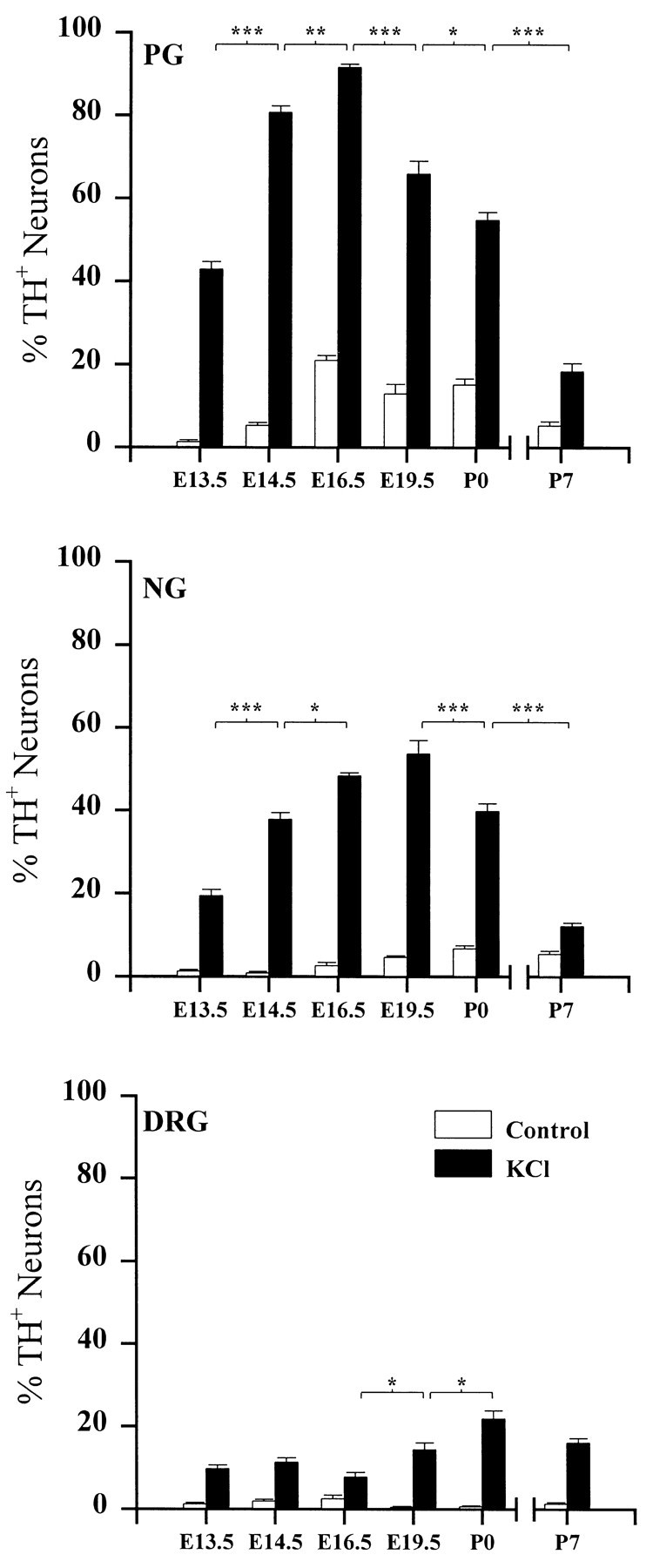
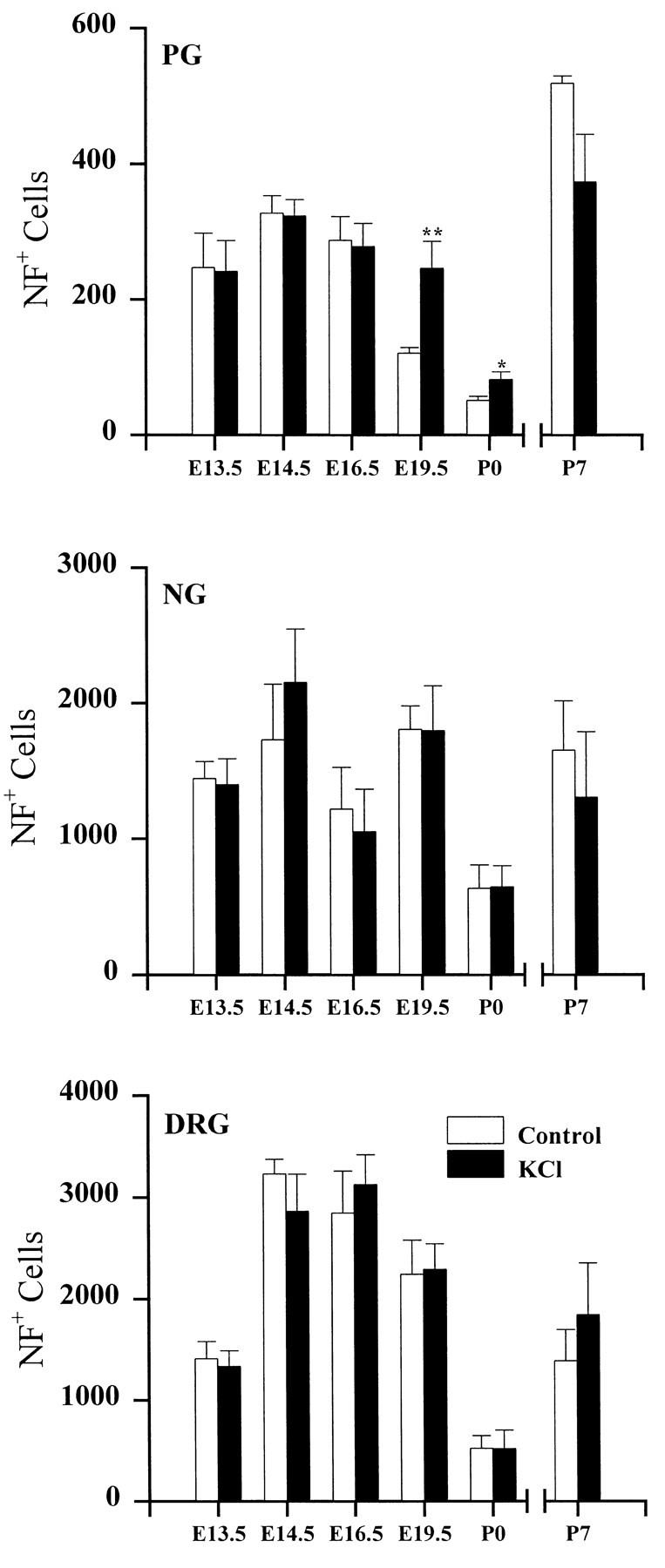
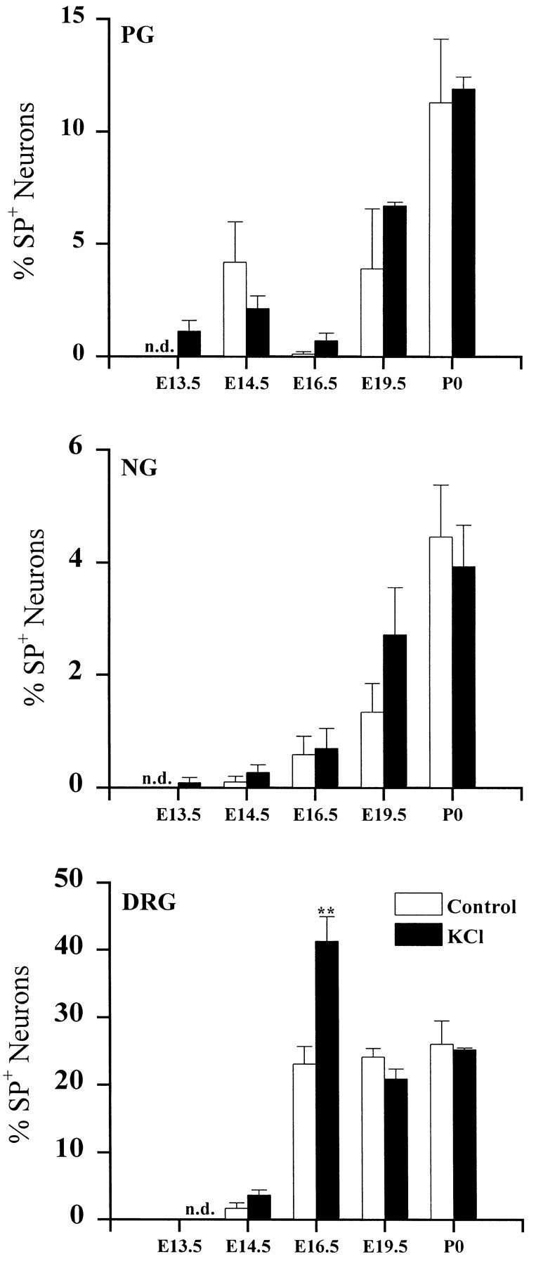
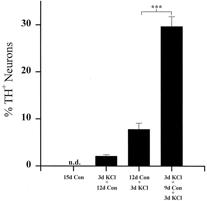
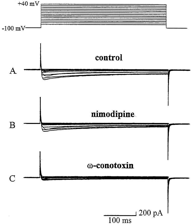
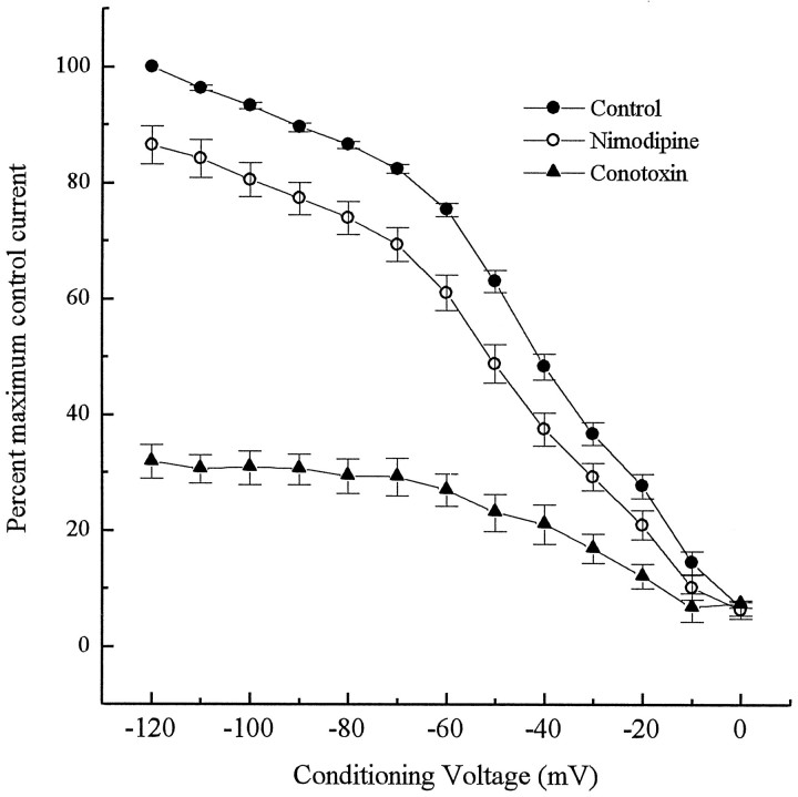
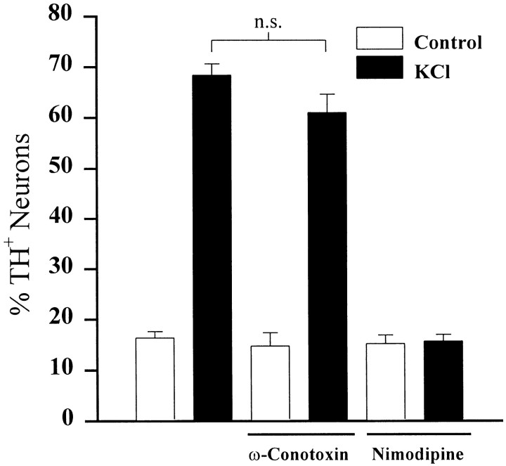
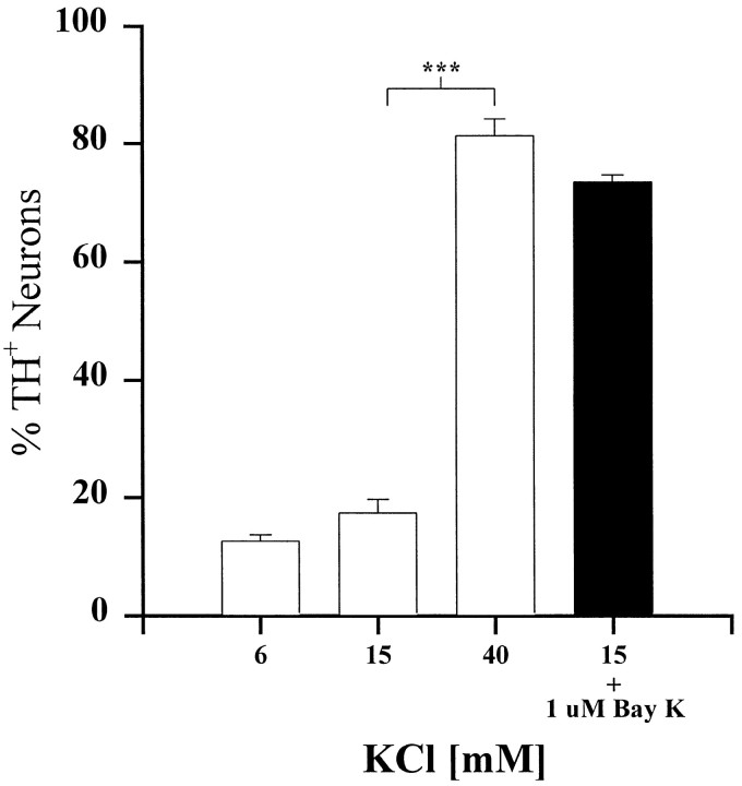
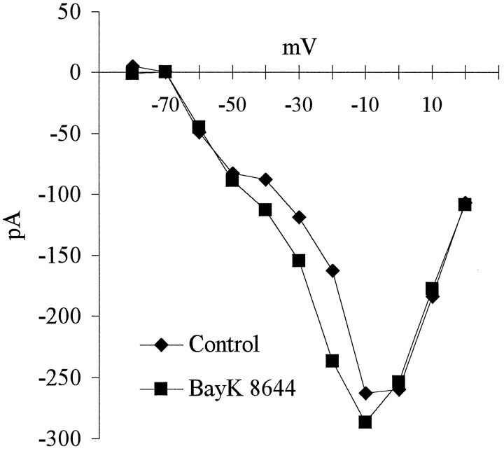
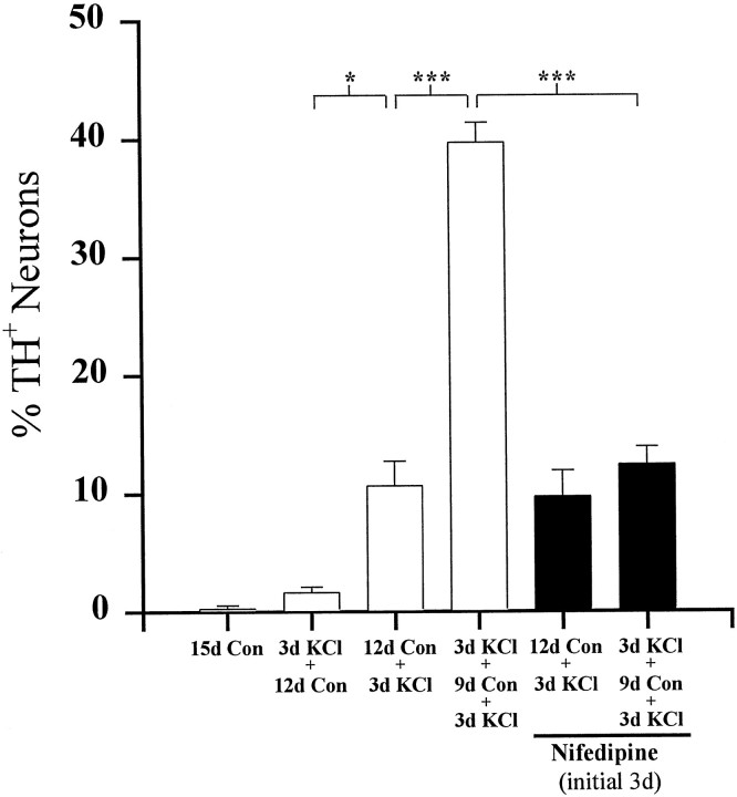
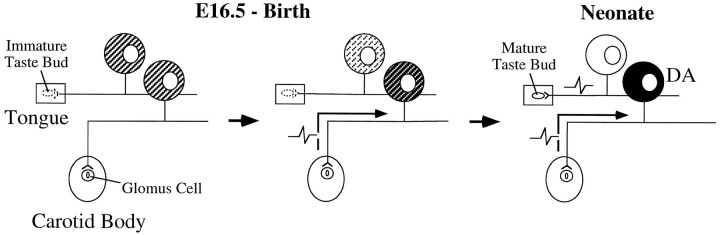
To examine the influence of activity-dependent cues on differentiation of primary afferent neurons, we investigated the short- and long-term effects of depolarization and calcium influx on expression of transmitter traits in sensory ganglion cell cultures. We focused on expression of tyrosine hydroxylase (TH), a marker for dopaminergic neurons, in developing petrosal ganglion (PG), nodose ganglion, and dorsal root ganglion neurons grown in the presence or absence of depolarizing concentrations of KCl. Exposure to 40 mM KCl increased the proportion of TH-immunoreactive neurons in all three ganglia in a developmentally regulated manner that corresponded to the temporal pattern of dopaminergic expression in vivo. PG neurons, for example, were most responsive to elevated KCl on embryonic day 16.5 (E16.5), the age at which the dopaminergic phenotype is first detectable in vivo. However, KCl was relatively ineffective at increasing TH expression in neonatal PG, indicating a critical period for induction of this phenotype by depolarization. Detailed analysis of TH induction in PG neurons demonstrated that, although N-type calcium channels carried the majority of the high voltage-activated calcium current, only L-type calcium channel blockade inhibited the effect of elevated KCl. Further studies revealed that after removal of high KCl, neurons remained sensitized to subsequent stimulation for >1 week. Specifically, cultures exposed to KCl beginning on E16.5 (the conditioning stimulus), then returned to control medium, and subsequently re-exposed to elevated KCl after 9 d (the test stimulus) contained fourfold more TH-positive neurons than did cultures exposed to the test stimulus alone. Moreover, blockade of L-type calcium channels during the conditioning stimulus completely abolished long-term potentiation of the TH response to elevated KCl. These findings demonstrate a novel role for L-type calcium channels in activity-dependent plasticity of transmitter expression in sensory neurons and indicate that exposure to depolarizing stimuli during early development may alter neuronal response properties at later ages.


Figures

Fig. 1.
Fig. 1.
The developmental time course of depolarization-induced TH expression in PG, NG, and DRG neurons. Ganglia were removed at the ages indicated and grown in dissociate cell culture for 3 d in the absence (Control) or presence (KCl) of 40 mm KCl. Eachbar indicates the percentage of neurons exhibiting TH immunoreactivity. Data are presented as the mean ± SEM. P7 ganglia were grown on Matrigel, whereas all others were grown on laminin (see Materials and Methods). Each KCl-treated group is significantly different from its corresponding control. Comparisons between ages were made using ANOVA followed by Scheffé’s test; *p < 0.05; **p < 0.01; ***p < 0.001.
Fig. 2.
Fig. 2.
Neuron survival in PG, NG, and DRG. Cultures were grown for 3 d in the absence (Control) or presence (KCl) of 40 mm KCl.Bars represent the mean number (± SEM) of neurofilament-positive cells per ganglion. Significant differences between KCl and corresponding control values were detected using ANOVA followed by Scheffé’s multiple comparison; *p < 0.05; **p < 0.01.
Fig. 3.
Fig. 3.
The developmental time course of depolarization-induced SP expression in PG, NG, and DRG neurons. Ganglia were removed at the ages indicated and grown in dissociate cell culture for 3 d in the absence (Control) or presence (KCl) of 40 mm KCl. Eachbar indicates the mean percentage (± SEM) of neurons exhibiting SP immunoreactivity; n.d., not detectable. Comparisons with corresponding controls were made using Student’st test; **p < 0.01.
Fig. 4.
Fig. 4.
Long-term potentiation of TH expression by KCl depolarization. Dissociated E16.5 PG neurons were cultured for a total of 15 d as indicated. Each bar shows the mean percentage (± SEM) of neurons exhibiting TH immunoreactivity at the end of the culture period. Con, Control;n.d., not detectable. Comparisons were made using ANOVA followed by Scheffé’s test; ***p < 0.001.
Fig. 5.
Fig. 5.
Representative recordings of inward calcium currents from an E16.5 PG neuron. Depolarizing voltage steps from −80 to +40 mV were applied in 10 mV increments from a holding potential of −100 mV. A, A recording from a neuron in the control bath solution. B, A recording from the same neuron in the presence of nimodipine (2 μm). C, A recording from the same neuron after washing with control bath solution to remove nimodipine and superfusing with ω-conotoxin (1 μm).
Fig. 6.
Fig. 6.
Voltage-dependent inactivation of high threshold calcium current in E16.5 PG neurons. Conditioning pulses ranging from −120 to 0 mV were applied for 2 sec before applying a test voltage of 0 mV. Points represent normalized data of percentages of maximum control current (± SEM; n = 5).Closed circles, Control inactivation curve;closed triangles, inactivation in the presence of ω-conotoxin (1 μm); open circles, inactivation in the presence of nimodipine (2 μm).
Fig. 7.
Fig. 7.
The effect of selective calcium channel antagonists on depolarization-induced TH expression. Dissociated E16.5 PG neurons were cultured for 2 d and subsequently exposed (KCl) or not exposed (Control) to 40 mm KCl in the presence or absence of either ω-conotoxin (1 μm) or nimodipine (2 μm) for 24 hr. Each barindicates the percentage (± SEM) of neurons exhibiting TH immunoreactivity; n.s., not significantly different; ANOVA followed by Scheffé’s test.
Fig. 8.
Fig. 8.
Selective activation of L-type calcium channels mimics the effect of 40 mm KCl. E16.5-dissociated PG cultures were grown for 3 d in medium containing 6 mm KCl (control), 15 mm KCl, 40 mmKCl, or 15 mm KCl plus 1 μm Bay K-8644 (black bar). Each bar indicates the mean percentage (± SEM) of neurons exhibiting TH immunoreactivity. Comparisons were made using ANOVA followed by Scheffé’s test; ***p < 0.001
Fig. 9.
Fig. 9.
Current–voltage relationship derived from recordings from an E16.5 PG neuron in the presence of Bay K-8644. Depolarizing voltage steps from −80 to +40 mV were applied in 10 mV increments from a holding potential of −100 mV in the absence (diamonds) or presence (squares) of 1 μm Bay K-8644. The effect of Bay K-8644 is greatest between −40 and −20 mV.
Fig. 10.
Fig. 10.
Inhibition of long-term potentiation of TH expression by L-type calcium channel blockade. Dissociated E16.5 PG neurons were grown for a total of 15 d as indicated. Eachbar shows the mean percentage (± SEM) of neurons exhibiting TH immunoreactivity at the end of the culture period.Black bars represent the percentage of TH-positive neurons in cultures exposed to 1 μm nifedipine during the initial 3 d of culture only. Comparisons were made using ANOVA followed by Scheffé’s test; *p< 0.05; ***p < 0.001.
Fig. 11.
Fig. 11.
Proposed model for activity-dependent development of the dopaminergic (DA) phenotype in carotid body afferent neurons. On E16.5, all PG neurons have the potential to express the DA phenotype (shading). However, during late fetal development (middle panel), this potential is gradually lost (light shading), except in those neurons that become sufficiently depolarized (dark shading). We hypothesize that a subpopulation of PG neurons that innervate the carotid body become active during this window of transmitter plasticity and, as a consequence, develop a stable DA phenotype. In contrast, gustatory afferents, whose receptors are not terminally differentiated until after birth, do not become sufficiently depolarized during this critical period to induce DA phenotypic traits.

References

    1. Altman J, Bayer SA. Development of the cranial nerve ganglia and related nuclei in the rat. Adv Anat Embryol Cell Biol. 1982;74:1–87. - PubMed
    1. Bading H, Ginty DD, Greenberg ME. Regulation of gene expression in hippocampal neurons by distinct calcium signaling pathways. Science. 1993;260:181–186. - PubMed
    1. Baker H, Farbman AI. Olfactory afferent regulation of the dopamine phenotype in the fetal rat olfactory system. Neuroscience. 1993;52:115–134. - PubMed
    1. Biscoe TJ, Purves MJ, Sampson SR. Types of nervous activity which may be recorded from the carotid sinus nerve in the sheep foetus. J Physiol (Lond) 1969;202:1–23. - PMC - PubMed
    1. Bisgard GE, Neubauer JA. Peripheral and central effects of hypoxia. In: Dempsey JA, Pack AI, editors. Lung biology in health and disease: regulation of breathing. Dekker; New York: 1995. pp. 617–668.

Publication types

MeSH terms

Substances

LinkOut - more resources