

Case Reports
doi: 10.3904/kjim.1997.12.2.232.
Idiopathic retroperitoneal fibrosis presented as an abdominal mass and nephrotic syndrome
Affiliations
- PMID: 9439160
- PMCID: PMC4531988
- DOI: 10.3904/kjim.1997.12.2.232
Item in Clipboard
Case Reports
Idiopathic retroperitoneal fibrosis presented as an abdominal mass and nephrotic syndrome
Korean J Intern Med.
1997 Jun.
Abstract
We present a 30-year-old male patient who was initially diagnosed as minimal change nephrotic syndrome, 5 years later, the patient developed a localized form of idiopathic retroperitoneal fibrosis (IRF). An elevated ESR and concomitant nephrotic syndrome in the patient suggested the immunologic nature of IRF, IRF has been reported in association with collagen diseases and rarely with proliferative and nonproliferative glomerulopathies. To our knowledge, the association between minimal change lesion (MC) and IRF has not been reported. Furthermore, the fact that IRF presented itself as an abdominal mass and lacked systemic symptoms was also unusual.
Figures

Light microscopy shows a slight increase in size of the glomeruli but otherwise no abnormal findings are observed (H&E ×100).

Postcontrast CT scan reveals that the mass is a mixture of highly attenuated solid portions and low-attenuated necrotic portions. The outer margin of the mass is discrete and no intra-abdominal lymph node enlargement is observed.
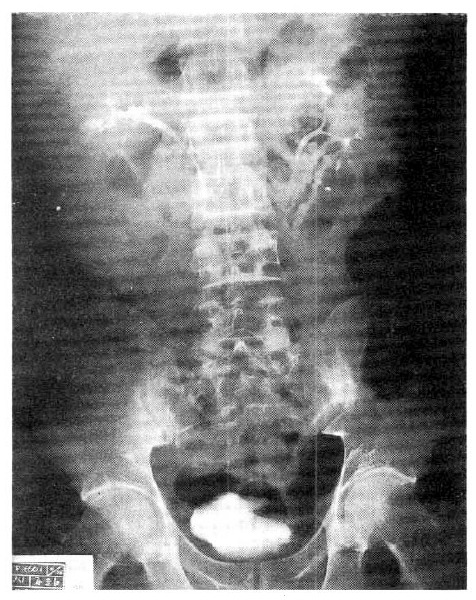
Anintravenous urography, the right kidney is rotated upward on the frontal plane, but no abnormal finding is seen in the shape of calyx, pelvis and ureter, nor the excretory function of both kidneys.

A cross surface of the removed mass shows it to be a fairly well-circumscribed mass with fascicles of fibrous tissue and two areas of cystic change.
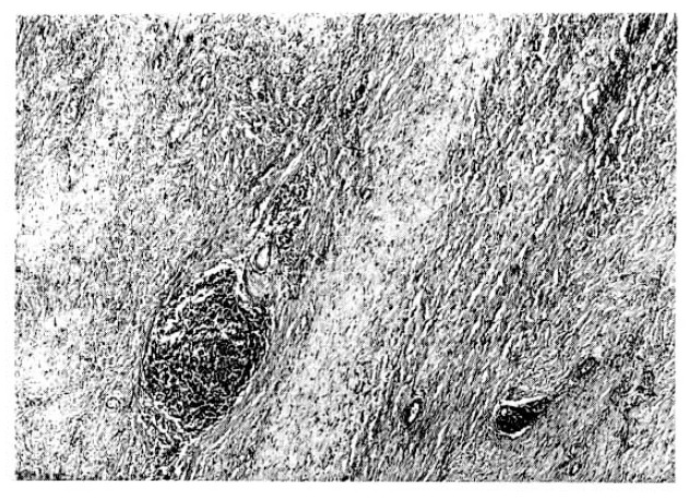
Photomicrographs of the mass show irregular fibrous bands and scattered inflammatory cell collections (H&H ×100).

At a higher magnification of the fibrous mass, the inflammatory cells consist of lymphocytes and some plasma cells (H&E ×350).
Similar articles
-
A Case of Idiopathic Retroperitoneal Fibrosis Associated With Sjögren's Syndrome.Mil Med. 2016 Oct;181(10):e1407-e1409. doi: 10.7205/MILMED-D-15-00567. Mil Med. 2016. PMID: 27753592
-
Idiopathic retroperitoneal fibrosis: implications for a systemic disorder.Clin Exp Rheumatol. 1983 Apr-Jun;1(2):157-60. Clin Exp Rheumatol. 1983. PMID: 6681136
-
Idiopathic retroperitoneal fibrosis and probable systemic lupus erythematosus.JAMA. 1966 Jun 13;196(11):1022-4. JAMA. 1966. PMID: 5952426 No abstract available.
-
Amyloid A gastrointestinal amyloidosis associated with idiopathic retroperitoneal fibrosis. Report of a rare autopsy case and review of the literature.Arch Pathol Lab Med. 2003 Jun;127(6):735-8. doi: 10.5858/2003-127-735-AAGAAW. Arch Pathol Lab Med. 2003. PMID: 12741901 Review.
-
Retroperitoneal fibrosis: single-centre experience from 1992 to 2010, current status of knowledge and review of the international literature.Scand J Urol. 2013 Oct;47(5):370-7. doi: 10.3109/00365599.2012.747564. Epub 2012 Dec 4. Scand J Urol. 2013. PMID: 23206245 Review.
Cited by
-
Acute renal vein thrombosis and nephrotic syndrome in the setting of retroperitoneal fibrosis.Oxf Med Case Reports. 2015 Jun 17;2015(6):309-10. doi: 10.1093/omcr/omv043. eCollection 2015 Jun. Oxf Med Case Reports. 2015. PMID: 26421156 Free PMC article.
References
-
- Amis E Stephen., Jr Retroperitoneal fibrosis. AJR. 1991 Aug;157:321–329. - PubMed
-
- GS Que, Mandema E. A case of idiopathic retro-peritoneal fibrosis presenting as a systemic collagen disease. Am J Med. 1964;36:320–329. - PubMed
-
- Richard L, Lipman B, Johnson G, Berg A, Shapiro P. Idiopathic retroperitoneal fibrosis and probable systemic lupus erythematosus. JAMA. 1966;196:204–206. - PubMed
-
- Kay RG. Retroperitoneal vasculitis with perivascular fibrosis. Br J Urol. 1963;35:284–292. - PubMed
-
- Katz SM, Bates O, Yudis M, Falkner B, Griffith E. Immune complex glomerulonephritis in a case of retroperitoneal fibrosis. Am J Clin Pathol. 1977;67:436–439. - PubMed
Publication types
MeSH terms
LinkOut - more resources
Full Text Sources
Miscellaneous