. 1998 Jan 26;140(2):391-401.
doi: 10.1083/jcb.140.2.391.

Cytoplasmic dynein and dynactin are required for the transport of microtubules into the axon

Affiliations

Cytoplasmic dynein and dynactin are required for the transport of microtubules into the axon

F J Ahmad et al. J Cell Biol. .

Abstract

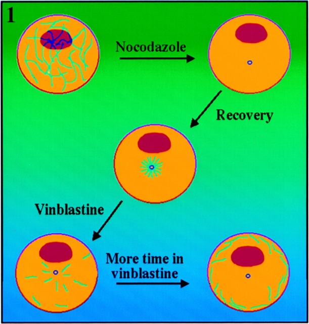
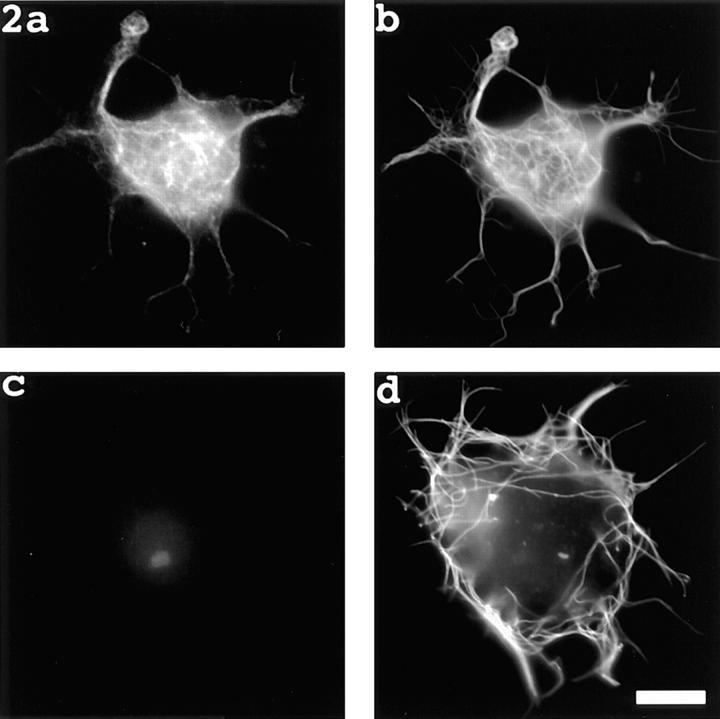
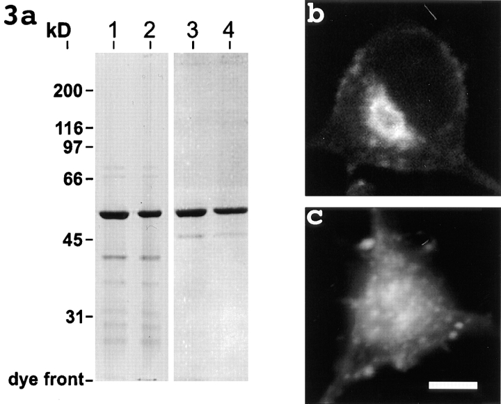
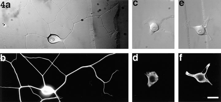
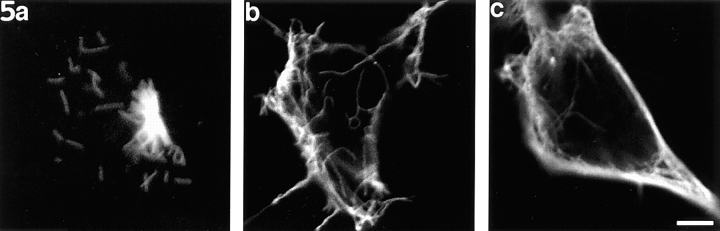
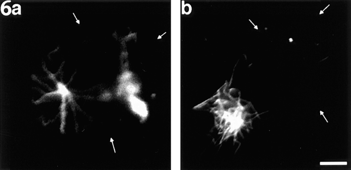
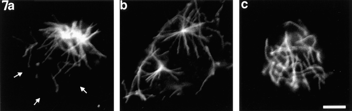
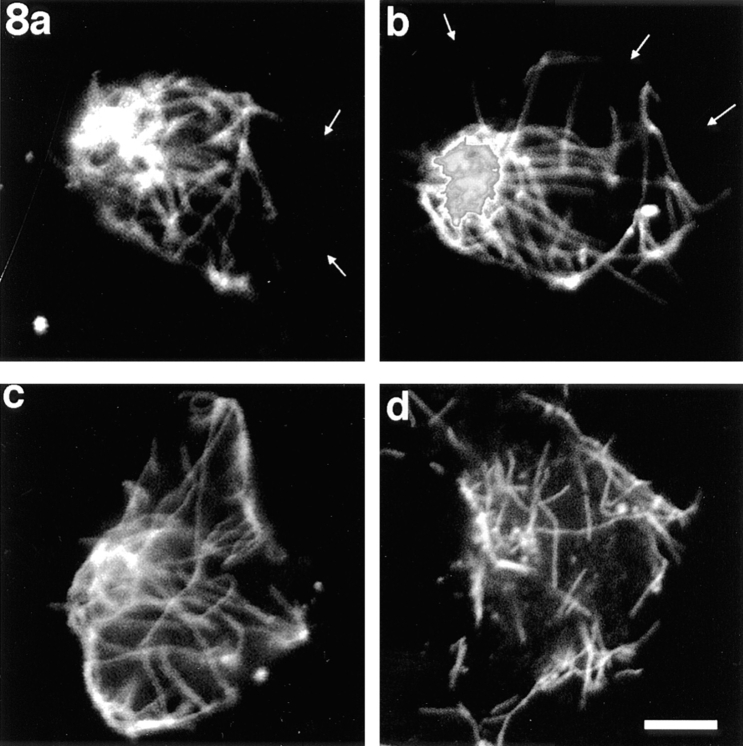
Previous work from our laboratory suggested that microtubules are released from the neuronal centrosome and then transported into the axon (Ahmad, F.J., and P.W. Baas. 1995. J. Cell Sci. 108: 2761-2769). In these studies, cultured sympathetic neurons were treated with nocodazole to depolymerize most of their microtubule polymer, rinsed free of the drug for a few minutes to permit a burst of microtubule assembly from the centrosome, and then exposed to nanomolar levels of vinblastine to suppress further microtubule assembly from occurring. Over time, the microtubules appeared first near the centrosome, then dispersed throughout the cytoplasm, and finally concentrated beneath the periphery of the cell body and within developing axons. In the present study, we microinjected fluorescent tubulin into the neurons at the time of the vinblastine treatment. Fluorescent tubulin was not detected in the microtubules over the time frame of the experiment, confirming that the redistribution of microtubules observed with the experimental regime reflects microtubule transport rather than microtubule assembly. To determine whether cytoplasmic dynein is the motor protein that drives this transport, we experimentally increased the levels of the dynamitin subunit of dynactin within the neurons. Dynactin, a complex of proteins that mediates the interaction of cytoplasmic dynein and its cargo, dissociates under these conditions, resulting in a cessation of all functions of the motor tested to date (Echeverri, C.J., B.M. Paschal, K.T. Vaughan, and R.B. Vallee. 1996. J. Cell Biol. 132: 617-633). In the presence of excess dynamitin, the microtubules did not show the outward progression but instead remained near the centrosome or dispersed throughout the cytoplasm. On the basis of these results, we conclude that cytoplasmic dynein and dynactin are essential for the transport of microtubules from the centrosome into the axon.


Figures

Figure 1
Figure 1
Schematic illustration of our pharmacological strategy for revealing the progression of microtubules outward from the neuronal centrosome (Ahmad and Baas, 1995; see Results for details).
Figure 2
Figure 2
Evidence that the pharmacological strategy reveals microtubule transport, not microtubule assembly. a and b show a neuron treated with nocodazole, rinsed free of the drug, immediately injected with fluorescent tubulin, and then prepared for immunofluorescence visualization of microtubules 30 min later. The rhodamine image (depicting fluorescent tubulin that had incorporated into microtubule polymer [a]) and the fluorescein image (immunofluorescence image of total microtubule mass [b]) are similar in appearance, indicating complete or near-complete incorporation of fluorescent tubulin into polymer. c and d show a neuron treated with nocodazole, rinsed free of the drug, simultaneously exposed to 50 nM vinblastine, injected with fluorescent tubulin (see Materials and Methods), and then prepared for immunofluorescence visualization of microtubules 30 min later. The immunofluorescence image shows the expected peripheral concentration of microtubules (d). The rhodamine image shows only faint background fluorescence and a fluorescent “scar” made by the injection (c). No fluorescent microtubule polymer was detected. Bar, 5 μm.
Figure 3
Figure 3
SDS-PAGE gel showing the purity of the recombinant dynamitin preparations (a) and micrographs showing Golgi distribution in a control neuron (b) and a neuron 6 h after microinjection with recombinant dynamitin (c). a shows Coomassie blue staining of native (lanes 1 and 3) and boiled (lanes 2 and 4) dynamitin preparations purified either by nickel-affinity (lanes 1 and 2) or by two-step chromatography (lanes 3 and 4). The analyses confirm the presence of a single major band of the expected size, 50 kD, common to both preparations. In b, the control neuron shows a discrete Golgi apparatus located near the nucleus. In c, the Golgi elements are dispersed throughout the injected cell. Bar, 5 μm.
Figure 4
Figure 4
Effects on axon outgrowth of recombinant dynamitin. (a) A neuron 7 h after plating. Hundreds of microns of axons have grown. (b) An immunofluorescence image showing the dense microtubule array of the same cell after extraction and fixation. (c) A typical neuron 9 h after microinjection with the recombinant dynamitin. The cell has extended a broad lamellipodium but no axons. (d) An immunofluorescence image of the microtubule array within this cell. The microtubules splayed into the lamellipodia but did not form dense bundles as they did within the axons of control neurons. (e) One of a small number of neurons injected with recombinant dynamitin that extended processes (shown 9 h after microinjection). In these cases, the processes were significantly shorter than the axons of control neuron and also had a broad flat appearance that was rather different from the thin cylindrical shape of the control axons. (f) An immunofluorescence image of the microtubule array within this cell. The microtubules within these processes are more spayed apart and less paraxial than those within the control axons. Bar, 10 μm.
Figure 5
Figure 5
Neurons at various stages of the pharmacological regime designed to reveal the outward progression of microtubules from the centrosome. The cells shown here were injected with the boiled recombinant dynamitin before rinsing out the nocodazole (see Results for details). The results were indistinguishable from those obtained on uninjected cells. (a) A neuron that had been treated with nocodazole, microinjected with the boiled protein, rinsed free of the drug for 3 min, and then prepared for immunofluorescence visualization of microtubules. A dense radial array of short microtubules is apparent at the centrosome, as are a few unattached microtubules of roughly the same length. (b) A neuron treated in similar fashion and then exposed to 50 nM vinblastine for 15 min. Microtubules are essentially absent from the central region of the cell body and are loosely packed beneath its periphery. (c) A neuron exposed to vinblastine for 30 min. The microtubules are more tightly packed at cell periphery. The microtubules vary somewhat in length from cell to cell, with some of the microtubules appearing longer in the cell shown in b than those in the cell shown in a. However, the differences are no greater than those observed among different cells before or after vinblastine treatment. Bar, 3 μm.
Figure 6
Figure 6
Examples of neurons treated with nocodazole, injected with recombinant dynamitin, rinsed free of the drug for 3 min, and then immunostained to reveal microtubules. (a) A neuron with a discrete site of nucleation similar to control neurons. The microtubules are fewer and somewhat longer than the microtubules in similarly treated control neurons. (b) A neuron in which the site of nucleation is somewhat more diffuse than in a similarly treated control (uninjected or injected with boiled dynamitin protein) neuron, but the level of microtubule reassembly is similar. Small white arrows indicate the perimeter of the cell body where it is not apparent from the immunofluorescence image. Bar, 3 μm.
Figure 7
Figure 7
Examples of neurons treated with nocodazole, injected with recombinant dynamitin, rinsed free of drug for 7.5 min, exposed to vinblastine for 15 min, and then immunostained to reveal microtubules. In no such case did the microtubules vacate the central region of the cell body and concentrate beneath the cell periphery, as was the case with similarly treated uninjected neurons or neurons injected with boiled protein. (a) A neuron in which most of the microtubules remained attached to their apparent nucleation site, which had become rather diffuse in appearance. (b) A neuron in which the nucleation site had split into two, and most of the microtubules remained attached to these sites. A few unattached microtubules were present at cell periphery, but the vast majority were not. (c) A neuron in which the microtubules were dispersed throughout the cytoplasm and not attached to any apparent nucleation sites. Small white arrows indicate the perimeter of the cell body where it is not apparent from the immunofluorescence image. Bar, 4 μm.
Figure 8
Figure 8
Examples of neurons treated with nocodazole, injected with recombinant dynamitin, rinsed free of drug for 7.5 min, and then exposed to vinblastine for 30 min. In no such case did the microtubules vacate the central region of the cell body and concentrate beneath the cell periphery, as was the case with similarly treated uninjected neurons or neurons injected with boiled protein. (a and b) Neurons in which some of the microtubules appear to be attached to a rather diffuse nucleation site. Other microtubules were dispersed. (c and d) Neurons in which the microtubules do not appear to be attached to discernible nucleation sites, but rather appear to be dispersed throughout the cytoplasm. Fluorescence intensity is saturating within the densest cluster of microtubules in b. Small white arrows indicate the perimeter of the cell body where it is not apparent from the immunofluorescence image. Bar, 4 μm.

Similar articles

Cited by

References

    1. Ahmad FJ, Baas PW. Microtubules released from the neuronal centrosome are transported into the axon. J Cell Sci. 1995;108:2761–2769. - PubMed
    1. Ahmad FJ, Joshi HC, Centonze VE, Baas PW. Inhibition of microtubule nucleation at the neuronal centrosome compromises axon growth. Neuron. 1994;12:271–280. - PubMed
    1. Allan V. Motor proteins: a dynamic duo. Curr Biol. 1996;6:630–633. - PubMed
    1. Baas PW. The neuronal centrosome as a generator of microtubules for the axon. Curr Top Dev Biol. 1996;33:281–298. - PubMed
    1. Baas PW. Microtubules and axonal growth. Curr Opin Cell Biol. 1997;9:29–36. - PubMed

Publication types