. 1998 Jan 26;140(2):409-18.
doi: 10.1083/jcb.140.2.409.

Bone morphogenetic protein signaling is required for maintenance of differentiated phenotype, control of proliferation, and hypertrophy in chondrocytes

Affiliations

Bone morphogenetic protein signaling is required for maintenance of differentiated phenotype, control of proliferation, and hypertrophy in chondrocytes

M Enomoto-Iwamoto et al. J Cell Biol. .

Abstract

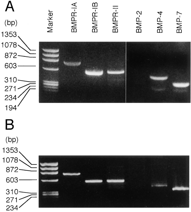
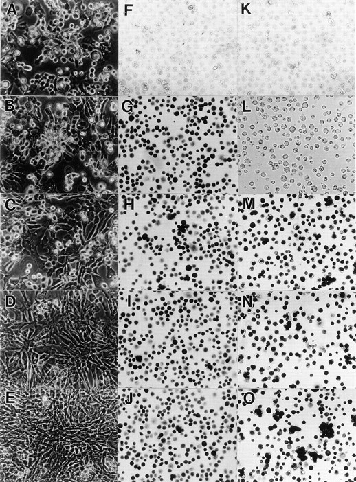
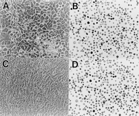
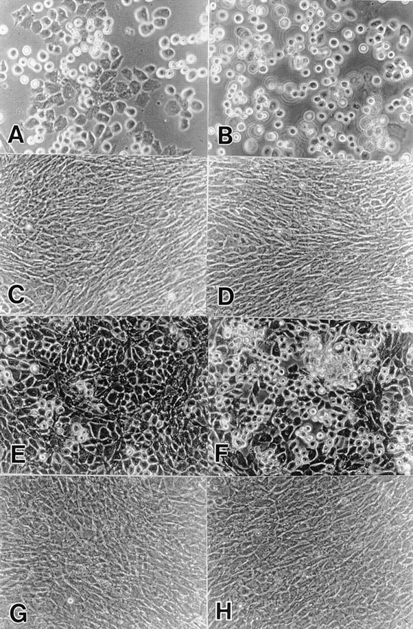
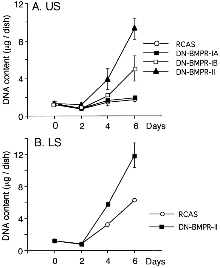
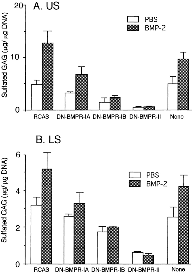
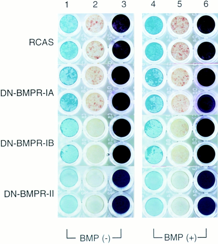
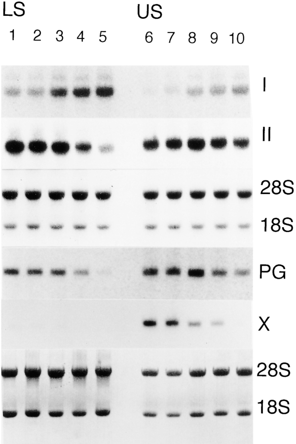
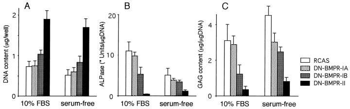
To examine the role of bone morphogenetic protein (BMP) signaling in chondrocytes during endochondral ossification, the dominant negative (DN) forms of BMP receptors were introduced into immature and mature chondrocytes isolated from lower and upper portions of chick embryo sternum, respectively. We found that control sternal chondrocyte populations expressed type IA, IB, and II BMP receptors as well as BMP-4 and -7. Expression of a DN-type II BMP receptor (termed DN-BMPR-II) in immature lower sternal (LS) chondrocytes led to a loss of differentiated functions; compared with control cells, the DN-BMPR- II-expressing LS chondrocytes proliferated more rapidly, acquired a fibroblastic morphology, showed little expression of type II collagen and aggrecan genes, and upregulated type I collagen gene expression. Expression of DN-BMPR-II in mature hypertrophic upper sternal (US) chondrocytes caused similar effects. In addition, the DN-BMPR-II-expressing US cells exhibited little alkaline phosphatase activity and type X collagen gene expression, while the control US cells produced both alkaline phosphatase and type X collagen. Both DN-BMPR-II-expressing US and LS chondrocytes failed to respond to treatment with BMP-2 . When we examined the effects of DN forms of types IA and IB BMP receptors, we found that DN-BMPR-IA had little effect, while DN-BMPR-IB had similar but weaker effects compared with those of DN-BMPR-II. We conclude that BMP signaling, particularly that mediated by the type II BMP receptor, is required for maintenance of the differentiated phenotype, control of cell proliferation, and expression of hypertrophic phenotype.


Figures

Figure 1
Figure 1
Expression of BMP receptors and BMPs in chondrocytes. Total RNA was isolated from (A) day 17 chicken embryo sterna and (B) upper sternal chondrocytes cultured for 3 d. RT-PCR for the indicated gene was performed as described in Materials and Methods. Amplified products were analyzed by 2% agarose gel electrophoresis.
Figure 2
Figure 2
Efficiency of infection in upper sternal chondrocyte cultures by DN-BMPR–encoding viruses. Freshly isolated US chondrocytes were infected with RCAS viruses encoding DN-BMPR-IA (C, H, and M), DN-BMPR-IB (D, I, and N), DN-BMPR-II (E, J, and O), or vector alone (B, G, K, and L) for 3 d in medium 199 containing 10% FBS. The medium was then changed to high glucose DME containing 10% FBS, and cells were cultured for an additional 4 d. On day 7, the cells were harvested and replated in 35-mm dishes at the density of 1.5 × 10⁵ cells/dish, and the microscopic photographs of live cells were taken after 5 d (A–E). Aliquots of the harvested cells were plated onto poly- l-lysine–coated dishes at the density of 2.0 × 10⁶/ml, fixed with 3.7% formaldehyde after 15 min from plating, and stained with antibodies to the RCAS virus gag protein (F–J) or c-myc peptide (L–O) as described in Materials and Methods. A and F represent uninfected cultures. K is a culture incubated with control nonimmune ascites.
Figure 3
Figure 3
Efficiency of infection in lower sternal chondrocyte cultures by DN-BMPR-II–encoding virus. Freshly isolated LS chondrocytes were infected with RCAS virus encoding DN-BMPR-II (C and D) or vector alone (A and B) in medium 199 containing 10% FBS for 3 d and were then processed as cells in Fig. 2. (A) Control cells; (B) control cells stained with viral gag antibody; (C) BMPR-II–expressing cells; (D) BMPR-II–expressing cells stained with viral gag antibody.
Figure 4
Figure 4
Effects of the exogenous BMP-2 on the cell morphology. Freshly isolated US (A–D) and LS (E–H) chondrocytes were infected with RCAS virus encoding DN-BMPR-II (C, D, G, and H) or vector alone (A, B, E, and F) in medium 199 containing 10% FBS for 3 d. The medium was then changed to high glucose DME containing 10% FBS. On day 7 (US) or 5 (LS), the cells were harvested and replated in 35-mm dishes at the density of 1.5 × 10⁵ cells/dish. 200 ng/ml of rhBMP-2 was added on the next day (B, D, F, and H). Photographs was taken after 5 d.
Figure 5
Figure 5
Effects of DN-BMPRs on cell proliferation. US (A) and LS (B) chondrocytes were infected with DN-BMPR viruses, harvested by trypsinization on day 7 and 5, respectively, and then replated in 35-mm dishes at the density of 1.5 × 10⁵ cells/dish. DNA contents were determined at the indicated times as described in Materials and Methods. Values represent average ± SD for three cultures, and similar results were obtained in three independent experiments.
Figure 6
Figure 6
Effects of DN-BMPRs on GAG synthesis in US and LS chondrocyte cultures. US (A) and LS (B) chondrocytes were infected with DN-BMPR viruses, harvested by trypsinization on day 7 (US) or day 5 (LS), and then replated in 24-well plates at the density of 5.0 × 10⁴ cells/dish. Sulfated GAG and DNA contents were determined on day 5 as described in Materials and Methods. rhBMP-2 was added from day 1 at the concentration of 200 ng/ml. Data represent average ± SD for three cultures, and similar results were obtained in two independent experiments.
Figure 7
Figure 7
Histochemical analysis of proteoglycan accumulation and APase activity in US chondrocytes infected by DN-BMPR viruses. US chondrocytes were infected with RCAS virus encoding DN-BMPRs or vector alone (RCAS) and replated on type I collagen–coated 96-well plates at the density of 2.0 × 10⁴ cells/ dish on day 7. Cells were cultured in the presence of 10 μg/ml of ascorbic acid with (BMP (+)) or without (BMP ()) rhBMP-2 (200 ng/ml) for 7 d. Cultures were then fixed with 3.7% formaldehyde and stained with Alcian blue (lanes 1 and 4) or crystal violet (lanes 3 and 6) as described in Materials and Methods. Lanes 2 and 5 show the results of histochemical detection of APase.
Figure 8
Figure 8
Changes in expression of extracellular matrix genes induced by DN-BMPRs. LS (lanes 1–5) and US (lanes 6–10) chondrocytes were infected with RCAS virus encoding DN-BMPR-IA (lanes 3 and 8), DN-BMPR-IB (lanes 4 and 9), DN-BMPR-II (lanes 5 and 10), or vector alone (lanes 2 and 7), harvested by trypsinization on day 7 for US and day 5 for LS, and then replated in 100-mm dishes at the density of 1.0 × 10⁶ cells/plate. 7 d after plating, whole cellular RNAs were extracted, separated on agarose gels, and blotted onto nylon membranes. Membranes were hybridized to 32P-labeled probes encoding type I collagen (I) and type X collagen (X) and then reblotted with the 32P-labeled probes encoding type II collagen (II ) and aggrecan (PG), respectively. Blots were prestained with methylene blue to mark the 28S and 18S rRNA subunits. Lanes 1 and 6 represent uninfected cultures.
Figure 9
Figure 9
Effects of DN- BMPRs on US chondrocytes in serum-free cultures. US chondrocytes were infected with RCAS virus encoding DN-BMPRs or vector alone (RCAS) and replated in type I collagen–coated 96-well plates at the density of 1.0 × 10⁴ cells/dish on day 7. Cells were then cultured in chemically defined medium as described in Materials and Methods for 7 d. The DNA content (A), sulfated GAG content (B), and APase activity (C) were measured as described in Materials and Methods. The data represent average ± SD for three cultures. * unit: One unit of APase activity corresponds to the hydrolysis of 1 nmol per min.

References

    1. Benya PD, Shaffer JD. Dedifferentiated chondrocytes reexpress the differentiated collagen phenotype when cultured in agarose gels. Cell. 1982;30:215–224. - PubMed
    1. Bessey OA, Lowry OH, Brock MJ. A method for the rapid determination of alkaline phosphatase with five cubic millimeters of serum. J Biol Chem. 1946;164:321–329. - PubMed
    1. Carey DE, Liu X. Expression of bone morphogenetic protein-6 messenger RNA in bovine growth plate chondrocytes of different size. J Bone Miner Res. 1995;10:401–405. - PubMed
    1. Chen P, Carrington JL, Hammonds RG, Reddi AH. Stimulation of chondrogenesis in limb bud mesoderm cells by recombinant human bone morphogenetic protein 2B (BMP-2B) and modulation by transforming growth factor β1 and β2. Exp Cell Res. 1991;195:509–515. - PubMed
    1. Dewulf N, Verschueren K, Lonnoy O, Moren A, Grimsby S, Vande-Spiegle K, Miyazono K, Huylebroeck D, Ten-Dijke P. Distinct spatial and temporal expression patterns of two type I receptors for bone morphogenetic proteins during mouse embryogenesis. Endocrinology. 1995;136:2652–2653. - PubMed

Publication types

MeSH terms

Substances