. 1998 Jan 26;140(2):419-30.
doi: 10.1083/jcb.140.2.419.

Mice that lack thrombospondin 2 display connective tissue abnormalities that are associated with disordered collagen fibrillogenesis, an increased vascular density, and a bleeding diathesis

Affiliations

Mice that lack thrombospondin 2 display connective tissue abnormalities that are associated with disordered collagen fibrillogenesis, an increased vascular density, and a bleeding diathesis

T R Kyriakides et al. J Cell Biol. .

Abstract

Thrombospondin (TSP) 2, and its close relative TSP1, are extracellular proteins whose functions are complex, poorly understood, and controversial. In an attempt to determine the function of TSP2, we disrupted the Thbs2 gene by homologous recombination in embryonic stem cells, and generated TSP2-null mice by blastocyst injection and appropriate breeding of mutant animals. Thbs2-/- mice were produced with the expected Mendelian frequency, appeared overtly normal, and were fertile. However, on closer examination, these mice displayed a wide variety of abnormalities. Collagen fiber patterns in skin were disordered, and abnormally large fibrils with irregular contours were observed by electron microscopy in both skin and tendon. As a functional correlate of these findings, the skin was fragile and had reduced tensile strength, and the tail was unusually flexible. Mutant skin fibroblasts were defective in attachment to a substratum. An increase in total density and in cortical thickness of long bones was documented by histology and quantitative computer tomography. Mutant mice also manifested an abnormal bleeding time, and histologic surveys of mouse tissues, stained with an antibody to von Willebrand factor, showed a significant increase in blood vessels. The basis for the unusual phenotype of the TSP2-null mouse could derive from the structural role that TSP2 might play in collagen fibrillogenesis in skin and tendon. However, it seems likely that some of the diverse manifestations of this genetic disorder result from the ability of TSP2 to modulate the cell surface properties of mesenchymal cells, and thus, to affect cell functions such as adhesion and migration.


Figures

Figure 1
Figure 1
Disruption of the Thbs2 gene and characterization of TSP2-null mice. (A) Targeting strategy. A schematic representation of the Thbs2 gene locus is shown on the first line. The targeting construct consists of a PGK-TK cassette, and 1.5 and 3.3 kb of genomic sequence (dotted vertical lines) flanking a PGK-Neo cassette. The directions of transcription of the TK and Neo genes are indicated by the bold arrows. In the targeted allele, the PGK-Neo cassette replaces 2.6 kb of genomic DNA, which contains exons 2 and 3 of the gene. Angled arrows indicate the start of transcription of Thbs2, and open and filled boxes represent untranslated and translated exons, respectively. B, BamHI; E, EcoRI; K, KpnI. (B) Southern analysis of BamHI digests of DNA from electroporated ES clones. The probe (A, top) detects a 6-kb fragment in a digest of a wild-type clone (lane 1), and bands of 6 kb and 4.8 kb in a digest of a targeted clone (lane 2). (C) Southern analysis of tail DNA from the progeny of Thbs2 +/− mice, analyzed as in B. The pattern of 6.0- and 4.8-kb bands indicates that the animals analyzed in lanes 1 and 2 are Thbs2 +/−, that in lane 3 is Thbs2 +/+, and that in lane 4 is Thbs2 −/−. (D) Northern blot analysis of RNA prepared from cultured embryonic fibroblasts. Hybridization was performed with a 32P-labeled 515-bp Thbs2 cDNA probe (nucleotides 834–1,348), or with a β-actin cDNA probe. Lanes 1 and 2 were loaded with 5 and 10 μg of fibroblast RNA, respectively, derived from a Thbs2 −/− embryo. Lanes 3 and 4 were loaded with 5 and 10 μg, respectively, of fibroblast RNA derived from a Thbs2 +/+ embryo. The faster migrating transcript has been observed by us and others in the past; its nature is unknown. (E) Immunoblot analysis of protein (50 μg) extracted from 17-d-old embryos. An anti-TSP2 antibody was used. Lane 1, protein from a Thbs2 +/+ embryo. A band migrating at the expected molecular mass 200 kD is observed. A faster migrating band is also present and is likely to represent a proteolytic fragment of TSP2 since its presence and intensity were variable. Lane 2, protein from a Thbs2 −/− embryo. The same samples were immunoblotted with anti-TSP1 antibodies and the presence of approximately equal levels of a TSP1-specific band was evident in both samples (data not shown).
Figure 2
Figure 2
Appearance of the TSP2-null mouse. Disruption of the Thbs2 gene does not alter the gross appearance of the mouse, but the increased flexibility of tail tendons and ligaments permits the tying of a knot in the tail. This manipulation is not possible in a normal mouse.
Figure 3
Figure 3
Histological analysis of skin and bone in the TSP2-null mouse. (A and B) Dermis from the back of a Thbs2 +/+ mouse (A) and a Thbs2 −/− mouse (B), stained with Verhoeff-van Gieson stain. Collagen fibers stain orange-red and epidermis, cells, and hair follicle stain yellow-brown. The collagen fibers in A form a wicker-like mesh but are largely oriented parallel to the epidermal surface. Collagen fibers in B appear disorganized and lack the predominant orientation parallel to the epidermal surface. Bar, 50 μm. (C and D) Mid-diaphyseal cross-section of the femur of a Thbs2 +/+ mouse (C) and a Thbs2 −/− mouse (D), stained with H & E. The increased cortical thickness and reduced marrow cavity of the femur from the mutant animal are evident. The less regularly oval contour, and tendency to form a ridge, is a feature of the abnormal bone. Bar, 500 μm.
Figure 4
Figure 4
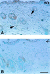
Immunohistochemical analysis for TSP-2 in back skin from (A) a Thbs2+/+ mouse and (B) a Thbs2 −/− mouse. The sections were counterstained with methyl green. The presence of TSP2 immunoreactivity, observed as brown-black color, is evident in A. The arrows indicate dermal cells that appear to be positive for TSP2. Immunoreactivity to the epidermis, sweat glands, and hair follicles is, in part, nonspecific. Bar, 50 μm.
Figure 5
Figure 5
Electron micrographs of collagen fibrils in reticular dermis from a Thbs2 +/+ mouse (A) and a Thbs2 −/− mouse (B). The presence of larger fibrils with irregular contours in the tissue from the mutant animal, in comparison with the more uniformly sized circular fibrils in the control tissue, is evident. Bar, 250 nm.
Figure 6
Figure 6
Distribution of collagen fibril diameters, as measured from electron micrographs of skin from Thbs2 +/+ (solid bars) and Thbs2 −/− (shaded bars) mice. The distribution is skewed in the direction of larger fibrils in the mutant tissue.
Figure 7
Figure 7
Measurements of tensile strength of skin from Thbs2 +/+, Thbs2 −/−, and decorin (Dcn) −/− mice. Force or load in newtons is plotted against displacement or elongation of skin in cm. A and B show experiments with tissues from different animals. In both, skin from TSP2-null mice ruptures at a lower load, and shows increased ductility (greater displacement per unit increase in load), than skin from control mice.
Figure 8
Figure 8
Electron micrographs of collagen fibrils in tendon from (A) a Thbs2 +/+ mouse, and (B) a Thbs2 −/− mouse. In B, larger fibrils are relatively more numerous than smaller fibrils and frequently display irregular contours. Small diameter fibrils in A represent tapered fibril ends and are much less frequent in B. Bar, 250 nm.
Figure 9
Figure 9
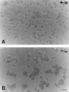
Appearance of neonatal skin fibroblasts isolated from Thbs2 +/+ mice (A) and Thbs2 −/− mice (B) 1 h after plating on bacteriologic plastic in DME. Control fibroblasts remain dispersed whereas fibroblasts from mutant mice form small aggregates that coalesce with time. Bar, 50 μm.
Figure 10
Figure 10
Fibroblast attachment assays. (A) Adult skin fibroblasts (2 × 10⁵/ml) were plated, in the presence of DME plus 10% serum, on untreated (control) wells or on wells coated with vitronectin or fibronectin in a 96-well tissue culture plate, and allowed to attach for 1 h. Attached cells were quantitated spectrophotometrically by adsorbed methylene blue. (B) In a separate experiment, cells that did not attach under conditions such as those described in A were retested for their ability to attach under the same conditions. Shaded bars, Thbs2 +/+ cells; open bars, Thbs2 −/− cells. All differences are significant (P ⩽ 0.05).
Figure 11
Figure 11
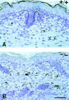
Immunohistochemical analysis for vWF of back skin from (A) a Thbs2 +/+ mouse and (B) a Thbs2 −/− mouse. The sections were counterstained with hematoxylin. These sections are representative of those reported in Table II and show the increased vascular density in TSP2-null mice. Bar, 50 μm.

References

    1. Adams JC. Formation of stable microspikes containing actin and the 55 kda actin bundling protein, fascin, is a consequence of cell adhesion to thrombospondin-1: implications for the anti-adhesive activities of thrombospondin-1. J Cell Sci. 1995;108:1977–1990. - PubMed
    1. Adams JC, Lawler J. Diverse mechanisms for cell attachment to platelet thrombospondin. J Cell Sci. 1993;104:1061–1071. - PubMed
    1. Andrikopoulos K, Liu X, Keene DR, Jaenisch R, Ramirez F. Targeted mutation in the Col5α2 gene reveals a regulatory role for type V collagen during matrix assembly. Nat Genet. 1995;9:31–36. - PubMed
    1. Aumailley M, Mann K, von der Mark H, Timpl R. Call attachment properties of collagen type VI and Arg-Gly-Asp dependent binding to its α2(VI) and α3(VI) chains. Exp Cell Res. 1989;181:463–474. - PubMed
    1. Barabino GA, Wise RJ, Woodbury VA, Zhang BB, Bridges KA, Hebbel RP, Lawler J, Ewenstein BM. Inhibition of sickle erythrocyte adhesion to immobilized thrombospondin by von willebrand factor under dynamic flow conditions. Blood. 1997;89:2560–2567. - PubMed

Publication types