Review
. 1997:13:457-512.
doi: 10.1146/annurev.cellbio.13.1.457.

The two-component signaling pathway of bacterial chemotaxis: a molecular view of signal transduction by receptors, kinases, and adaptation enzymes

Affiliations
Review

The two-component signaling pathway of bacterial chemotaxis: a molecular view of signal transduction by receptors, kinases, and adaptation enzymes

J J Falke et al. Annu Rev Cell Dev Biol. 1997.

Abstract

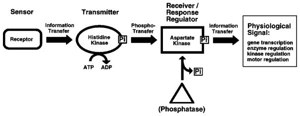
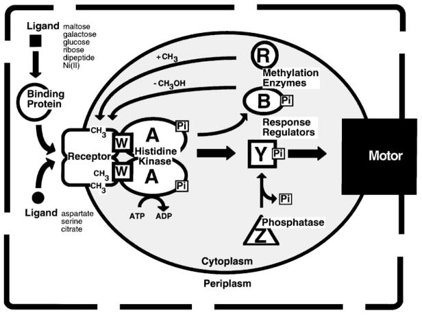
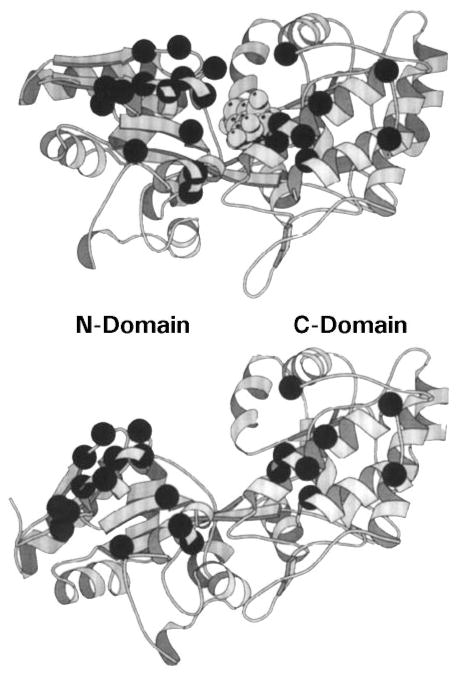
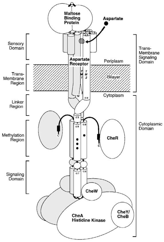
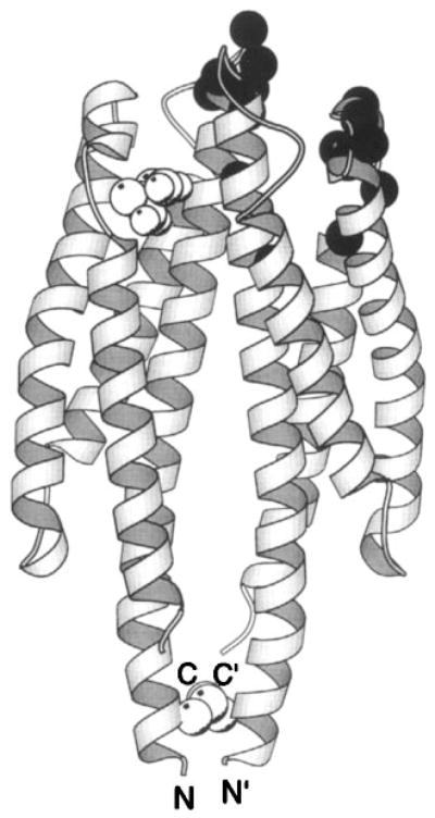
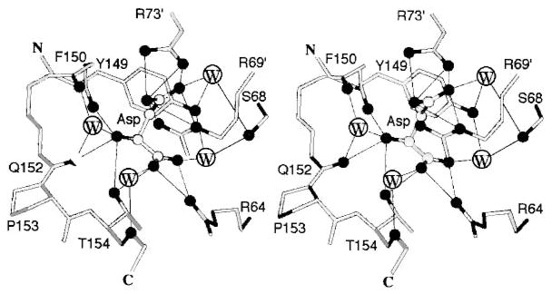
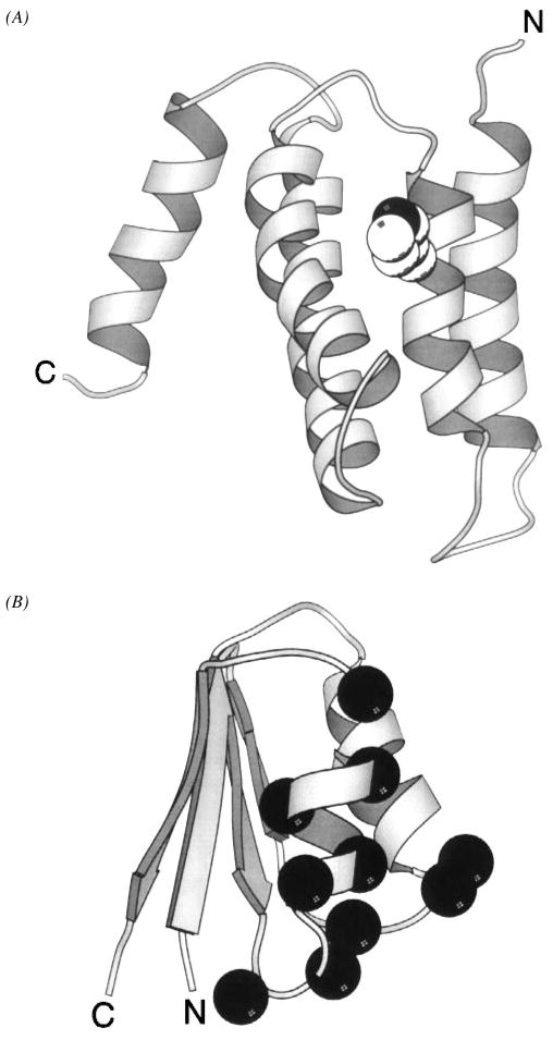
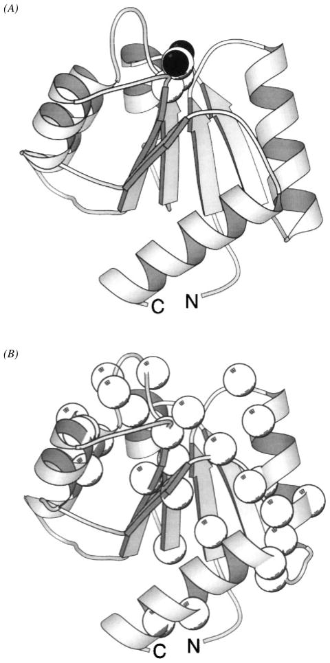
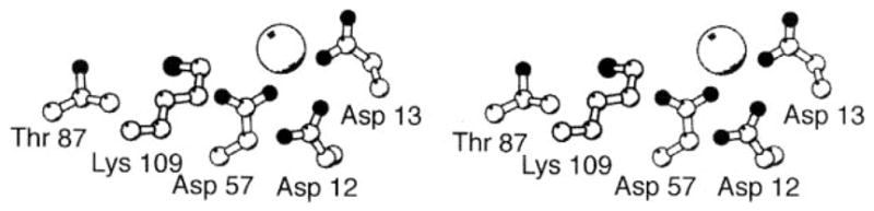
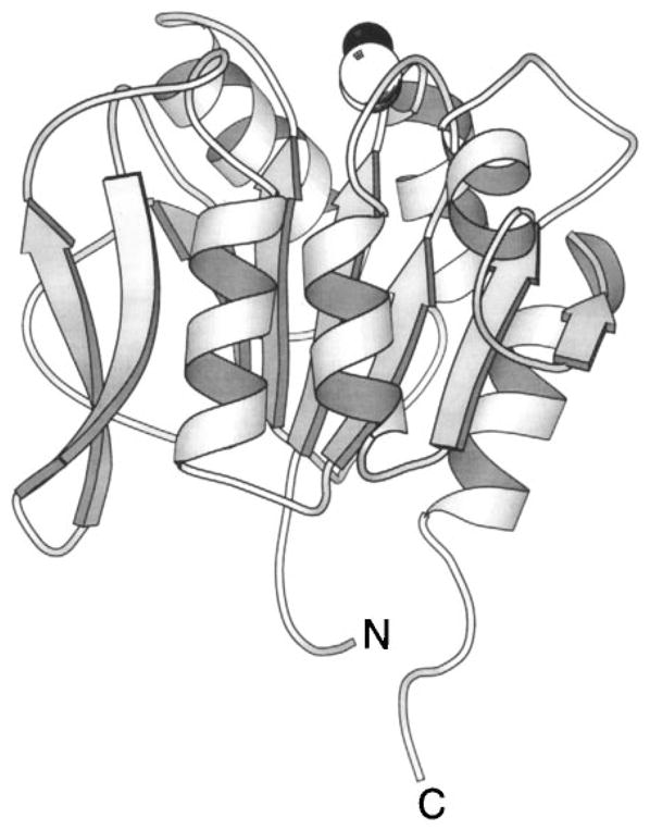
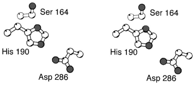
The chemosensory pathway of bacterial chemotaxis has become a paradigm for the two-component superfamily of receptor-regulated phosphorylation pathways. This simple pathway illustrates many of the fundamental principles and unanswered questions in the field of signaling biology. A molecular description of pathway function has progressed rapidly because it is accessible to diverse structural, biochemical, and genetic approaches. As a result, structures are emerging for most of the pathway elements, biochemical studies are elucidating the mechanisms of key signaling events, and genetic methods are revealing the intermolecular interactions that transmit information between components. Recent advances include (a) the first molecular picture of a conformational transmembrane signal in a cell surface receptor, (b) four new structures of kinase domains and adaptation enzymes, and (c) significant new insights into the mechanisms of receptor-mediated kinase regulation, receptor adaptation, and the phospho-activation of signaling proteins. Overall, the chemosensory pathway and the propulsion system it regulates provide an ideal system in which to probe molecular principles underlying complex cellular signaling and behavior.


Figures

Figure 1
Figure 1
Information flow through a two-component signaling pathway. Shown are the standard prokaryotic and eukaryotic signaling components including (a) the sensor module, typically a transmembrane receptor with two putative membrane-spanning helices; (b) a transmitter histidine kinase that is regulated by the receptor and catalyzes autophosphorylation on histidine; and (c) a receiver or response regulator whose active site catalyzes phosphotransfer from the transmitter, thereby yielding autophosphorylation on aspartate. The response regulator can catalyze its own dephosphorylation, but some pathways require a separate phosphatase to generate more rapid dephosphorylation, or to provide additional pathway regulation. Different pathways display highly specialized assemblages of the modular elements; e.g. the sensor, transmitter, and response regulator modules can be separate proteins or can be fused together in various combinations (see text for references).
Figure 2
Figure 2
Self-organized swarm pattern generated by chemotaxing E. coli. Shown is the negative image of a bacterial culture in which growth was initiated at the center of a semisolid medium of defined composition. The pattern was formed over a period of three days as the cells swarmed outward from the center, migrating in response to self-generated gradients of chemical attractants. Dark areas indicate regions of high cell density (Budrene & Berg 1995).
Figure 3
Figure 3
Domain organization of chemosensory pathway components. Confirmed, proteolytically sensitive interdomain linkers that release stable isolated domains are indicated as horizontal bars; structural or functional subdomains are separated by vertical bars. The aspartate receptor is composed of a sensory ligand-binding and transmembrane-signaling domain, coupled to a cytoplasmic kinase regulation domain (TM, transmembrane; MH, methylation). The transmitter histidine kinase CheA is composed of four functionally distinct domains involved in phosphotransfer (P1), response regulatory docking (P2), dimerization, and histidine autophosphorylation and receptor coupling. CheY and CheB share homologous aspartate kinase receiver domains; CheB also possesses a separate methylesterase domain. Residues shown in bold indicate phosphorylation sites on CheA, CheY, and CheB. Ongoing studies are mapping the domain structures of CheZ and FliM (see text for references).
Figure 4
Figure 4
The chemosensory two-component pathway of E. coli and S. typhimurium. Arrows indicate the action of one component on another. Attractants and repellents in the periplasm bind to specific transmembrane receptors or to soluble binding proteins that in turn bind to transmembrane receptors. The transmembrane receptors are coupled by a scaffolding protein (CheW) to a cytoplasmic histidine kinase (CheA), which in turn regulates two response regulators (CheB and CheY). Phosphorylation of CheB modulates the adaptation system in which CheR methylates specific regulatory glutamate side chains on the cytoplasmic surface of the receptor, whereas phospho-CheB hydrolyzes these modifications. The steady state level of receptor methylation provided by the opposing CheR and CheB reactions enables the pathway to adapt to background stimuli and also provides a simple chemical memory. Phosphorylation of CheY modulates the rotary flagellar motor as phospho-CheY docks to the motor switch apparatus, thereby controlling the direction of motor rotation and the swimming behavior of the cell. Although CheY can catalyze its own dephosphorylation, the rate of phospho-signal inactivation is enhanced by a phosphatase activity (CheZ) (see text for references).
Figure 5
Figure 5
Ligand-induced cleft closure in a periplasmic binding protein. Shown are crystal structures of the maltose binding protein (MBP) in its sugar-occupied (upper panel) and apo (lower panel) states (Scharff et al 1992). The ligand-binding site lies in a deep cleft separating the two domains. Bound ligand stabilizes the closed conformation of the cleft, whereas the apo cleft can open by at least 35° and also exhibits an 8° hinge twist. The structural and dynamic differences between these two states regulates the docking of binding proteins to their specific transmembrane receptors (Careaga & Falke 1992). Dark α-carbon spheres denote the genetically defined receptor-docking surface (Zhang et al 1992).
Figure 6
Figure 6
A typical receptor-kinase signaling complex illustrated by the aspartate receptor. The transmembrane receptor provides the architectural framework of the super-molecular signaling complex (Borkovich et al 1989, Ninfa et al 1991, Gegner et al 1992, Schuster et al 1993, Wu et al 1996). Most of the chemosensory pathway components are associated with this complex, either stably or transiently. The kinetically stable core ternary complex is composed of the dimeric receptor (illustrated as a collection of helices), the coupling protein CheW, and the dimeric histidine kinase CheA. Other components are believed to be in rapid equilibrium between bound and soluble forms, including periplasmic binding proteins, the methyltransferase CheR, the methylesterase CheB, and the motor response regulator CheY.
Figure 7
Figure 7
The periplasmic sensory domain of the transmembrane aspartate receptor. The crystal structure of this water-soluble, isolated domain reveals a homodimer of identical four-helix bundles (Milburn et al 1991, Yeh et al 1996). The engineered interdomain disulfide bond (CPK, open sphere, bottom) stabilizes native interactions present in the full-length receptor (Falke & Koshland 1987, Chervitz et al 1995), wherein the membrane-spanning helices would continue in a downward direction. Ligand binding occurs at the opposite, extreme periplasmic end of the domain. Shown is the single molecule of bound aspartate observed in the crystal structure (CPK, open sphere, upper), as well as the genetically defined docking surface for a single molecule of MBP comprised by residues on both receptor subunits (α-carbon, filled sphere; Gardina et al 1997).
Figure 8
Figure 8
The aspartate-binding site of the transmembrane aspartate receptor. The dimeric receptor possesses two aspartate-binding sites that are symmetric in the apo dimer. The first molecule of aspartate binds with high affinity to one of these sites, which has been characterized crystallographically as shown (Milburn et al 1991, Yeh et al 1996). Highlighted are the protein residues and four water molecules that provide direct and indirect aspartate coordination, as well as the Ser68 residue implicated in negative cooperativity between the two sites (Kolodziej et al 1996). Owing to this negative cooperativity, the first aspartate binding event substantially weakens or completely prevents the second binding event (Biemann & Koshland 1994, Danielson et al 1994, Yeh et al 1996).
Figure 9
Figure 9
The aspartate-induced displacement of the transmembrane signaling helix. Shown are the periplasmic regions of the four membrane-spanning helices, two provided by each subunit. When the apo and aspartate-occupied crystal structures (Milburn et al 1991) are superimposed using their static B subunits as a guide, aspartate is observed to displace only the α4/TM2 transmembrane helix in subunit A, termed the signaling helix (Chervitz & Falke 1996). This displacement consists of a vertical, 1.6 Å piston component directed down toward the cytoplasm as well as a subtle 5° helix tilt (difficult to visualize in this perspective). The kink or notch near the upper N-terminal end of the signaling helix is generated by conserved Pro153. This proline creates an indentation in the signaling helix complementary to the shape of the bound ligand, thereby controlling the vertical position the helix. Gray and black helices represent the apo and aspartate-occupied structures, respectively; cross-sectional shapes specify helices from subunits A (elliptical) and B (square), also denoted by primes.
Figure 10
Figure 10
Engineered cysteine pairs that yield lock-on and lock-off disulfide bonds in the full-length, aspartate receptor-kinase complex. Shown are the periplasmic ends of the four transmembrane helices in the dimer (Milburn et al 1991), two of which have been extended by modeling into the bilayer region (Chervitz & Falke 1996). A disulfide formed between cysteines Cys25/Cys197 or between Cys39/Cys183 locks the kinase on and decreases aspartate affinity. At the other extreme, a disulfide linkage between cysteines Cys176/Cys43 or between Cys179/Cys39 locks kinase activity off and increases the aspartate affinity (Chervitz & Falke 1996). These properties mirror those expected for the native on and off states of the receptor-kinase complex, respectively, in which aspartate binding causes kinase inactivation. Lock-on disulfides trap upward vertical displacements of the signaling helix; lock-off disulfides (analogous to aspartate binding) trap downward displacements toward the cytoplasm.
Figure 11
Figure 11
Model for the cytoplasmic domain of the transmembrane receptors. A secondary-structure analysis of aligned sequences from over 56 related receptors suggests that each subunit of the homodimeric domain contains five amphiphilic helices (α5 to α9) and a short region of β-strand (β1) (LeMoual & Koshland 1996, Danielson 1997). Functionally, the domain is divided into the linker region, which provides the interface to the transmembrane signaling helix; the methylation region, which contains the sites of adaptive methylation (large black circles); and the signaling domain, which promotes CheW and CheA binding (see text for references). Also shown are the locations of lock-on and lock-off mutations in the serine receptor (white and black small circles, respectively), as well as second site suppressors of the inhibitory A19K mutation in the first transmembrane helix of the aspartate receptor (white small squares) (Ames et al 1988, Oosawa & Simon 1986). Both sets of mutations identify critical regulatory regions.
Figure 12
Figure 12
Structure of the CheR methyltransferase enzyme. The CheR protein uses S-adenosyl-methionine as a substrate for methyl transfer to the adaptation sites of transmembrane chemosensory receptors. This crystal structure reveals two distinct domains connected by a long, single-strand hinge (Djordjevic & Stock 1997). The N-terminal domain is an assembly of perpendicular helices; the C-terminal domain exhibits the α/β folding motif. The bound S-adenosyl-homocysteine molecule (CPK, sphere), a product of the methylation reaction, identifies the location of the active site region between the two domains. (Black spheres indicate oxygen atoms.)
Figure 13
Figure 13
Two domains of the CheA histidine kinase. (A) The N-terminal phosphotransfer domain, termed P1, provides the phospho-histidine used as a substrate during phosphotransfer from CheA to response regulators. The NMR solution structure of the phosphotransfer domain reveals a bundle of five helices (Zhou et al 1995, Zhou & Dahlquist 1997). The site of phosphorylation is the Nε2 nitrogen atom (black) of the His48 imidazole ring (CPK, sphere), located on the surface of helix α2. Phosphorylation yields a local structural change limited to the immediate environment of the phospho-histidine. (B) The solution structure of the response regulator docking domain, designated P2, displays an open-faced β-sandwich folding motif (McEvoy et al 1995, 1996). The residues implicated in CheY binding (α-carbon, black sphere) are clustered to a distinct docking surface.
Figure 14
Figure 14
Structure of the response regulator CheY, illustrating the phospho-induced conformational regulation of three docking surfaces. (A) The CheY molecule serves as a receiver of signals from CheA and as the response regulator for motor switching. This crystal structure, which includes a bound catalytic Mg2+ ion, displays the α/β folding motif of unphosphorylated CheY (Stock et al 1993). The site of phosphorylation is Asp57 located at the upper edge of the parallel β-sheet (CPK, side chain). Other crystallographic and NMR structures have yielded the same overall backbone fold (see references in text). (B) View of CheY from the same perspective showing residues (α-carbon, sphere) perturbed by the phosphorylation-induced global conformational change, as revealed by aromatic side chain (Drake et al 1993) or backbone (Lowry et al 1994) NMR frequency changes. The large, phospho-regulated surface is seen to cover most of the protein. In addition, smaller backbone frequency changes are observed throughout the molecule, indicating that the conformational change is global (Lowry et al 1994). (C) Same perspective, illustrating a CheA docking surface defined by NMR (α-carbon, black sphere, McEvoy et al 1995, 1996) and surfaces implicated by genetic studies as important to motor switch docking (α-carbon, white sphere; Roman et al 1992, Sockett et al 1992) or CheZ interactions (α-carbon, gray sphere; Sanna et al 1995). The three regions are largely distinct, and none of the interfaces directly overlaps the phosphorylation site (Asp57 is indicated as a ball-and-stick side chain). Some overlap exists between the CheA and motor docking surfaces (Shukla & Matsumura 1995). Phospho-activation of CheY generates a global conformational change that alters the conformation of these docking regions (Drake et al 1993, Lowry et al 1994).
Figure 14
Figure 14
Structure of the response regulator CheY, illustrating the phospho-induced conformational regulation of three docking surfaces. (A) The CheY molecule serves as a receiver of signals from CheA and as the response regulator for motor switching. This crystal structure, which includes a bound catalytic Mg2+ ion, displays the α/β folding motif of unphosphorylated CheY (Stock et al 1993). The site of phosphorylation is Asp57 located at the upper edge of the parallel β-sheet (CPK, side chain). Other crystallographic and NMR structures have yielded the same overall backbone fold (see references in text). (B) View of CheY from the same perspective showing residues (α-carbon, sphere) perturbed by the phosphorylation-induced global conformational change, as revealed by aromatic side chain (Drake et al 1993) or backbone (Lowry et al 1994) NMR frequency changes. The large, phospho-regulated surface is seen to cover most of the protein. In addition, smaller backbone frequency changes are observed throughout the molecule, indicating that the conformational change is global (Lowry et al 1994). (C) Same perspective, illustrating a CheA docking surface defined by NMR (α-carbon, black sphere, McEvoy et al 1995, 1996) and surfaces implicated by genetic studies as important to motor switch docking (α-carbon, white sphere; Roman et al 1992, Sockett et al 1992) or CheZ interactions (α-carbon, gray sphere; Sanna et al 1995). The three regions are largely distinct, and none of the interfaces directly overlaps the phosphorylation site (Asp57 is indicated as a ball-and-stick side chain). Some overlap exists between the CheA and motor docking surfaces (Shukla & Matsumura 1995). Phospho-activation of CheY generates a global conformational change that alters the conformation of these docking regions (Drake et al 1993, Lowry et al 1994).
Figure 15
Figure 15
The aspartate kinase active site of CheY. Shown is the Mg2+-occupied structure of the unphosphorylated active site (Stock et al 1993), illustrating the highly conserved catalytic residues. Asp57 serves as the site of phosphorylation, and the aspartate triad (Asp12, Asp13, Asp57) provides both direct and indirect Mg2+ coordination, the latter via solvent. Lys109 and Thr87 act as acid-base catalysts. The Mg2+ ion serves as an essential cofactor in both the autocatalytic phosphorylation and dephosphorylation reactions. (For additional references, see text.)
Figure 16
Figure 16
The methylesterase domain of CheB. The C-terminal domain of CheB is a methylesterase that hydrolyzes the regulatory methyl esters and amides of the receptor adaptation sites. In the full-length protein this activity is regulated by phosphorylation of the N-terminal receiver domain (not shown). The crystal structure of the isolated methylesterase domain (West et al 1995) displays an α/β folding motif coupled to a β-hairpin (extreme left). The highlighted Ser164 side chain (CPK, sphere), located on one edge of the β-sheet, acts as the nucleophile in ester and amide hydrolysis.
Figure 17
Figure 17
The methyltransferase active site of CheB. Shown are the catalytic residues, including the Ser164 residue essential for catalytic activity and proposed to act as the nucleophile in the methyl ester and amide hydrolysis (West et al 1995). Together the Ser164, His190 and Asp286 side chains form a novel catalytic triad that is functionally, but not structurally, analogous to the catalytic triads of serine proteases.

References

    1. Alex LA, Borkovich KA, Simon MI. Hyphal development in Neurospora crassa. Involvement of a 2-component histidine kinase. Proc Natl Acad Sci USA. 1996;93:3416–21. - PMC - PubMed
    1. Ames P, Chen J, Wolff A, Parkinson JS. Structure-function studies of bacterial chemosensors. Cold Spring Harbor Symp Quant Biol. 1988;53:59–66. - PubMed
    1. Ames P, Parkinson JS. Constitutively signaling fragments of Tsr, the Escherichia coli serine chemoreceptor. J Bacteriol. 1994;176:6340–48. - PMC - PubMed
    1. Ames P, Yu AL, Parkinson JS. Methylation segments are not required for chemotactic signaling by cytoplasmic fragments of Tsr, the methyl-accepting serine chemoreceptor of Escherichia coli. Mol Microbiol. 1996;19:737–46. - PubMed
    1. Appleby JL, Parkinson JS, Bourret RB. Signal transduction via the multistep phosphorelay. Not necessarily a road less traveled. Cell. 1996;86:845–48. - PubMed

Publication types

LinkOut - more resources