. 1998 Feb;72(2):934-42.
doi: 10.1128/JVI.72.2.934-942.1998.

Nitric oxide inhibits rhinovirus-induced cytokine production and viral replication in a human respiratory epithelial cell line

Affiliations

Nitric oxide inhibits rhinovirus-induced cytokine production and viral replication in a human respiratory epithelial cell line

S P Sanders et al. J Virol. 1998 Feb.

Abstract

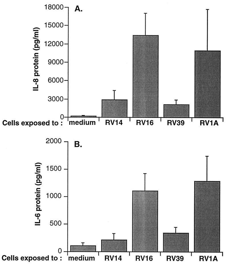
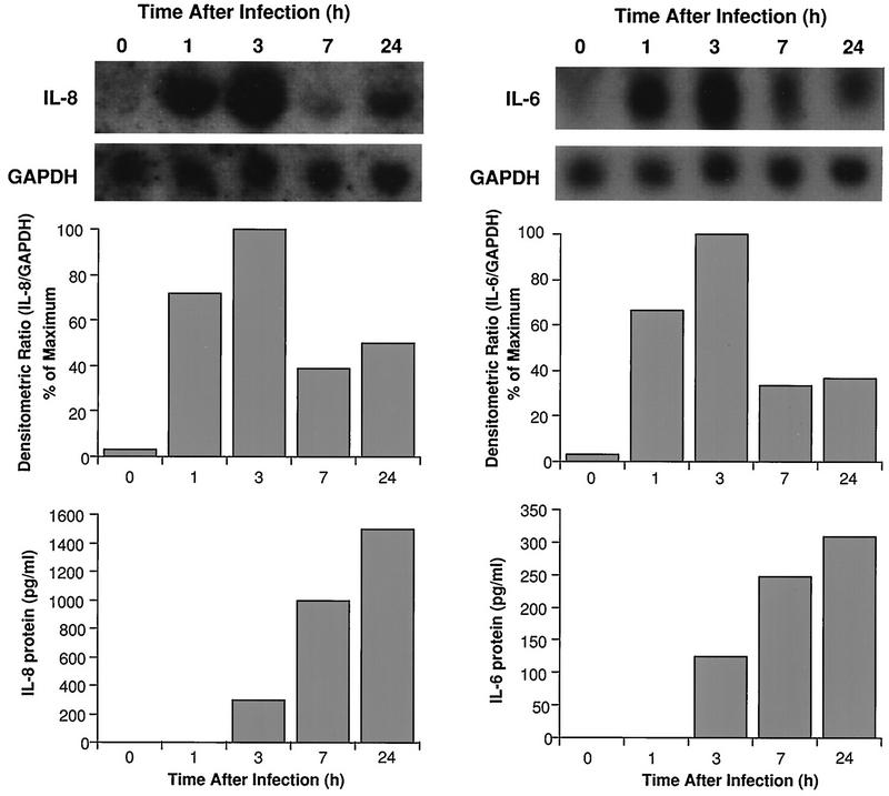
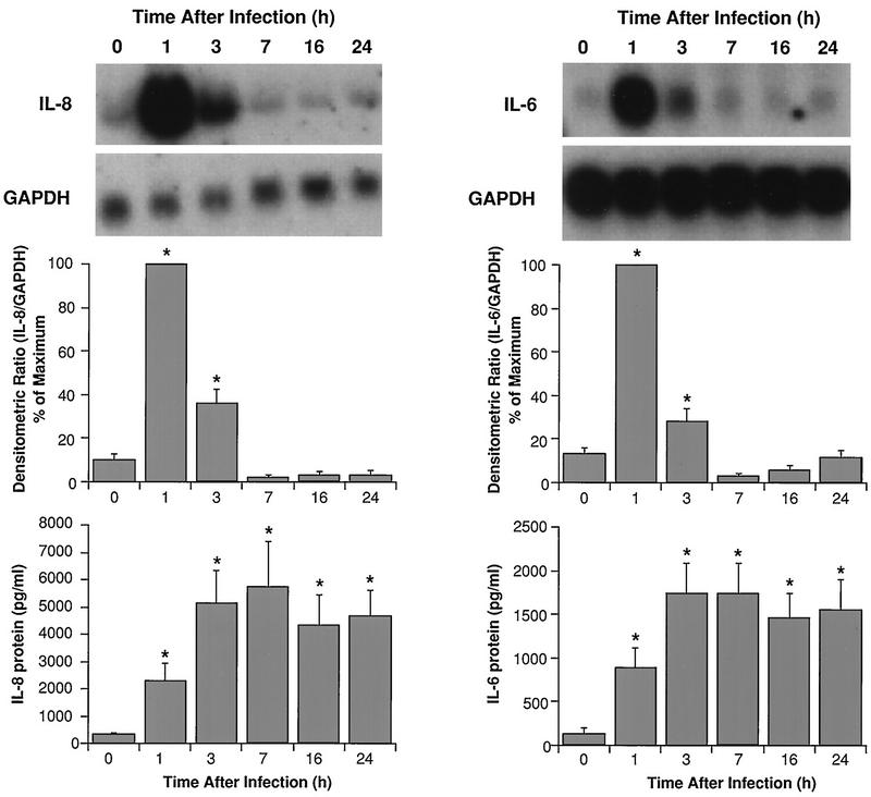
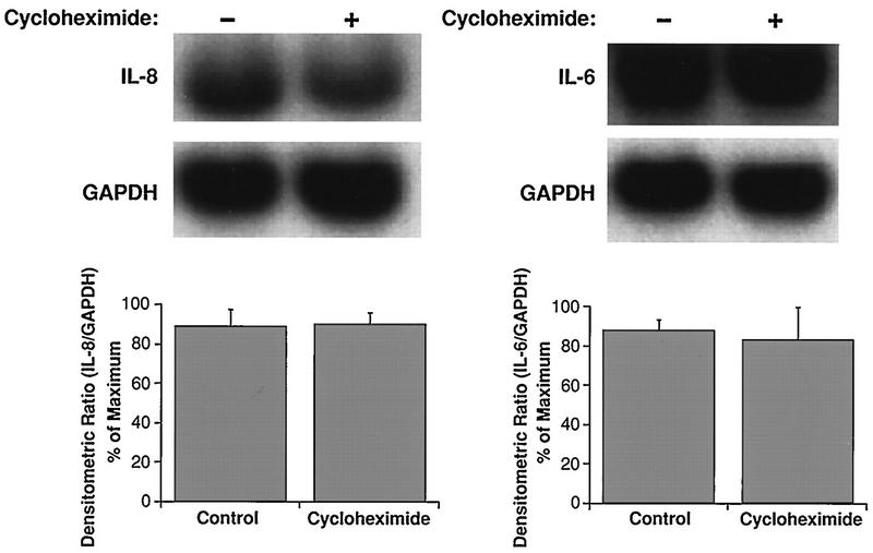
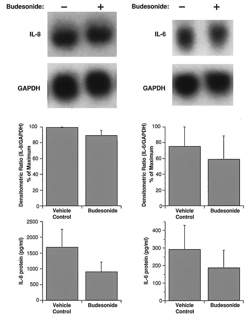
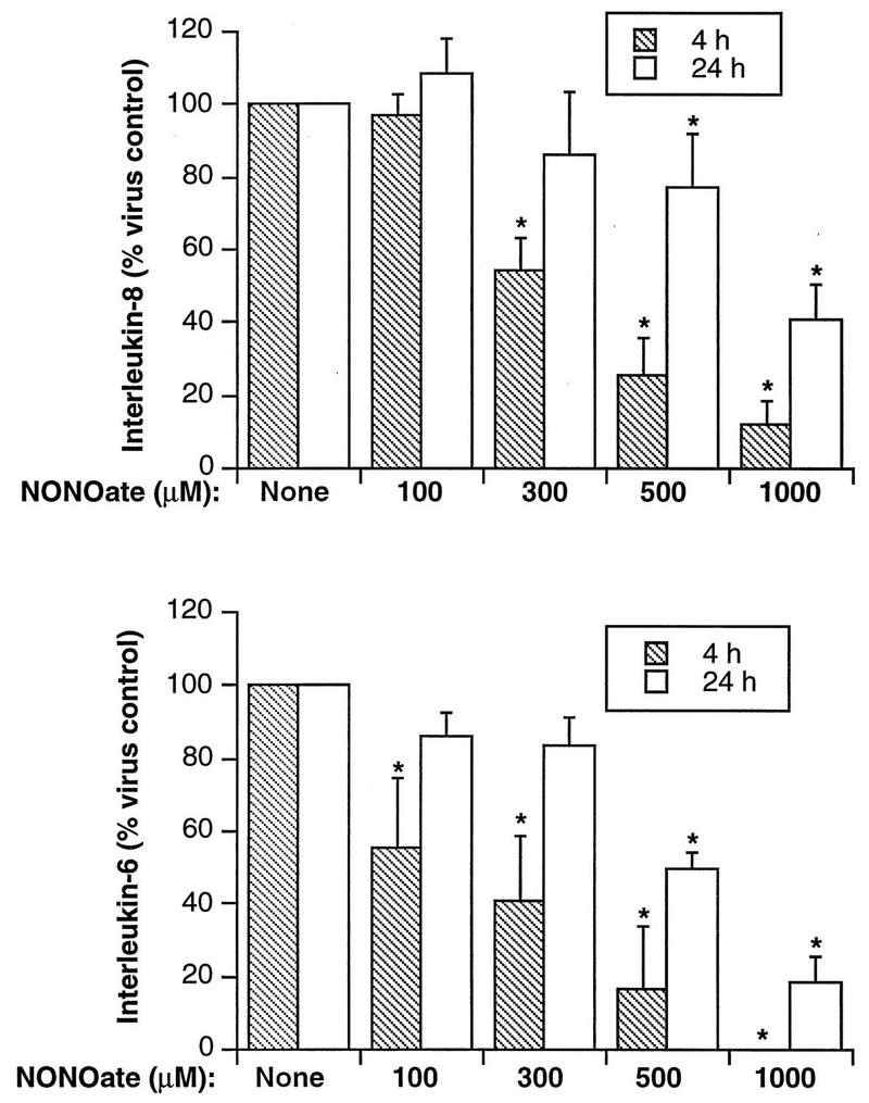
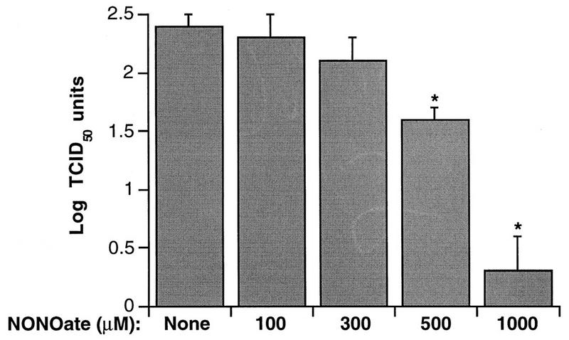
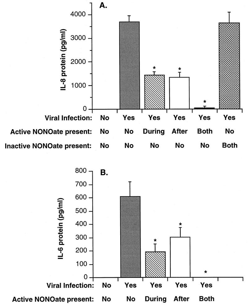
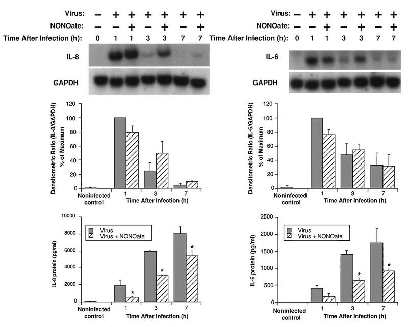
To better understand the early biochemical events that occur in human rhinovirus (HRV) infections, we examined the kinetics and mechanisms of interleukin-8 (IL-8) and IL-6 production from infected epithelial cells. Several HRV strains caused IL-8 and IL-6 production, but HRV-16 induced maximal IL-8 and IL-6 mRNA expression and protein production more rapidly than did HRV-14, despite similar rates of replication of the two viral strains. Viral induction of cytokine mRNA does not require new protein synthesis, since it was unaffected by cycloheximide treatment. The potent glucocorticoid budesonide did not affect viral replication or cytokine mRNA induction but modestly inhibited cytokine protein production. Interestingly, the nitric oxide donor 3-(2-hydroxy-2-nitroso-1-propylhydrazino)-1-propanamine (NONOate) inhibited both rhinovirus replication and cytokine production in a dose-dependent fashion without reducing levels of cytokine mRNA. The NONOate effects were due to release of nitric oxide, because NONOate that had been depleted of its nitric oxide content had no effect. Thus, nitric oxide may play an important anti-inflammatory and antiviral role in colds and nitric oxide donors may represent a novel therapeutic approach.


Figures

FIG. 1
FIG. 1
Cytokine production from BEAS-2B cells 24 h after infection with each of several strains of HRV. Data represent the mean plus SEM from three experiments. (A) Production of IL-8; (B) production of IL-6.
FIG. 2
FIG. 2
Time course of induction of steady-state mRNA levels and proteins for IL-8 (left) and IL-6 (right) from HRV-14-infected BEAS-2B cells. The upper panels show Northern blots for each cytokine and for the housekeeping gene product, GAPDH. The center panels show densitometric ratios, while the lower panels show protein levels produced at each of the indicated time points. Data are from a representative experiment (n = 3).
FIG. 3
FIG. 3
Time course of induction of steady-state mRNA levels and protein for IL-8 (left) and IL-6 (right) from HRV-16-infected BEAS-2B cells. The upper panels show representative Northern blots for each cytokine and for the housekeeping gene product, GAPDH. The center panels show the means plus SEM of densitometric ratios for four experiments. The lower panels show the means plus SEM of amounts of protein produced for four experiments at each time point. Asterisks indicate significant increases in each parameter relative to the zero time control (P < 0.05 in each case).
FIG. 4
FIG. 4
Cycloheximide does not alter steady-state mRNA levels for IL-8 (left) and IL-6 (right) measured 1 h after infection with HRV-16. The upper panels show representative Northern blots, while the lower panels show the means plus SEM of densitometric ratios for four experiments.
FIG. 5
FIG. 5
Effects of budesonide (10−7 M) on steady-state mRNA levels and proteins for IL-8 (left) and IL-6 (right) from HRV-16-infected BEAS-2B cells. The upper panels show representative Northern blots with mRNA extracted 1 h after infection. The center panels show the means plus SEM of densitometric ratios from three experiments. The lower panels show the means plus SEM of amounts of protein produced 7 h after infection in three experiments.
FIG. 6
FIG. 6
Dose-dependent inhibition of cytokine production from HRV-16-infected BEAS-2B cells by NONOate. The upper panel shows the means plus SEM from four experiments for IL-8 production at 4 and 24 h after HRV-16 infection, while the lower panel shows data for IL-6 production. The asterisks indicate significant inhibition compared to levels produced at the same time after infection in the absence of NONOate (P < 0.05 in each case).
FIG. 7
FIG. 7
Dose-dependent inhibition by NONOate of HRV-16 titers in BEAS-2B supernatants recovered 24 h after viral exposure. Data represent the means plus SEM of values from four experiments. The asterisks indicate significant inhibition compared to levels produced in the absence of NONOate (P < 0.05 in each case).
FIG. 8
FIG. 8
Comparison of the effects of inactive NONOate and of active NONOate added at different times during the infection procedure on HRV-16-induced cytokine production from BEAS-2B cells. NONOate was used at a final concentration of 1,000 μM, and protein levels were measured 4 h after infection. (A) Means plus SEM of values for IL-8 production from three experiments; (B) data for IL-6. Asterisks indicate significant inhibition compared to levels produced by virus alone (P < 0.05 in each case).
FIG. 9
FIG. 9
Effects of NONOate (500 μM) on steady-state mRNA levels and proteins for IL-8 (left) and IL-6 (right) at different times after infection with HRV-16. The upper panels show representative Northern blots for each cytokine and for the housekeeping gene product, GAPDH. The center panels show the means plus SEM of densitometric ratios for three experiments. The lower panels show the means plus SEM of amounts of protein produced for three experiments at each time point. The asterisks indicate significant inhibition by NONOate compared to levels produced by virus alone (P < 0.05 in each case).

Similar articles

Cited by

References

    1. Akiro S, Hirano T, Taga T, Kishimoto T. Biology of multifunctional cytokines: IL 6 and related molecules (IL 1 and TNF) FASEB J. 1990;4:2860–2867. - PubMed
    1. Arnold R, Humbert B, Werchau H, Gallati H, König W. Interleukin-8, interleukin-6, and soluble tumour necrosis factor receptor type I release from a human pulmonary epithelial cell line (A549) exposed to respiratory syncytial virus. Immunology. 1994;82:126–133. - PMC - PubMed
    1. Arola M, Ziegler T, Puhakka H, Lehtonen O P, Ruuskanen O. Rhinovirus in otitis media with effusion. Ann Otol Rhinol Laryngol. 1990;99:451–453. - PubMed
    1. Asano K, Chee C B E, Gaston B, Lilly C M, Gerard C, Drazen J M, Stamler J S. Constitutive and inducible nitric oxide synthase gene expression, regulation, and activity in human lung epithelial cells. Proc Natl Acad Sci USA. 1994;91:10089–10093. - PMC - PubMed
    1. Baggiolini M, Walz A, Kunkel S L. Neutrophil-activating peptide-1/interleukin 8, a novel cytokine that activates neutrophils. J Clin Invest. 1989;84:1045–1049. - PMC - PubMed

Publication types

LinkOut - more resources