. 1998 Feb;72(2):965-74.
doi: 10.1128/JVI.72.2.965-974.1998.

Attenuation of the vaccine Oka strain of varicella-zoster virus and role of glycoprotein C in alphaherpesvirus virulence demonstrated in the SCID-hu mouse

Affiliations

Attenuation of the vaccine Oka strain of varicella-zoster virus and role of glycoprotein C in alphaherpesvirus virulence demonstrated in the SCID-hu mouse

J F Moffat et al. J Virol. 1998 Feb.

Abstract

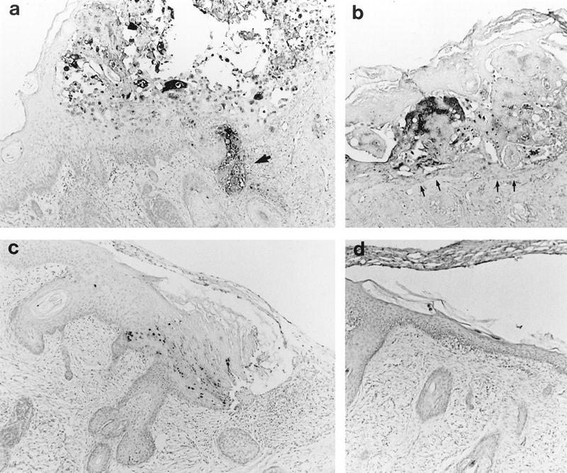
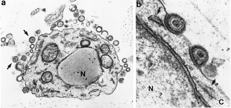
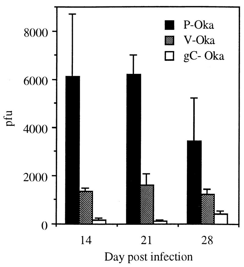
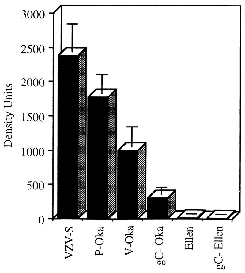
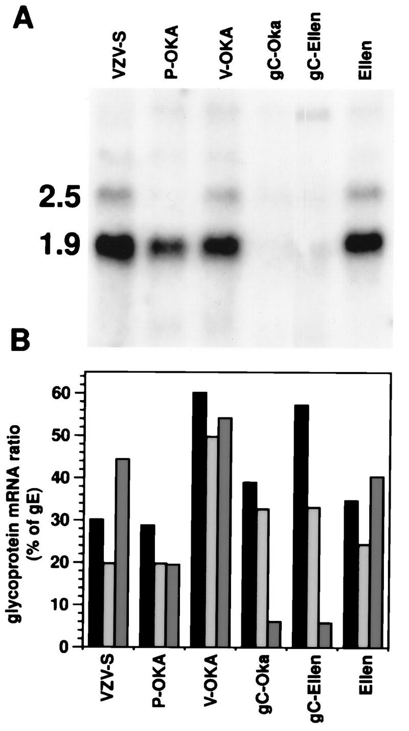
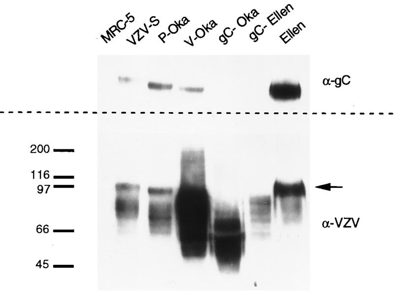
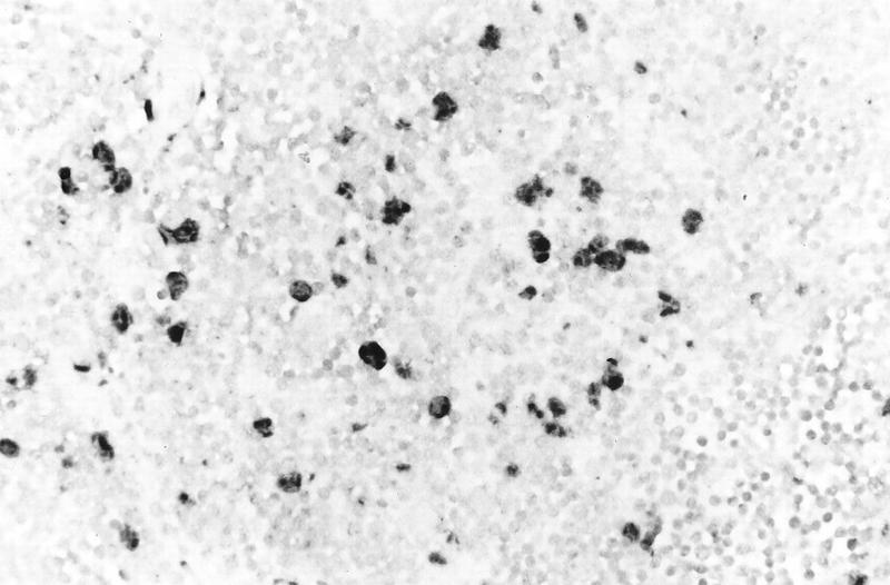
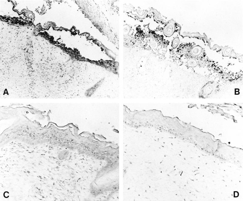
The SCID-hu mouse implanted with human fetal tissue is a novel model for investigating human viral pathogenesis. Infection of human skin implants was used to investigate the basis for the clinical attenuation of the varicella-zoster virus (VZV) strain, V-Oka, from which the newly licensed vaccine is made. The pathogenicity of V-Oka was compared with that of its parent, P-Oka, another low-passage clinical isolate, strain Schenke (VZV-S), and VZV-Ellen, a standard laboratory strain. The role of glycoprotein C (gC) in infectivity for human skin was assessed by using gC-negative mutants of V-Oka and VZV-Ellen. Whereas all of these VZV strains replicated well in tissue culture, only low-passage clinical isolates were fully virulent in skin, as shown by infectious virus yields and analysis of implant tissues for VZV DNA and viral protein synthesis. The infectivity of V-Oka in skin was impaired compared to that of P-Oka, providing the first evidence of a virologic basis for the clinical attenuation of V-Oka. The infectivity of V-Oka was further diminished in the absence of gC expression. All strains except gC-Ellen retained some capacity to replicate in human skin, but cell-free virus was recovered only from implants infected with P-Oka or VZV-S. Although VZV is closely related to herpes simplex virus type 1 (HSV-1) genetically, experiments in the SCID-hu model revealed differences in tropism for human cells that correlated with differences in VZV and HSV-1 disease. VZV caused extensive infection of epidermal and dermal skin cells, while HSV-1 produced small, superficial lesions restricted to the epidermis. As in VZV, gC expression was a determinant for viral replication in skin. VZV infects human CD4+ and CD8+ T cells in thymus/liver implants, but HSV-1 was detected only in epithelial cells, with no evidence of lymphotropism. These SCID-hu mouse experiments show that the clinical attenuation of the varicella vaccine can be attributed to decreased replication of V-Oka in skin and that tissue culture passage alone reduces the ability of VZV to infect human skin in vivo. Furthermore, gC, which is dispensable for replication in tissue culture, plays a critical role in the virulence of the human alphaherpesviruses VZV and HSV-1 for human skin.


Figures

FIG. 1
FIG. 1
Histological analysis of VZV-infected skin implants. Subcutaneous skin implants infected with P-Oka (a), V-Oka (b), or gC-Oka (c) or mock infected (d) were fixed in paraformaldehyde, paraffin embedded, and cut into 3-μm sections before in situ hybridization. Darkly stained cells indicate VZV DNA; tissue was counterstained lightly with hematoxylin. At day 21 postinfection, gC-Oka lesions appeared in the epidermis (c). V-Oka lesions were larger; however, the basement membrane remained intact (arrows), and virus did not penetrate into the dermis (b). P-Oka skin lesions were largest (a), and VZV DNA was detected deep within dermal fibroblasts (arrow). Mock-infected skin had a normal appearance (d), and no DNA was detected in the implant. Histology shown is representative of four implants. Magnification, ×83.
FIG. 2
FIG. 2
VZV release from skin cells, determined by transmission electron microscopy of skin cells 21 days after inoculation with VZV-S. (a) Enveloped virons containing dense cores have dispersed from the cell (arrows). Vacuoles from which particles egressed are visible adjacent to the cell membrane. Nucleocapsids are not visible in the nucleus (N) by this staining method. Magnification, ×24,000. (b) Higher-magnification view of virions with intact lipid bilayer on cell surface. Remnants of cytoplasmic vacuoles which carried virions to plasma membrane are indicated by arrows. N, nucleus; C, cytoplasm. Magnification, ×108,000.
FIG. 3
FIG. 3
Infectious virus in VZV-infected skin implants. Implants were inoculated with P-Oka, V-Oka, or gC-Oka and harvested 14, 21, or 28 days postinfection. Cell-associated virus was measured in an infectious focus assay, and PFU per implant was calculated. Error bars indicate the standard error of the mean. On day 21, all differences between strains were statistically significant (P ≤ 0.02, Student’s t test).
FIG. 4
FIG. 4
VZV protein synthesis in skin implants. Implants were inoculated with VZV and harvested on day 21 postinfection. Protein was extracted from infected implants, separated by SDS-PAGE, and transferred to nylon membranes. Western blot analysis was performed; VZV proteins were detected with a high-titer human polyclonal serum and ECL on a phosphorimager. The concentration of VZV protein in the range of 70 to 120 kDa was measured digitally in density units. The means and standard error are shown for each VZV strain. All paired VZV strains are statistically different from each other (P ≤ 0.04, Student’s t test) except (i) VZV-S and P-Oka and (ii) VZV-Ellen and gC-Ellen.
FIG. 5
FIG. 5
VZV glycoprotein mRNA ratios and expression of gC mRNA in vitro. VZV-S, P-Oka, V-Oka, gC-Oka, gC-Ellen, and VZV-Ellen were grown to equivalent CPE in MeWo cells, and total cell mRNA was prepared. RNA was electrophoresed, transferred to membranes, and hybridized with 32P-labeled probes specific for gB, gC, gE, and gH transcripts. Northern blots were visualized, and band intensities were quantitated by phosphorimager analysis. (A) Northern blot showing the 2.5- and 1.9-kb gC mRNA transcripts. Transcription of gC from gC-Oka and gC-Ellen was markedly decreased. (B) gB/gE (black bars), gH/gE (gray bars), and gC/gE (dark gray bars) ratios were calculated as a percentage of the gE transcript for each strain. Transcription of gC was deficient in gC-Oka and gC-Ellen, whereas gB and gH transcription was not altered. VZV-S, P-Oka, V-Oka, and VZV-Ellen had normal glycoprotein profiles.
FIG. 6
FIG. 6
Western blot analysis of gC expression in vitro. Protein was extracted from VZV-infected or uninfected MRC-5 cells, separated by SDS-PAGE, and transferred to a polyvinylidene difluoride membrane. On duplicate membranes, VZV proteins were detected with a polyclonal human immune serum (lower panel) and gC was detected with a monospecific human serum directed against a vaccinia virus-gC recombinant (upper panel). Bound antibodies were detected with horseradish peroxidase conjugated anti-human immunoglobulin G and visualized by ECL. Typical broad bands of VZV proteins were detected in all lanes of infected MRC-5 cells and not in uninfected cells. The approximately 105-kDa gC band, indicated by the arrow, can be seen on the blot probed with anti-VZV serum in all strains except gC-Oka and gC-Ellen, and the corresponding bands are missing on the blot probed with anti-gC serum. Molecular masses (in kilodaltons) are shown on the left.
FIG. 7
FIG. 7
Histological analysis of an HSV-infected thy/liv implant inoculated with HSV-1 KOS and harvested on day 2 after infection. The implant was fixed in paraformaldehyde, paraffin embedded, and cut into 3-μm sections before in situ hybridization was performed. Darkly stained cells indicate where HSV-1 DNA was detected in cortical epithelial cells. Uninfected T cells were lightly counterstained with hematoxylin. Magnification, ×131.
FIG. 8
FIG. 8
Histological analysis of HSV-infected skin implants. Subcutaneous skin implants were inoculated with HSV-1 KOS (A), HSV-1 ΔgC2-3rev (B), or HSV-1 ΔgC2-3 (C) or mock infected (D) and harvested on day 6 postinfection. Implants were fixed in paraformaldehyde, paraffin embedded, and cut into 3-μm sections before in situ hybridization was performed. HSV-1 DNA was detected in the equivalent epidermal lesions produced by KOS and ΔgC2-3rev (A and B). No HSV-1 DNA was detected in the epidermis of ΔgC2-3- or mock-infected implants (C and D). Hematoxylin counterstain revealed tissue histology. Magnifications: A and B, ×61; C and D, ×122.

Similar articles

Cited by

References

    1. Aldrovandi G M, Feuer G, Gao L, Jamieson B, Kristeva M, Chen I S Y, Zack J A. The SCID-hu mouse as a model for HIV-1 infection. Nature. 1993;363:732–736. - PubMed
    1. Arvin A M. Varicella-zoster virus. In: Fields B N, Knipe D N, Howley P M, editors. Virology. Philadelphia, Pa: Lippincott-Raven Publishers; 1996. pp. 2547–2585.
    1. Arvin A M, Koropchak C M, Williams B R G, Grumet F C, Foung S K. Early immune response in healthy and immunocompromised subjects with primary varicella-zoster virus infection. J Infect Dis. 1986;154:422–429. - PubMed
    1. Auwaerter P G, Kaneshima H, McCune J M, Wiegand G, Griffin D E. Measles virus infection of thymic epithelium in the SCID-hu mouse leads to thymocyte apoptosis. J Virol. 1996;70:3734–3740. - PMC - PubMed
    1. Bergen R E, Diaz P S, Arvin A M. The immunogenicity of the Oka/Merck varicella vaccine in relation to infectious varicella-zoster virus and relative viral antigen content. J Infect Dis. 1990;162:1049–1054. - PubMed

Publication types

Substances