. 1998 Feb;72(2):994-1004.
doi: 10.1128/JVI.72.2.994-1004.1998.

Construction of retroviral vectors with improved safety, gene expression, and versatility

Affiliations

Construction of retroviral vectors with improved safety, gene expression, and versatility

S H Kim et al. J Virol. 1998 Feb.

Abstract

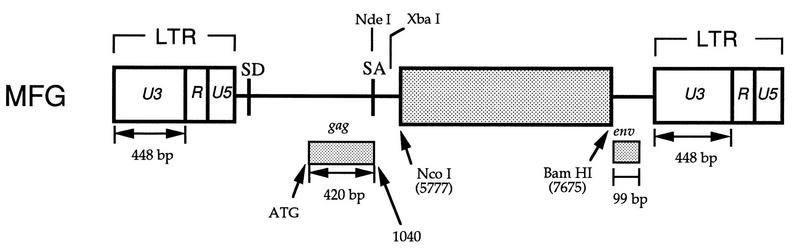
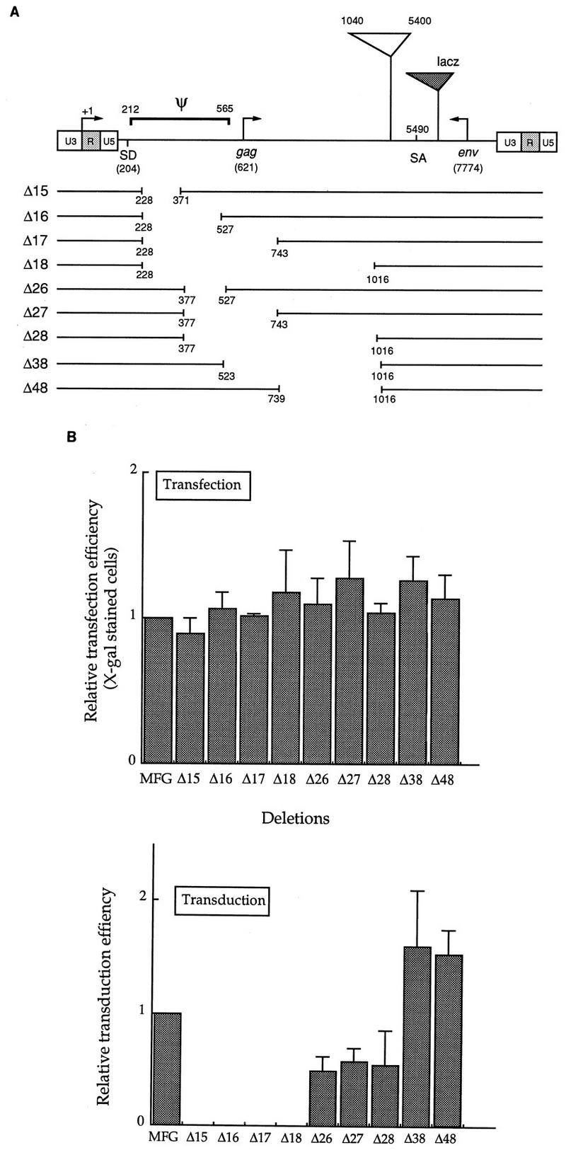
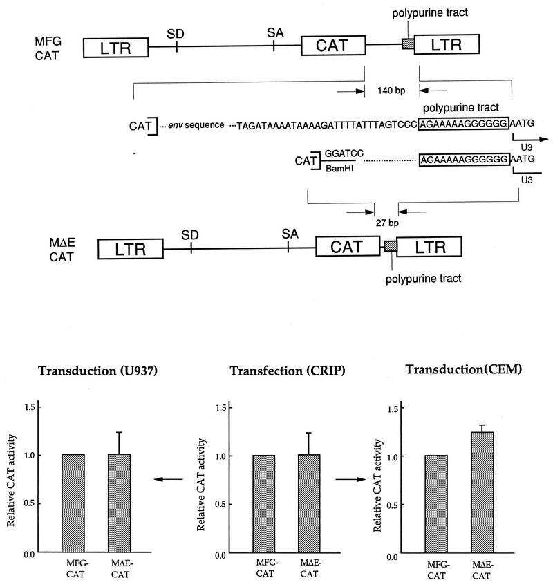
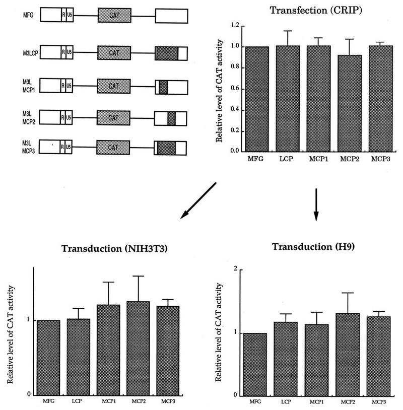
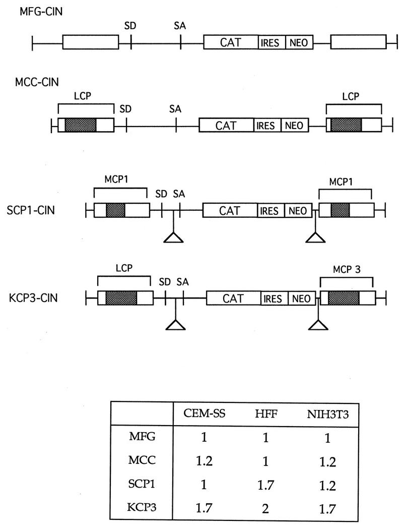
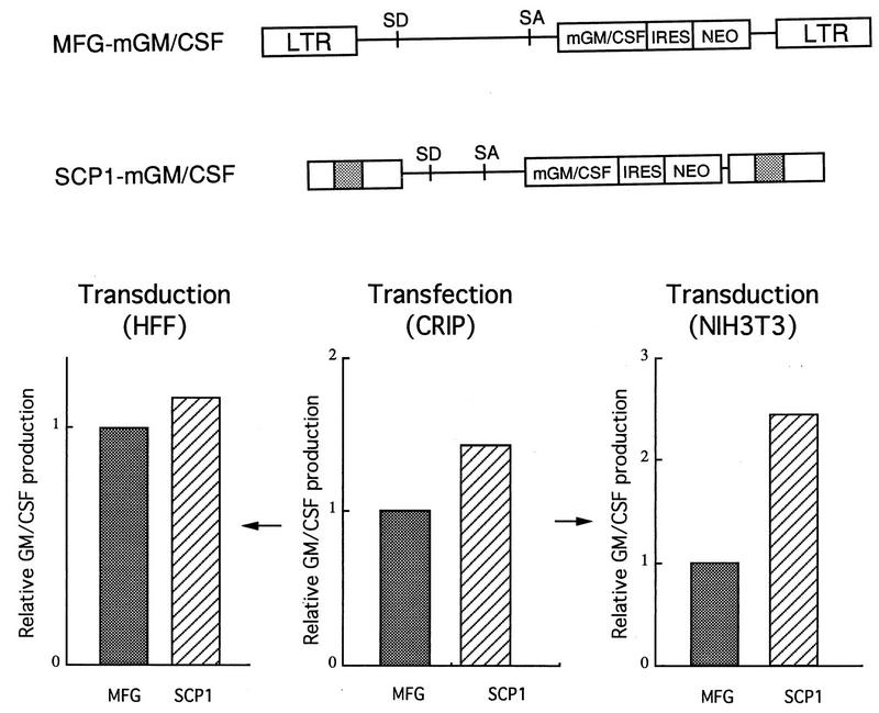
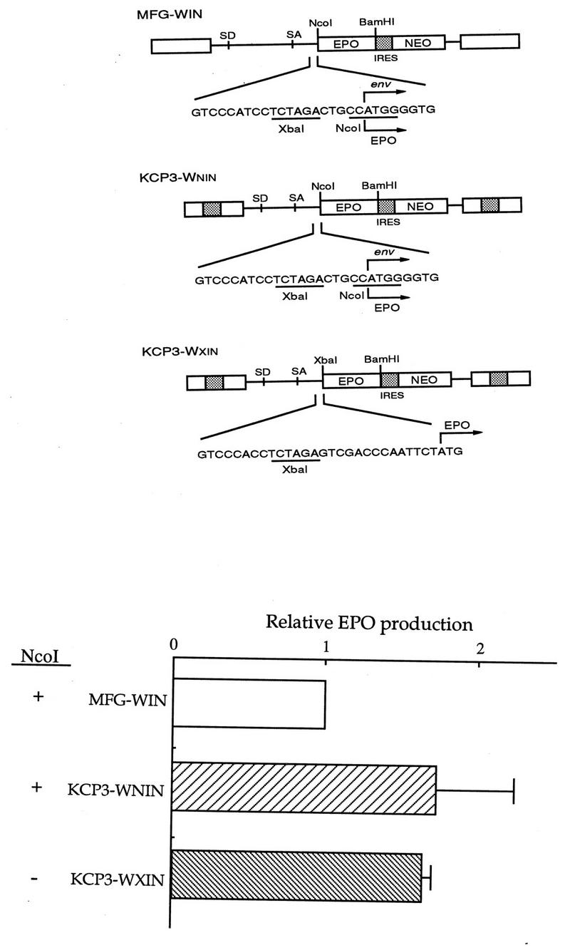
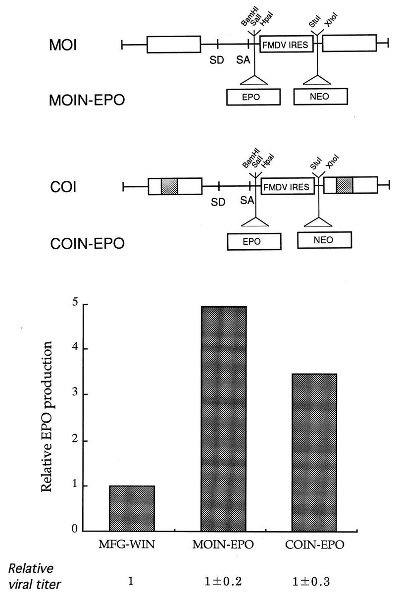
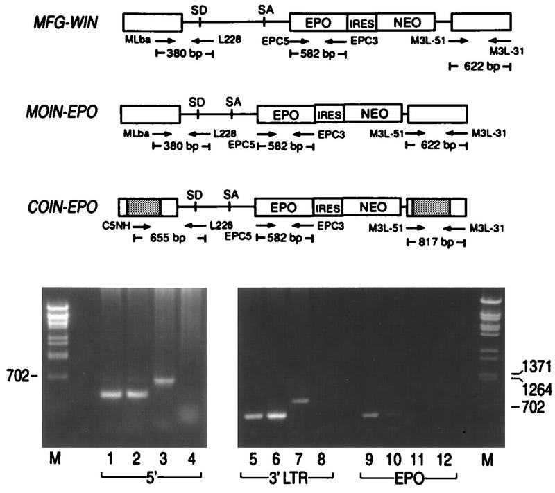
Murine leukemia virus (MLV)-based retroviral vectors are the most frequently used gene delivery vehicles. However, the current vectors are still not fully optimized for gene expression and viral titer, and many genetic and biochemical features of MLV-based vectors are poorly understood. We have previously reported that the retroviral vector MFG, where the gene of interest is expressed as a spliced mRNA, is superior in the level of gene expression with respect to other vectors compared in the study. As one approach to developing improved retroviral vectors, we have systematically performed mutational analysis of the MFG retroviral vector. We demonstrated that the entire gag coding sequence, together with the immediate upstream region, could be deleted without significantly affecting viral packaging or gene expression. To our knowledge, this region is included in all currently available retroviral vectors. In addition, almost the entire U3 region could be replaced with the heterologous human cytomegalovirus immediately-early promoter without deleterious effects. We could also insert internal ribosome entry sites (IRES) and multicloning sites into MFG without adverse effects. Based on these observations, we have constructed a series of new, improved retroviral constructs. These vectors produced viral titers comparable to MFG, expressed high levels of gene expression, and stably transferred genes to the target cells. Our vectors are more convenient to use because of the presence of multicloning sites and IRESs, and they are also more versatile because they can be readily converted to various applications. Our results have general implications regarding the design and development of improved retroviral vectors for gene therapy.


Figures

FIG. 1
FIG. 1
Schematic representation of the retroviral vector MFG. In MFG, the gene of interest (dotted box) is cloned into the NcoI site, containing the start codon in it, and expressed as a spliced mRNA. MFG contains the 420- and 99-bp coding sequences for gag and env, respectively. U3 of Moloney MLV is 448 bp long. ATG, start codon of gag.
FIG. 2
FIG. 2
Localization of the packaging signal sequence. (A) Summary of deletions. Nine deletions were constructed as described in Materials and Methods. Ψ indicates the packaging sequence previously defined by Mann et al. (25), which includes the gag coding region as well as the entire sequence between SD and the start codon for gag. The numbering system is based on that of Shinnick et al. (38). The region between positions 1040 and 5400 includes gag and pol coding sequences and is missing from MFG. lacZ was used as a reporter gene in this study, and its relative position is shown as a dotted triangle. Note that the vector is not drawn to scale. (B) Effects of deletion on gene expression and viral titers. Deletion constructs, together with the parental vector MFG-lacZ, were transfected to the packaging line BING or CRIP. (In this figure, only the result for BING is shown.) After 3 days, culture supernatants were filtered through 0.45-μm-pore-size filters, while cells were stained with X-Gal to measure transfection efficiency. Duplicate dishes were also prepared for some constructs and subjected to the ONPG assay for β-galactosidase activity. Cell-free viral supernatants were used to transduce NIH 3T3 cells, and after 3 days the cells were stained with X-Gal to determine the viral titer. The transfection and transduction efficiency of MFG were set to 1, and those of others were normalized to it. More than five transfections and transductions were performed at separate times. In one independent experiment, four to six transfections and transductions were carried for each mutant.
FIG. 3
FIG. 3
Effect of deletion of the residual env coding sequence. (A) In MΔE-CAT, 113 bp, including the entire env coding sequence (99 bp), was deleted but the polypurine tract remained intact. In this experiment, the bacterial CAT sequence was used as a reporter gene. The vector is not drawn to scale. (B) Effect on gene expression. MFG-CAT and MΔE-CAT constructs were transfected to the packaging line CRIP, cell-free viral supernatants were used to transduce the human promonocytic line U937 and T-lymphoid line CEM-SS, and all the cells were subjected to the CAT assay. Other experimental conditions are as described in the legend to Fig. 2. The expression of MFG was set to 1.
FIG. 4
FIG. 4
Construction of chimeric U3. (A) Four HCMV MIEP fragments were used to substitute the U3 sequence. They are CR, CCI, CCII, and CP. (B) Schematic diagram of chimeric LTRs. Four restriction sites (NheI, PvuII, XbaI, and SacI) are naturally present in U3, and their coordinates are shown in parentheses. These sites were used to clone the four HCMV MIEP fragments. The relative positions of the CAAT and TATA boxes of U3 are indicated. The numbers shown above the LTR are the lengths of U3 (unshaded) or HCMV MIEP (shaded) that replaced a part of U3, while those in parentheses are the coordinates of MLV based on the numbering of Shinnick et al. (38). Note that the promoter is not drawn to scale.
FIG. 5
FIG. 5
Effect of the chimeric 3′ LTR on gene expression. The five constructs, including the parental MFG-CAT, were transfected to the packaging line CRIP, and cell-free viral supernatant was used to transduce NIH 3T3 cells and the human T-lymphoid line H9. Both transfected and transduced cells were subjected to CAT analysis. Expression of MFG-CAT was set to 1, and the others were normalized to it.
FIG. 6
FIG. 6
Comparison of retroviral vectors containing IRES and neo. Retroviral vectors were constructed to contain chimeric U3 at both the 5′ and 3′ LTRs, deletions in gag and env coding sequences, and the selectable marker NEO gene linked to CAT through IRES. Again, for each cell line, expression of MFG-CAT was set to 1 and those of the other constructs were normalized to it. Because the assay conditions were different for the different cell lines, direct comparison between cell lines based on the above numbers should be avoided. Transductions were performed at least three to five times for each line at separate times. Here the result from one representative experiment is shown.
FIG. 7
FIG. 7
Comparison of expression levels of mGM-CSF between MFG and SCP1. The experimental conditions were identical to the others, except that one of the target cells was the human skin fibroblast cell line and the level of mGM-CSF, instead of CAT, was measured. The two retroviral vectors expressing mGM-CSF were transfected to CRIP cells. NIH 3T3 and human foreskin fibroblasts were transduced and then selected in the presence of G418. The same number of drug-resistant cells were plated on 6-cm plates, grown for another 3 days, and subjected to enzyme-linked immunosorbent assay. Expression of MFG–mGM-CSF was set to 1.
FIG. 8
FIG. 8
Effect of removal of NcoI in MFG. (A) Retroviral vector construction. MFG-WIN and KCP3-WNIN contain the NcoI site, while KCP3-WXIN does not. The nucleotide sequence around the NcoI site is shown. This sequence is identical in MFG-WIN and KCP3-WNIN. The human EPO cDNA sequence was used as a reporter gene. (B) Effect of gene expression. The three constructs were transfected to CRIP cells, cell-free viral supernatant was harvested to transduce NIH 3T3 cells, and the cells were selected in the presence of G418. The same number of drug-resistant cells were plated on 6-cm plates, grown for another 3 days, and subjected to enzyme-linked immunosorbent assay. Only the results from transduction assays are shown. Expression of MFG-WIN was set to 1, and those of the others were normalized to it.
FIG. 9
FIG. 9
Construction of improved retroviral vectors. Based on the results shown from Fig. 2 to 8, improved vectors were constructed and tested for their performance with EPO. In this example, the two constructs COI and MOI are shown. The former has a chimeric U3, identical to that of LCP at the 5′ LTR and MCP3 at the 3′ LTR, while the latter contains the original U3 from MLV. Both vectors have deletions around the gag region (like Δ38 in Fig. 2), no env coding sequence (Fig. 3), the convenient restriction site for the gene of interest, and IRES and NEO as selectable markers. The NEO sequence was added to the XhoI site of MOI and COI, resulting in MOIN and COIN, respectively. The EPO cDNA sequence was subsequently cloned into the BamHI site of MOIN and COIN, generating MOIN-EPO and COIN-EPO, respectively. The three vectors, including the parental construct MFG-WIN, were transfected into CRIP or BING cells, and cell-free viral supernatants were harvested to transduce NIH 3T3 cells. Viral titer were determined 3 days posttransduction as described by Byun et al. (9). Cells were selected in the presence of G418 to be close to the actual situation. Drug-resistant populations were obtained, and identical numbers of cells were plated on 6-cm culture plates. After 3 days, the levels were determined. To compare viral titers between vectors, G418-resistant cultures were obtained after transfection of PA317 cells with the above vectors and grown to similar densities on 10-cm culture plates. Viral titers were determined by the conventional and new methods (9, 10).
FIG. 10
FIG. 10
Test for preservation of retroviral sequences in transduced cells. Total cellular DNAs were prepared from G418-resistant NIH 3T3 cells transduced with MFG-EPO, MOIN-EPO, and COIN-EPO. The three pairs of oligonucleotide primers were used to amplify the regions indicated in the figure. SD, splice donor; SA, splice acceptor.

Similar articles

Cited by

References

    1. Adam M A, Miller A D. Identification of a signal murine retrovirus that is sufficient for packaging of nonretroviral RNA into virions. J Virol. 1988;62:3802–3806. - PMC - PubMed
    1. Alford R L, Honda S, Lawrence C B, Belmont J T. RNA secondary structure analysis of the packaging signal for Moloney murine leukemia virus. Virology. 1991;183:611–619. - PubMed
    1. Ali M, Lemoine N R, Ring C J A. The use of DNA viruses as vectors for gene therapy. Gene Ther. 1994;1:367–384. - PubMed
    1. Armentano D, Yu S-F, Kantoff P W, von Ruden T, Anderson W F, Gilboa E. Effect of internal viral sequences on the utility of retroviral vectors. J Virol. 1987;61:1647–1650. - PMC - PubMed
    1. Bandara G, Muller G M, Galea-Lauri J, Georgescu H I, Suchanek M K, Hung G L, Glorioso J C, Robbins P D, Evans C H. Intraarticular expression of biologically active interleukin 1-receptor-antagonist protein by ex vivo gene transfer. Proc Natl Acad Sci USA. 1993;90:10764–10768. - PMC - PubMed

Publication types