. 1998 Feb;72(2):1020-7.
doi: 10.1128/JVI.72.2.1020-1027.1998.

A spontaneous low-pathogenic variant of Theiler's virus contains an amino acid substitution within the predominant VP1(233-250) T-cell epitope

Affiliations

A spontaneous low-pathogenic variant of Theiler's virus contains an amino acid substitution within the predominant VP1(233-250) T-cell epitope

B S Kim et al. J Virol. 1998 Feb.

Erratum in

  • J Virol 1998 Aug;72(8):6965

Abstract

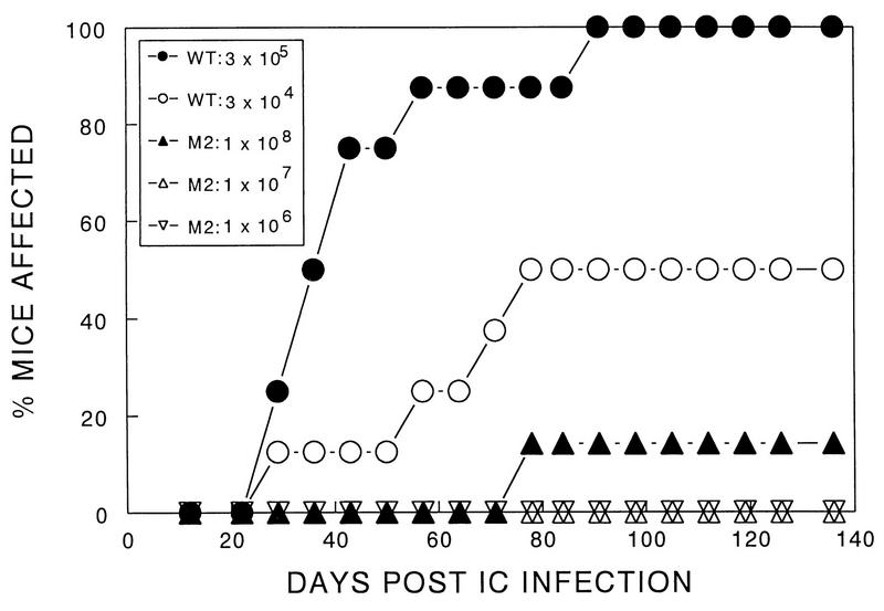
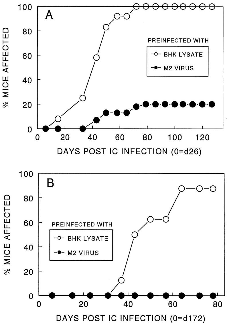
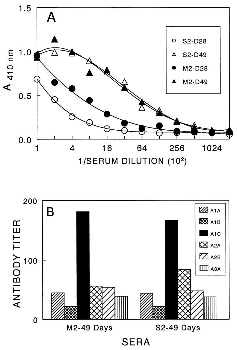
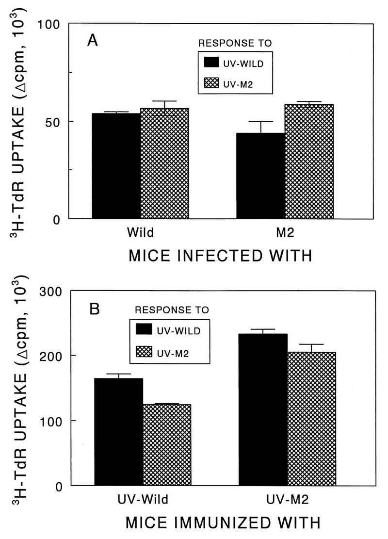
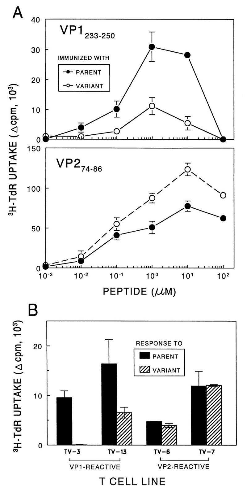
Theiler's murine encephalomyelitis virus (TMEV) induces immune-mediated demyelination after intracerebral inoculation of the virus into susceptible mouse strains. We isolated from a TMEV BeAn 8386 viral stock, a low-pathogenic variant which requires greater than a 10,000-fold increase in viral inoculation for the manifestation of detectable clinical signs. Intracerebral inoculation of this variant virus induced a strong, long-lasting, protective immunity from the demyelinating disease caused by pathogenic TMEV. The levels of antibodies to the whole virus as well as to the major linear epitopes were similar in mice infected with either the variant or wild-type virus. However, persistence of the variant virus in the central nervous system (CNS) of mice was significantly lower than that of the pathogenic virus. In addition, the T-cell response to the predominant VP1 (VP1(233-250)) epitope in mice infected with the variant virus was significantly weaker than that in mice infected with the parent virus, while similar T-cell responses were induced against another predominant epitope (VP2(74-86)). Further analyses indicated that a change of lysine to arginine at position 244 of VP1, which is the only amino acid difference in the P1 region, is responsible for such differential T-cell recognition. Thus, the difference in the T-cell reactivity to this VP1 region as well as the low level of viral persistence in the CNS may account for the low pathogenicity of this spontaneous variant virus.


Figures

FIG. 1
FIG. 1
Determination of the level of pathogenicity of TMEV variant M2. SJL mice were infected with different numbers of PFU of the parental (wild-type [wt]) or variant virus. Development of clinical signs associated with TMEV-induced demyelination in susceptible SJL was monitored for 136 days. The parent or M2 variant stock was inoculated intracerebrally (IC) into separate groups (8 to 12 mice per group) of SJL/J mice. The clinical signs were assessed as described in Materials and Methods. The unpaired nonparametric test indicates that the difference between the pathogenicity of 108 PFU of M2 and either 3 × 10⁵ or 3 × 10⁴ PFU of parent stock is very significant (P < 0.0001).
FIG. 2
FIG. 2
Histopathologic examination of SJL/J mice infected with either parent or M2 virus. At least 10 cross sections of each spinal cord from three mice per group were examined for histopathology 136 days after viral infection. A representative micrograph for each group is shown. One-micrometer-thick, Epon-embedded sections were stained with toluidine blue. Bar = 50 μm. (A) Spinal cord section from a mouse infected with the pathogenic parent virus showing severe white matter inflammation accompanied by axonal and myelin degeneration. Numerous macrophages are seen in the field. (B) Spinal cord section from a clinically healthy mouse after infection with 108 PFU of M2 variant virus demonstrates normal white matter. No inflammation or demyelination is seen in the field. (C) Spinal cord section from a clinically affected mouse after infection with 108 PFU of M2 variant virus shows mild to moderate white matter involvement by inflammation and demyelination. This field represents the maximum severity observed in animals infected with the variant virus.
FIG. 3
FIG. 3
Induction of long-lasting protective immunity after inoculation with M2 virus. (A) Fifteen SJL/J mice were inoculated intracerebrally (IC) with M2 variant virus (107 PFU). After 26 days, these mice were infected intracerebrally with pathogenic parent virus (106 PFU), and then the development of clinical signs was observed. As a control group, 12 mice injected intracerebrally with BHK lysates instead of M2 virus were subsequently infected with pathogenic virus. (B) Groups of SJL/J mice (eight mice per group) were initially inoculated with either BHK lysates or M2 virus and then infected with pathogenic parent virus after 172 days. The protection was extremely significant (P < 0.0001) in both experiments.
FIG. 4
FIG. 4
Viral persistence in the CNS following intracerebral (IC) infection of SJL/J mice with pathogenic parent and low-pathogenic variant viruses. (A) Determination of viral message levels in the spinal cords by RT-PCR. Two spinal cords per time point were pooled, and the presence of viral message was assessed by RT-PCR at 7, 14, 24, 35, and 55 days after intracerebral virus inoculation. The sense primer used for amplification represents the 5′ end of the leader coding sequence, and the antisense primer represents the 3′ end of the VP4 coding sequence. (B) Determination of PFU recovered from the spinal cords of virus-infected SJL/J mice. Three separate spinal cords were assessed at each time point of 35 and 78 days after viral inoculation.
FIG. 5
FIG. 5
Antibody responses to TMEV in SJL/J mice (five mice per group) infected with either pathogenic or low-pathogenic variant virus. Twofold serial dilutions of pooled sera containing equal volume from individual mice were assessed by ELISA. (A) Reactivity to purified TMEV by antibodies in sera from SJL/J mice infected with either pathogenic (S2 stock) or low-pathogenic (M2) virus at days 28 and 49 postinfection. (B) Reactivity to the major linear antibody epitopes by the same sera at day 49 postinfection.
FIG. 6
FIG. 6
Comparison of T-cell proliferation responses of SJL/J mice infected intracerebrally with live pathogenic or variant viruses and of SJL/J mice immunized subcutaneously with UV-inactivated pathogenic or variant viruses. (A) T-cell proliferative responses of spleens from SJL/J mice. SJL/J mice were infected intracerebrally with either parent (106 PFU) or M2 (106 PFU) virus. Three weeks after viral infection, spleens were pooled from two mice and subsequently cultured in triplicate (5 × 10⁵/well) in the presence of 25 μg of either UV-BeAn or UV-M2 per ml. (B) Proliferative response of T cells from BeAn- or M2-immunized mice to UV-inactivated parent and variant viruses. SJL/J mice were immunized at the base of the tail with 50 μg of UV-BeAn or UV-M2 emulsified in CFA. Nine days later, lymph node cells were pooled from two mice and subsequently cultured in triplicate (5 × 10⁵/well) with 25 μg of either UV-BeAn or UV-M2 per ml. All cultures were pulsed with [3H]TdR approximately 18 h before harvesting. A peptide containing the amino acids 5 to 19 of HEL was used as a negative control for both experiments. The background levels were less than 5,000 cpm and subtracted from the experimental values.
FIG. 7
FIG. 7
Assessment of the T-cell proliferative responses to TMEV and predominant epitopes. (A) T-cell proliferative responses of parent- and M2-immunized mice against the major VP1 and VP2 epitopes. SJL/J mice were immunized at the base of the tail with 50 μg of UV-BeAn or UV-M2 emulsified in CFA. Nine days later, lymph node cells were pooled from two mice and subsequently cultured in triplicate (5 × 10⁵/well) with various molar concentrations of VP1233–250 or VP274–86 for 4 days. A peptide containing the amino acids 5 to 19 of HEL was used as a negative control. Cultures were pulsed with [3H]TdR approximately 18 h before harvesting. Results are expressed as (mean cpm from peptide-stimulated cultures − mean cpm from HEL5–19-stimulated cultures) ± standard error of the mean. (B) Stimulation of CNS-derived T-cell lines specific for VP1233–250 and VP274–86 with the parent or M2 virus. Either VP1- or VP2-specific T-cell lines (2 × 10⁴/well) were stimulated with 12.5 μg of UV-inactivated parent or M2 virus per ml in the presence of irradiated, syngeneic splenocytes (5 × 10⁵/well) for 4 days as described in the text. TV-3 and TV-13 are specific for VP1233–250, and TV-6 and TV-7 are specific for VP274–86.
FIG. 8
FIG. 8
Strategy for sequencing the P1 region of M2 virus and identification of a codon leading to a single amino acid change within the VP1 T-cell epitope region. (A) Schematic presentation of the strategy for sequencing the P1 region of the M2 variant. Arrows indicate the specific primers used in sequencing, and the length represents the size of the sequence analyzed. UTR, untranslated region. (B) M2 virus contains a point mutation (A→G) at nucleotide position 3733 (indicated by the arrow) of VP1 that results in a lysine-to-arginine change at amino acid residue 244 of the VP1 capsid protein. The nucleotide sequences of parent and M2 viruses coding for the VP1244 region are shown.

Similar articles

Cited by

References

    1. Bae Y S, Yoon J W. Determination of diabetogenicity attributable to a single amino acid, Ala776, on the polyprotein of encephalomyocarditis virus. Diabetes. 1993;42:435–443. - PubMed
    1. Bahk Y Y, Kappel C A, Rasmussen G, Kim B S. Association between susceptibility to Theiler’s virus-induced demyelination and T-cell receptor Jβ1-Cβ1 polymorphism rather than Vβ deletion. J Virol. 1997;71:4181–4185. - PMC - PubMed
    1. Bureau J F, Montagutelli X, Lefebvre S, Guenet J L, Pla M, Brahic M. The interaction of two groups of murine genes determines the persistence of Theiler’s virus in the central nervous system. J Virol. 1992;66:4698–4704. - PMC - PubMed
    1. Calenoff M A, Faaberg K S, Lipton H L. Genomic regions of neurovirulence and attenuation in Theiler murine encephalomyelitis virus. Proc Natl Acad Sci USA. 1990;87:978–982. - PMC - PubMed
    1. Chen H H, Kong W P, Zhang L, Ward P L, Roos R P. A picornaviral protein synthesized out of frame with the polyprotein plays a key role in a virus-induced immune-mediated demyelinating disease. Nat Med. 1995;1:927–931. - PubMed

Publication types

Substances

Associated data

LinkOut - more resources