. 1998 Feb;72(2):1043-51.
doi: 10.1128/JVI.72.2.1043-1051.1998.

Resistance to virus infection conferred by the interferon-induced promyelocytic leukemia protein

Affiliations

Resistance to virus infection conferred by the interferon-induced promyelocytic leukemia protein

M K Chelbi-Alix et al. J Virol. 1998 Feb.

Abstract

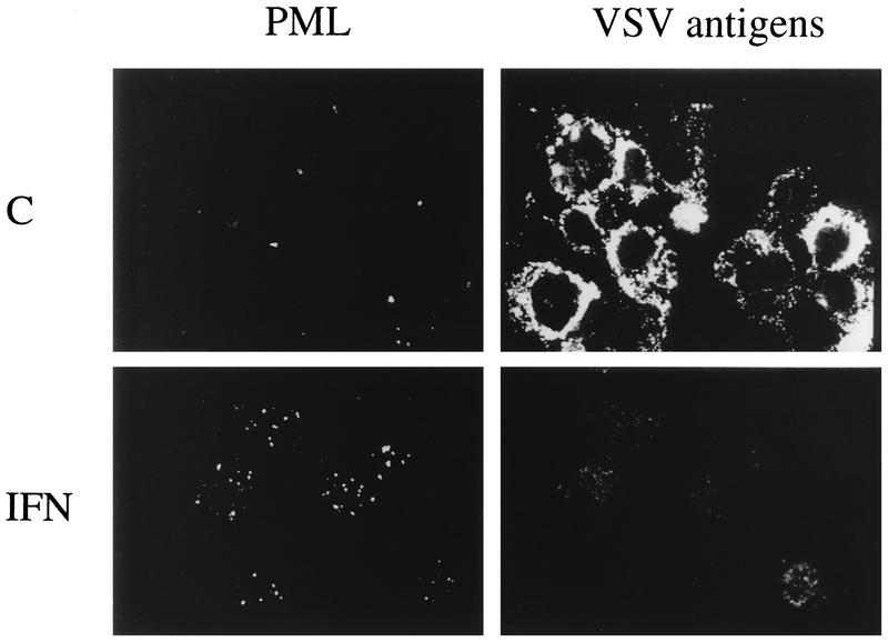
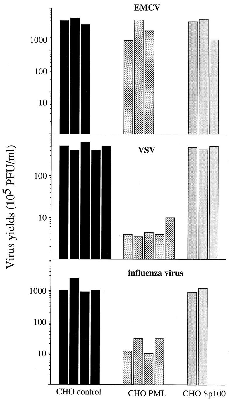
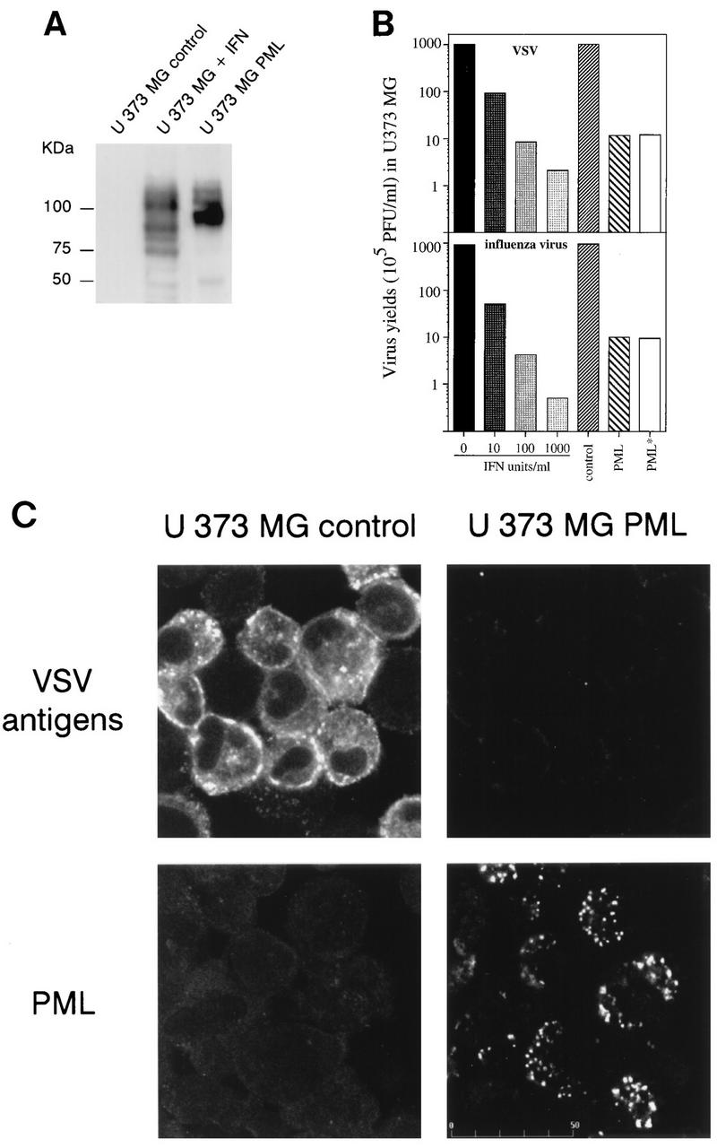
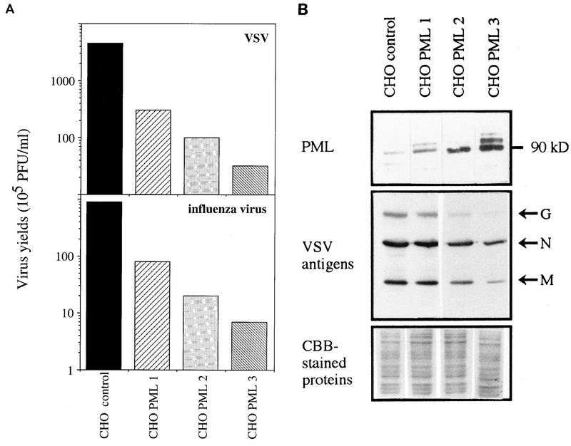
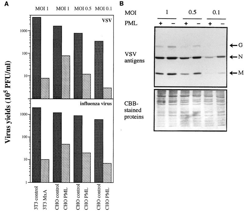
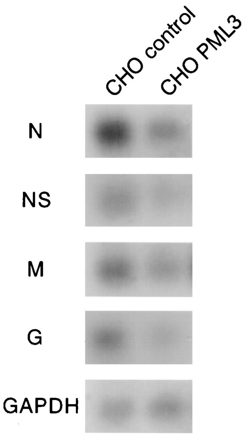
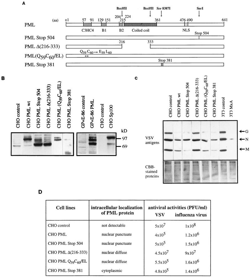
The interferon (IFN)-induced promyelocytic leukemia (PML) protein is specifically associated with nuclear bodies (NBs) whose functions are yet unknown. Two of the NB-associated proteins, PML and Sp100, are induced by IFN. Here we show that overexpression of PML and not Sp100 induces resistance to infections by vesicular stomatitis virus (VSV) (a rhabdovirus) and influenza A virus (an orthomyxovirus) but not by encephalomyocarditis virus (a picornavirus). Inhibition of viral multiplication was dependent on both the level of PML expression and the multiplicity of infection and reached 100-fold. PML was shown to interfere with VSV mRNA and protein synthesis. Compared to the IFN mediator MxA protein, PML had less powerful antiviral activity. While nuclear body localization of PML did not seem to be required for the antiviral effect, deletion of the PML coiled-coil domain completely abolished it. Taken together, these results suggest that PML can contribute to the antiviral state induced in IFN-treated cells.


Figures

FIG. 1
FIG. 1
IFN-β induces PML protein synthesis and inhibits VSV antigen expression in the human monocytic cell line U937. U937 cells were treated with 1,000 U of human IFN-β per ml. After 48 h at 37°C, control (C) and IFN-treated cells were infected with VSV at a MOI of 0.1. Double immunofluorescence were performed 24 h postinfection with mouse anti-PML antibodies visualized with Texas red and rabbit anti-VSV antibodies followed by FITC labelling.
FIG. 2
FIG. 2
Overexpression of PML, and not of Sp100, confers resistance against infections by VSV and influenza virus. CHO control (transfected with the empty vector), CHO PML3, or CHO Sp100 cells were infected with EMCV, VSV, or influenza A virus at an MOI of 0.1. After 16 h, viral titers were determined as described in Materials and Methods.
FIG. 3
FIG. 3
(A) PML level in IFN-treated U373 MG cells and U373 MG PML. U373 MG cells were treated for 48 h with 1,000 U of IFN-β per ml. Samples (50 μg) of extracts of control IFN-treated cells and U373 MG PML were analyzed by Western blotting and revealed with rabbit anti-PML antibodies. Note that all bands visualized by anti-PML antibodies are likely to be isoforms derived from alternative splicing of unique gene. Molecular size markers are indicated on the left. (B) Inhibition of virus replication in IFN-treated U373 MG cells and U373 MG PML. One series of U373 MG cells was treated for 48 h with 10, 100 or 1,000 U of IFN-β per ml. The second series of cells, U373 MG control (transfected with the empty vector), U373 MG PML, and U373 MG PML* (+ anti-IFN-α/β/γ antibodies [see Materials and Methods]) was seeded at 37°C for 5 h. The two series were then infected with VSV or influenza A virus at an MOI of 0.1. After 16 h, viral titers were determined as described in Materials and Methods. (C) Expression of PML in U373 MG cells inhibits the expression of VSV antigens (Top) Expression of VSV antigens in infected U373 MG control cells (transfected with the empty vector) and U373 MG PML. Immunofluorescence with rabbit anti-VSV antibodies was performed 13 h after infection with VSV at an MOI of 0.1 and revealed by FITC labelling. (Bottom) Expression of PML in U373 MG and U373 MG PML cells revealed by immunofluorescence with mouse anti-PML antibodies visualized with Texas red.
FIG. 4
FIG. 4
2′5′ A synthetase activity in IFN-treated cells and cells overexpressing PML. 2′5′ A synthetase activity was determined in cells extracts from CHO control cells (lane 1). CHO PML3 (lane 2), GP+E−86 control cells (lane 3), GP+E−86 PML (lane 4), U373 MG control cells (lane 5), U373 MG PML (lane 6), and U373 MG cells treated for 48 h with 10 U of IFN-β per ml (lane 7). All control cells are cells transfected with the empty vector. The 2′5′ A synthetase activity was determined by chromatographic analysis of the reaction substrate (ATP) and the products, 2′,5′-oligoadenylates (dimer and trimer), as previously described (7).
FIG. 5
FIG. 5
(A) PML levels parallel inhibition of virus multiplication. CHO control cells (transfected with the empty vector) and CHO PML 1, 2, and 3 clones were infected with VSV or influenza A virus at an MOI of 0.1. After 16 h, viral titers were determined as described in Materials and Methods. (B) PML levels parallel the inhibition of VSV antigen expression. CHO control cells (transfected with the empty vector) and CHO PML 1, 2, and 3 clones were infected for 13 h with VSV at an MOI of 0.1. Western blot analysis of the extracts of these cells was done as described in Materials and Methods. (Top) Revealed with rabbit anti-PML antibodies; (middle) revealed with anti-VSV antibodies (VSV antigens are indicated at the right); (bottom) Coomassie brilliant blue (CBB)-stained proteins.
FIG. 6
FIG. 6
Resistance of PML-expressing clones to VSV and influenza virus is MOI dependent. CHO control cells (transfected with the empty vector) and CHO PML 3 were infected with VSV or influenza A virus at different MOIs as indicated in the figure. Swiss 3T3 control and Swiss 3T3 MxA were infected at an MOI of 1 with VSV or influenza virus. (A) After 16 h, the cells were used for the determination of the viral titers. Antiviral activities are the means of three independent experiments. (B) CHO control cells and CHO PML 3 were infected with VSV for 10 h at different MOIs as indicated in the figure. The results of Western blot analysis are revealed with anti-VSV antibodies. (Top) revealed with anti-VSV antibodies (VSV antigens are indicated at the right); (bottom) Coomassie brilliant blue (CBB)-stained proteins.
FIG. 7
FIG. 7
CHO control cells (transfected with the empty vector) and CHO PML3 were infected with VSV at an MOI of 0.5 for 4 h. Total RNA was extracted as described in Materials and Methods. Samples (20 μg of RNA per lane) were analyzed for the presence of VSV N, NS, M, and G. GΔPDH, glyceraldehyde-3-phosphate dehydrogenase.
FIG. 8
FIG. 8
(A) Description of PML mutants. The structures of PML (including the C3HC4 zinc finger motif, the two B boxes and the coiled-coil) and of four PML mutants, the C-terminal PML mutant (PML Stop 504), the coiled-coil PML mutant [PMLΔ(216–333)], the RING finger PML mutant (Q59C60/EL), and the cytoplasmic PML mutant (Stop 381), are shown. aa, amino acids; NLS, nuclear localization signal. (B) Western blot analysis of PML in stably transfected CHO and GP+E−86 cells as well as the four PML mutants; also shown is the overexpression of Sp100 in CHO cells. (C) Analysis of PML domains involved in the inhibition of VSV antigens. CHO control (transfected with the empty vector), CHO PML wt (CHO PML3), and the four mutants of PML were infected with VSV for 13 h at an MOI of 0.1. Swiss 3T3 neo and 3T3 MxA were infected with VSV under the same conditions and used as control. Western blot analysis of the extracts of these cells was done as described in Materials and Methods. (Top) Revealed with anti-VSV antibodies (VSV antigens are indicated at the right); (bottom) Coomassie brilliant blue (CBB)-stained proteins. (D) Description of subcellular distributions in stably transfected CHO cells and the antiviral potentials of wild-type and mutant forms of human PML protein. Cells were infected with VSV at an MOI of 0.1. After 16 h, viral titers were determined as described in Materials and Methods.

Similar articles

Cited by

References

    1. Alber D, Staeheli P. Partial inhibition of vesicular stomatitis virus by the interferon-induced human 9-27 protein. J Interferon Cytokine Res. 1996;16:375–380. - PubMed
    1. Arnheiter H, Meier E. Mx proteins: antiviral proteins by chance or by necessity. New Biol. 1990;2:851–857. - PubMed
    1. Banerjee A K, Chattopadhyay D. Structure and function of the RNA polymerase of vesicular stomatitis virus. Adv virus Res. 1990;38:99–124. - PubMed
    1. Barlow P N, Luisi B, Milner A, Elliott M, Everett R. Structure of the C3HC4 domain by 1H-nuclear magnetic resonance spectroscopy. A new structural class of zinc-finger. J Mol Biol. 1994;237:201–211. - PubMed
    1. Blondel D, Harmison G G, Schubert M. Role of matrix protein in cytopathogenesis of vesicular stomatitis virus. J Virol. 1990;64:1716–1725. - PMC - PubMed

Publication types