. 1998 Feb;72(2):1195-202.
doi: 10.1128/JVI.72.2.1195-1202.1998.

Retroviral recombination rates do not increase linearly with marker distance and are limited by the size of the recombining subpopulation

Affiliations

Retroviral recombination rates do not increase linearly with marker distance and are limited by the size of the recombining subpopulation

J A Anderson et al. J Virol. 1998 Feb.

Abstract

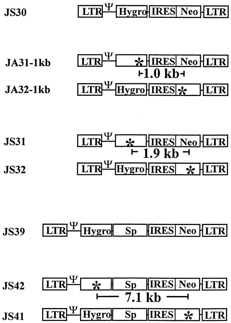
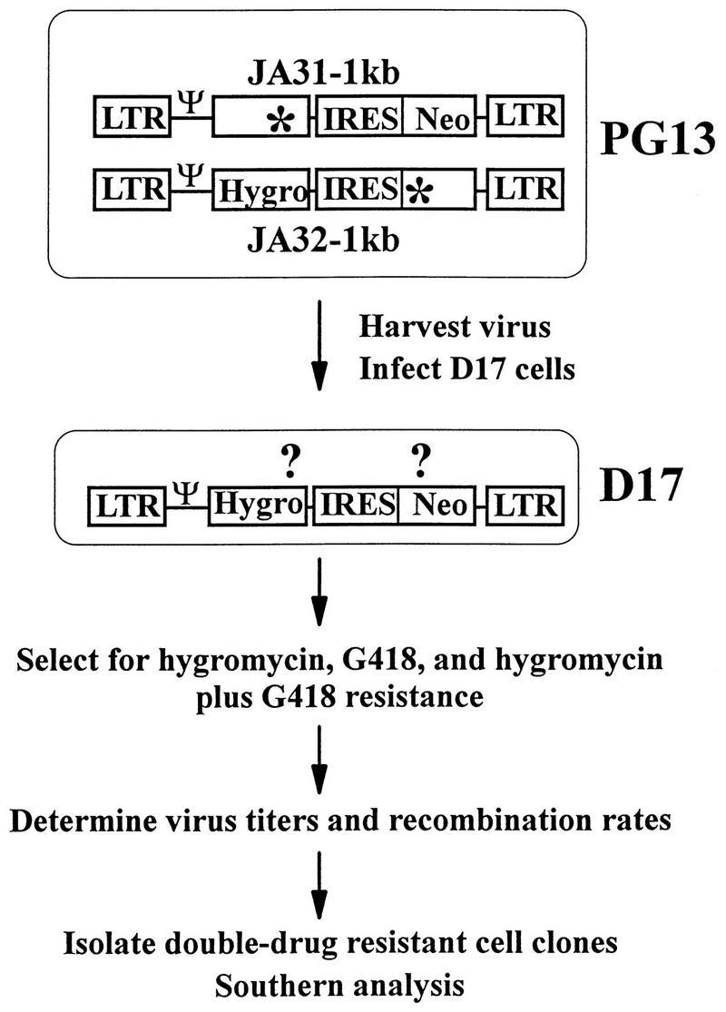
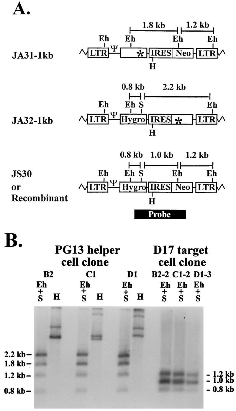
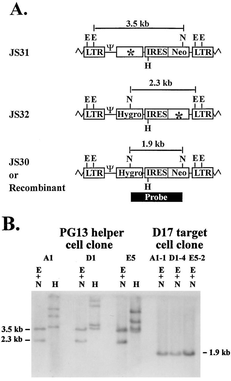
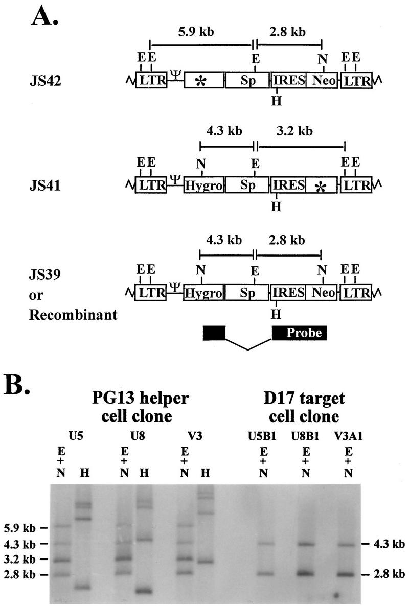
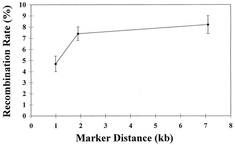
Recombination occurs at high frequencies in all examined retroviruses. The previously determined homologous recombination rate in one retroviral replication cycle is 4% for markers 1.0 kb apart in spleen necrosis virus (SNV). This has often been used to suggest that approximately 30 to 40% of the replication-competent viruses with 7- to 10-kb genomes undergo recombination. These estimates were based on the untested assumption that a linear relationship exists between recombination rates and marker distances. To delineate this relationship, we constructed three sets of murine leukemia virus (MLV)-based vectors containing the neomycin phosphotransferase gene (neo) and the hygromycin phosphotransferase B gene (hygro). Each set contained one vector with a functional neo and an inactivated hygro and one vector with a functional hygro and an inactivated neo. The two inactivating mutations in the three sets of vectors were separated by 1.0, 1.9, and 7.1 kb. Recombination rates after one round of replication were 4.7, 7.4, and 8.2% with markers 1.0, 1.9, and 7.1 kb apart, respectively. Thus, the rate of homologous recombination with 1.0 kb of marker distance is similar in MLV and SNV. The recombination rate increases when the marker distance increases from 1.0 to 1.9 kb; however, the recombination rates with marker distances of 1.9 and 7.1 kb are not significantly different. These data refute the previous assumption that recombination is proportional to marker distance and define the maximum recombining population in retroviruses.


Figures

FIG. 1
FIG. 1
MLV vectors used to measure the recombination rates at marker distances of 1.0, 1.9, and 7.1 kb. Ψ, packaging signal; Hygro, hygromycin phosphotransferase B gene; Neo, neomycin phosphotransferase gene; Sp, spacer DNA; ✻, inactivating frameshift mutation.
FIG. 2
FIG. 2
Experimental protocol to measure rates of recombination. Abbreviations and symbols are defined in the legend to Fig. 1.
FIG. 3
FIG. 3
Proviral structures of JA31-1kb, JA32-1kb, and JS30. Recombinants with two functional drug resistance genes have the same structure as JS30 does. (A) Predicted proviral structures. Eh, EheI; S, SacII, H, HindIII. Zigzag lines represent host cell sequences. A 1.9-kb DNA fragment from JS30 (the black box labeled probe) was used for random-priming reaction to generate a probe for Southern hybridization analysis. (B) Southern hybridization analysis of the proviral structures in helper and target cell clones. B2, C1, and D1 are PG13 helper cell clones infected with JA31-1kb and JA32-1kb. B2-2, C1-2, and D1-3 are double-drug-resistant D17 cell clones infected with viruses harvested from B2, C1, and D1, respectively. Other abbreviations and symbols are defined in the legend to Fig. 1.
FIG. 4
FIG. 4
Proviral structures of JS31, JS32, and JS30 (recombinant). (A) Predicted proviral structures. E, EcoRV; N, NcoI. Although each LTR contains two EcoRV sites, only one site in each LTR is shown for simplicity. (B) Southern hybridization analysis of the proviral structures in helper and target cell clones. A1, D1, and E5 are PG13 helper cell clones infected with JS31 and JS32. A1-1, D1-4, and E5-2 are double-drug-resistant D17 cell clones infected with viruses harvested from A1, D1, and E5, respectively. Other abbreviations and symbols are defined in the legends to Fig. 1 and 3.
FIG. 5
FIG. 5
Proviral structures of JS41, JS42, and JS39 (recombinant). (A) Predicted proviral structures. E, EcoRV; N, NcoI. Although each LTR contains two EcoRV sites, only one site in each LTR is shown for simplicity. (B) Southern hybridization analysis of the proviral structures in helper and target cell clones. U5, U8, and V3 are PG13 helper cell clones infected with JS41 and JS42, whereas U5B1, U8B1, and V3A1 are double-drug-resistant D17 cell clones infected with viruses harvested from the above-described helper cell clones, respectively. Other abbreviations and symbols are defined in the legends to Fig. 1 and 3.
FIG. 6
FIG. 6
Nonlinear relationship between recombination rate and marker distance. The means ± SE at the three marker distances are indicated.

References

    1. Adam M A, Ramesh N, Miller A D, Osborne W R A. Internal initiation of translation in retroviral vectors carrying picornavirus 5′ nontranslated regions. J Virol. 1991;65:4985–4990. - PMC - PubMed
    1. Anderson J A, Teufel II R J, Yin P D, Hu W-S. Correlated template-switching events during minus-strand DNA synthesis: a mechanism for high negative interference during retroviral recombination. J Virol. 1998;72:1186–1194. - PMC - PubMed
    1. Clavel F, Hoggan M D, Willey R L, Strebel K, Martin M A, Repaske R. Genetic recombination of human immunodeficiency virus. J Virol. 1989;63:1455–1459. - PMC - PubMed
    1. Coffin J M. Structure, replication, and recombination of retrovirus genomes: some unifying hypotheses. J Gen Virol. 1979;42:1–26. - PubMed
    1. Coffin J M. Structure and classification of retroviruses. In: Levy J A, editor. The retroviridae. New York, N.Y: Plenum Press; 1992. pp. 19–49.

Publication types

LinkOut - more resources