. 1998 Feb;72(2):1235-43.
doi: 10.1128/JVI.72.2.1235-1243.1998.

Virus-induced cell motility

Affiliations

Virus-induced cell motility

C M Sanderson et al. J Virol. 1998 Feb.

Abstract

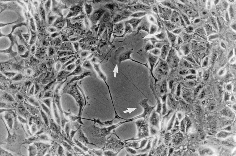
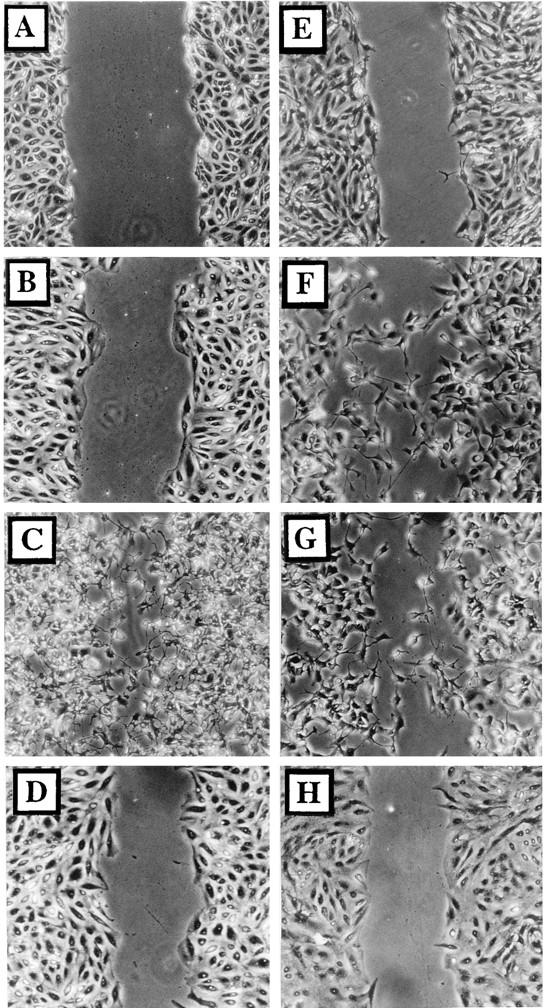
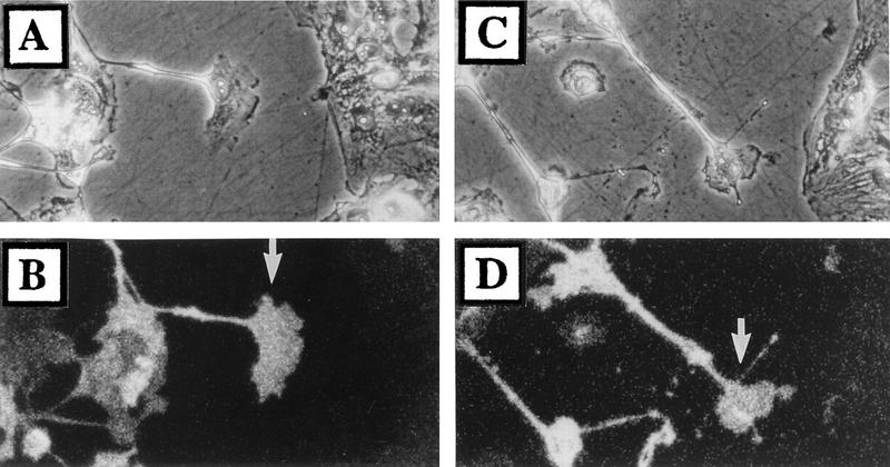
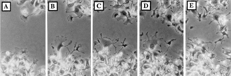
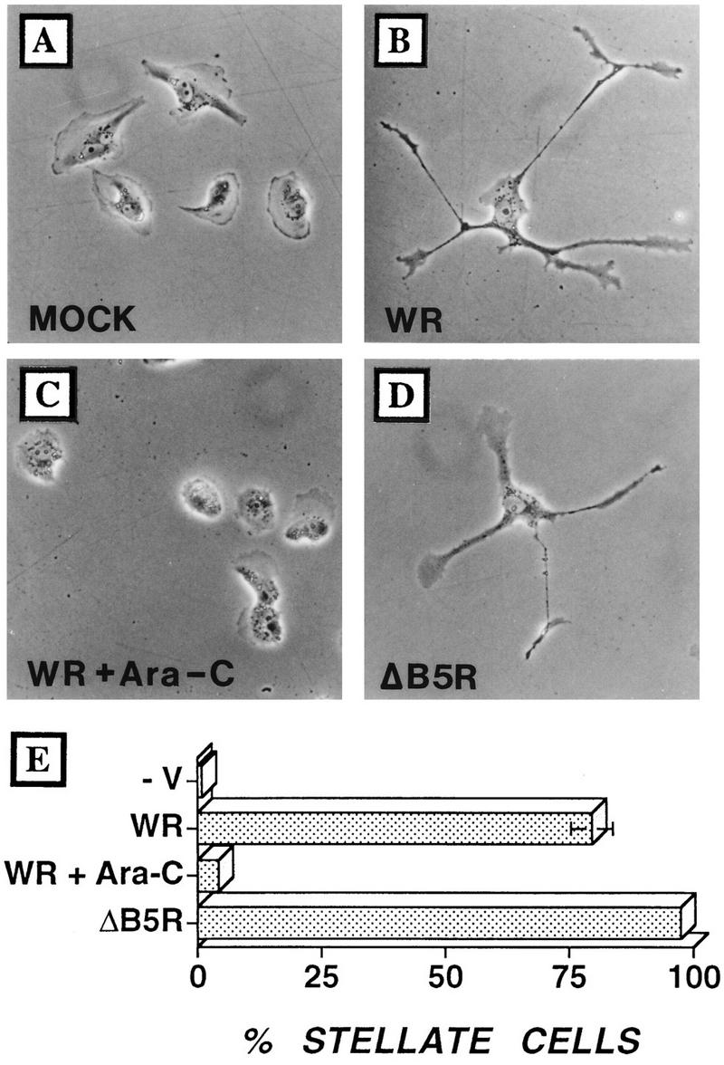
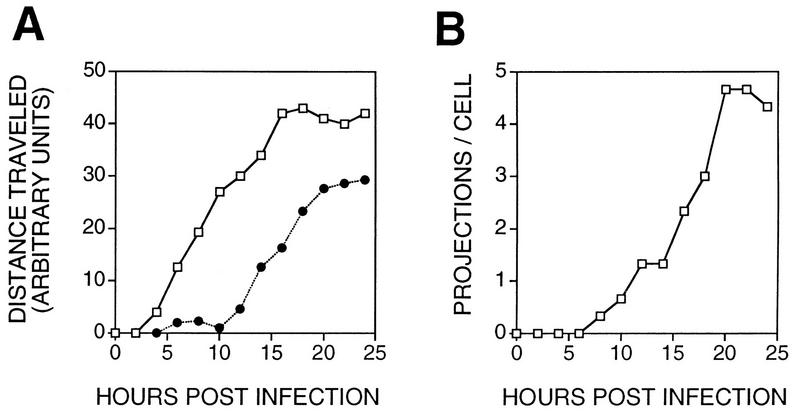
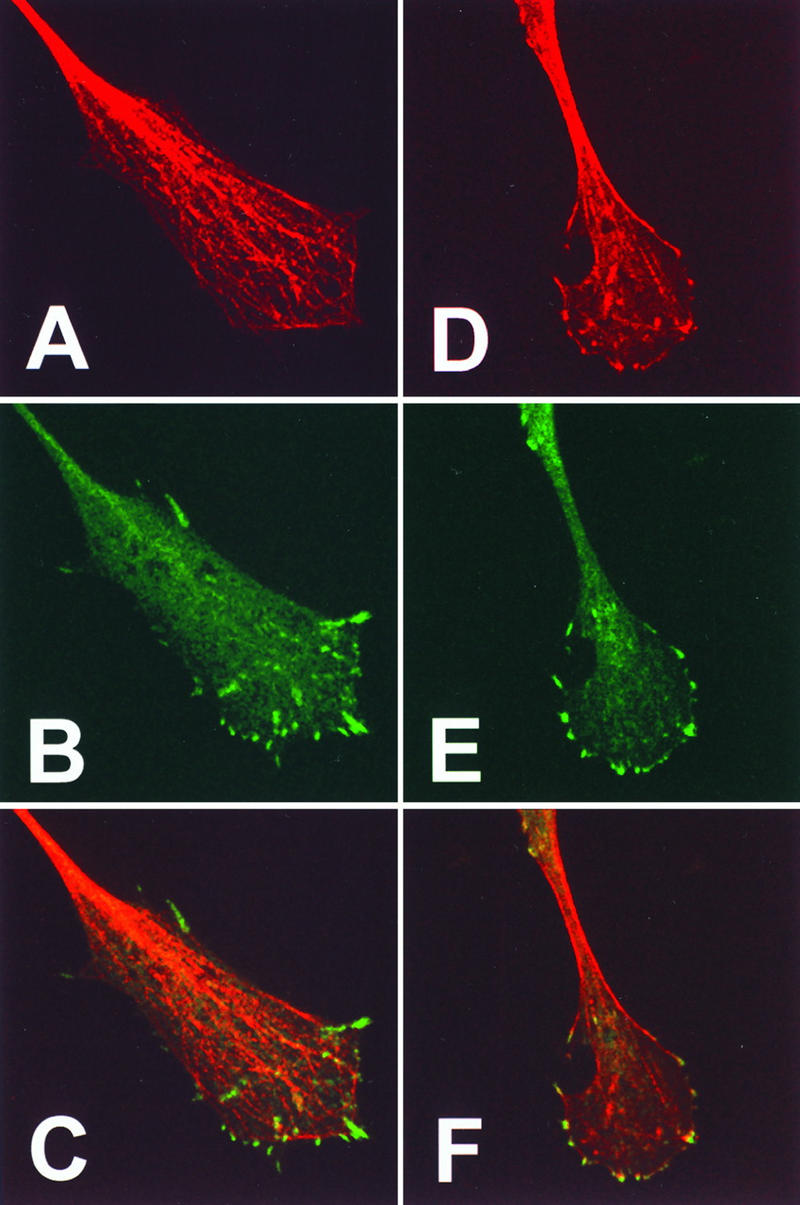
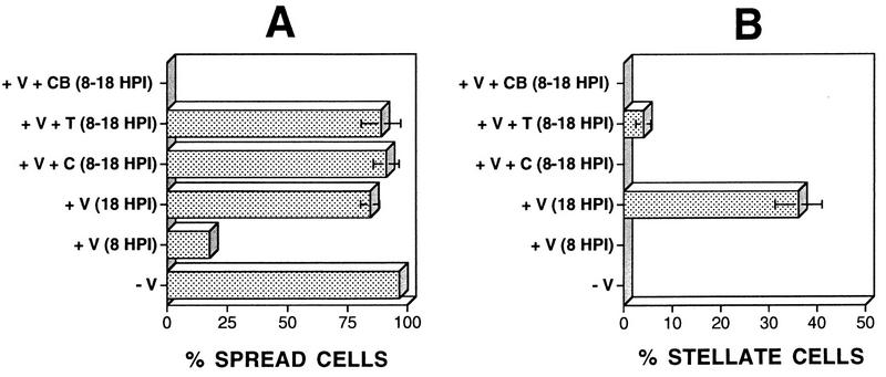
Many viruses induce profound changes in cell metabolism and function. Here we show that vaccinia virus induces two distinct forms of cell movement. Virus-induced cell migration was demonstrated by an in vitro wound healing assay in which infected cells migrated independently into the wound area while uninfected cells remained relatively static. Time-lapse microscopy showed that the maximal rate of migration occurred between 9 and 12 h postinfection. Virus-induced cell migration was inhibited by preinactivation of viral particles with trioxsalen and UV light or by the addition of cycloheximide but not by addition of cytosine arabinoside or rifampin. The expression of early viral genes is therefore necessary and sufficient to induce cell migration. Following migration, infected cells developed projections up to 160 microm in length which had growth-cone-like structures and were frequently branched. Time-lapse video microscopy showed that these projections were formed by extension and condensation of lamellipodia from the cell body. Formation of extensions was dependent on late gene expression but not the production of intracellular enveloped (IEV) particles. The requirements for virus-induced cell migration and for the formation of extensions therefore differ from each other and are distinct from the polymerization of actin tails on IEV particles. These data show that poxviruses encode genes which control different aspects of cell motility and thus represent a useful model system to study and dissect cell movement.


Figures

FIG. 1
FIG. 1
VV-induced plaques contain cells with motile features. Monolayers of BS-C-1 cells were infected with VV strain WR at 0.01 PFU/cell, and the plaque morphology was examined at 36 hpi by phase-contrast microscopy. Arrows indicate cells with a ruffled edge.
FIG. 2
FIG. 2
Infected cells become motile. Wounded BS-C-1 monolayers were photographed under phase contrast directly after wounding (A) or 24 h later (B to H). Cells were either mock infected (B), infected with VV at 5 PFU/cell (C, D, F, and G), or infected with VV which had been inactivated with trioxsalen and UV light at 5 PFU/cell (E). Cells shown in panel H were incubated in VV-conditioned medium for 24 h. Ara-C (40 μg/ml) (F), rifampin (100 μg/ml) (G), or cycloheximide (300 μg/ml) (D) was included throughout infection.
FIG. 3
FIG. 3
Only infected cells are motile. Wounded monolayers of BS-C-1 cells were infected with virus strain WR at 0.05 PFU/cell. At 24 hpi, cells were fixed and stained with rabbit anti-VV serum as described in Materials and Methods (B and D). Also shown are phase-contrast images (A and C) of matching epifluorescence photographs (B and D). Arrows indicate infected cells within the wound area.
FIG. 4
FIG. 4
Kinetics of VV-induced cell migration. A wounded monolayer of BS-C-1 cells was infected with VV at 5 PFU/cell and photographed at 9 (A), 12 (B), 15 (C), 18 (D), and 21 (E) hpi.
FIG. 5
FIG. 5
Infected cells develop multiple branched projections. BS-C-1 cells were seeded at low density to obtain isolated cells and then infected with VV at 5 PFU/cell. (A) Mock infection; (B) infection with VV; (C) infection with strain WR plus Ara-C; (D) infection with ΔB5R. Cells were photographed by phase-contrast microscopy at 24 hpi. (E) In different experiments (n = 4), between 100 and 120 cells were analyzed at 24 hpi and the proportion of cells showing three or more projections was calculated. Standard error bars are shown for each sample but are visible only for WR-infected cells.
FIG. 6
FIG. 6
Kinetics of cell movement and projection formation. BS-C-1 cells were grown and infected with VV as described for Fig. 3. The morphology and location of cells were recorded every 2 h until 24 hpi. (A) Distance moved for the leading edge of the cell (open squares) or the cell nucleus (closed circles) (n = 3). (B) Number of projections per cell (n = 3).
FIG. 7
FIG. 7
Video microscopy of projection formation. Shown is the formation of virus-induced cell extensions. BS-C-1 cells were grown and infected with VV strain WR as described for Fig. 4. From 13 to 16 hpi, cell morphology was monitored by video microscopy at 3-min intervals. (A to C) Single cell morphology at 13, 13.75, and 14.75 hpi, respectively. (D to I) Formation of the extension marked by arrows in panels A to C. The photograph shown in panel D was taken at 13 h and 9 min postinfection, and each subsequent photograph was taken 27 min after that shown in the previous panel (panel K = 16 h and 9 min postinfection). Magnification, ca. ×135 (A and B), ca. ×106 (C), and ca. ×451 (D to K).
FIG. 8
FIG. 8
Distribution of microtubules, actin, and paxillin within the growth cone of virus-induced projections. BS-C-1 cells were infected with VV at 10 PFU/cell and at 18 hpi were fixed and processed for immunofluorescence microscopy. Cells were stained with antitubulin (A) or with antipaxillin (B and E). Filamentous actin was visualized with phalloidin (D). (C and F) Merged images of panels A and B or D and E, respectively. Bar in panel F, 10 μm.
FIG. 9
FIG. 9
Involvement of actin filaments or microtubules in recovery from cell rounding and formation of cell projections. BS-C-1 cells were seeded at low density to obtain isolated cells and were then infected with VV at 5 PFU/cell. The number of spread cells was determined 8 hpi for mock-infected (−V) or WR strain-infected (+V) cells. Cytochalasin B (2 μM; CB), taxol (1 μM; T), or colchicine (50 μM; C) was added to infected cells at 8 hpi, and the number of spread cells (A) or stellate cells (B) was determined at 18 hpi.

References

    1. Alcamí A, Smith G L. A soluble receptor for interleukin-1β encoded by vaccinia virus: a novel mechanism of virus modulation of the host response to infection. Cell. 1992;71:153–167. - PubMed
    1. Allen W E, Jones G E, Pollard J W, Ridley A J. Rho, Rac and Cdc42 regulate actin organization and cell adhesion in macrophages. J Cell Sci. 1997;110:707–720. - PubMed
    1. Bablanian R, Baxt B, Sonnabend J A, Esteban M. Studies on the mechanisms of vaccinia virus cytopathic effects. II. Early cell rounding is associated with virus polypeptide synthesis. J Gen Virol. 1978;39:403–413. - PubMed
    1. Behrens J, Vakaet L, Friis R, Winterhager E, Van Roy F, Mareel M M. Loss of epithelial differentiation and gain of invasiveness correlates with tyrosine phosphorylation of the E-cadherin/β-catenin complex in cells transformed with a temperature-sensitive v-SRC gene. J Cell Biol. 1993;120:757–766. - PMC - PubMed
    1. Blasco R, Cole N B, Moss B. Sequence analysis, expression, and deletion of a vaccinia virus gene encoding a homolog of profilin, a eukaryotic actin-binding protein. J Virol. 1991;65:4598–4608. - PMC - PubMed

Publication types

LinkOut - more resources