. 1998 Feb;72(2):1334-44.
doi: 10.1128/JVI.72.2.1334-1344.1998.

Differential tropism and chemokine receptor expression of human immunodeficiency virus type 1 in neonatal monocytes, monocyte-derived macrophages, and placental macrophages

Affiliations

Differential tropism and chemokine receptor expression of human immunodeficiency virus type 1 in neonatal monocytes, monocyte-derived macrophages, and placental macrophages

W R Fear et al. J Virol. 1998 Feb.

Abstract

Laboratory-adapted (LA) macrophage-tropic (M-tropic) human immunodeficiency virus type 1 (HIV-1) isolates (e.g., HIV-1(Ba-L)) and low-passage primary (PR) isolates differed markedly in tropism for syngeneic neonatal monocytes, monocyte-derived macrophages (MDMs), and placental macrophages (PMs). Newly adherent neonatal monocytes and cultured PMs were highly refractory to infection with PR HIV-1 isolates yet were permissive for LA M-tropic isolates. Day 4 MDMs were also permissive for LA M-tropic isolates and additionally, were permissive for over half the PR isolates tested. Qualitative differences in PR HIV-1 infection of monocytes/MDMs could not be correlated with CD4 levels alone, and in all three cell types the block to PR HIV-1 strain replication preceded reverse transcription. Neonatal monocyte susceptibility to PR HIV-1 strains correlated with increasing CCR-5 expression during maturation. CCR-5 could not be detected on newly adherent (day 1) neonatal monocytes, in contrast to adult monocytes (H. Naif et al., J. Virol. 72:830-836, 1998), but was readily detectable after 4 to 7 days of culture. However, moderate CCR-5 mRNA levels were present in day 1 neonatal monocytes and remained constant during monocyte maturation. CCR-5 was not detectable on the surface of PMs, yet the receptor was present within permeabilized cells. Notably, two brain-derived PR HIV-1 isolates from a single patient, differing in their V3 loops, were discordant in their abilities to infect neonatal monocytes/MDMs and PMs, yet both isolates could infect newly adherent adult monocytes. Together these data strongly suggest that LA HIV-1 isolates are able to infect neonatal monocytes at earlier stages of maturation and lower-level expression of CCR-5 than PR isolates. The differences between neonatal and adult monocytes in susceptibility to PR isolates may also be related to the level of CCR-5 expression.


Figures

FIG. 1
FIG. 1
HIV-1 infection of fresh cord blood monocytes. (A) Kinetics of HIV-1 p24 antigen (Ag) production. Cord blood monocytes from three different donors were infected on the day of their isolation (day 0) with a panel of two LA (HIV-1JR-FL and HIV-1Ada-M) and seven PR (WM-1039, -1044, -1061, -1063, -1067, -1068, and -1076) HIV-1 isolates as described in Materials and Methods. Representative data from three identical experiments are presented. Supernatant samples were collected and assayed by ELISA for HIV-1 p24 antigen as indicated. For clarity, the curves for only two PR isolates are shown, as these produced kinetic patterns identical to those of all other PR isolates in the panel. (B) DNA PCR analysis of the long terminal repeat-gag region. On day 16 postinfection the cultures were terminated, DNA was extracted from the cells in situ, and DNA PCR was performed with primer pair M667-FG1 as described in Materials and Methods. The DNA was subsequently Southern blotted and hybridized with the oligonucleotide probe PFI. Positive results indicate complete or nearly complete viral RT. The negative control reaction tube (−ve) contained H2O in lieu of template DNA, while the positive control template (+ve) was DNA extracted from the HIV-1LAI-infected 8E5 cell line (24).
FIG. 2
FIG. 2
HIV infection of cord blood MDMs. (A) Kinetics of HIV-1 p24 antigen (Ag) production. Seven-day-old plastic-adherent MDMs were infected with HIV-1 isolates, and supernatants were sampled as indicated for ELISA determination of HIV-1 p24 antigen concentration. The MDMs were syngeneic with the monocytes described in the legend to Fig. 1, and all conditions of the experiment were the same as used for monocytes. The WM-1068 and WM-1076 data are representative of PR isolates WM-1039 and -1044 (productive infections; kinetic curves for the latter isolates were between those for WM-1076 and -1068). In contrast, kinetic curves for isolates WM-1063 and -1067 were very similar to the curve shown for WM-1061 (no infection). (B) PCR analysis of the long terminal repeat-gag region. Cell cultures were terminated on day 17 postinfection, and PCR was performed as described for Fig. 1.
FIG. 3
FIG. 3
HIV infection of PMs. (A) Kinetics of HIV-1 p24 antigen (Ag) production. Seven-day-old plastic-adherent PMs were infected with a panel of HIV-1 isolates, and supernatants were sampled for p24 antigen as described in the legend to Fig. 1. The PMs were syngeneic with the cord blood monocytes and MDMs described in the legends to Fig. 1 and 2. The kinetic curves for PR isolates WM-1039, -1063, -1067, -1068, and -1076 all fell between the curves shown for the representative PR isolates WM-1044 and -1061. (B) PCR analysis of the long terminal repeat-gag region. Cell cultures were terminated on day 17 postinfection, and PCR was performed as described for Fig. 1.
FIG. 4
FIG. 4
Comparison of HIV-1 replication in neonatal monocytes, MDMs, PMs, and adult monocytes. Cells (2 × 10⁵ to 5 × 10⁵) were infected with each HIV-1 isolate at a multiplicity of infection of 0.1. Monocytes/MDMs were incubated with HIV for 2 h, and PMs were incubated for 16 h. Results are representative of three experiments. Ag, antigen.
FIG. 5
FIG. 5
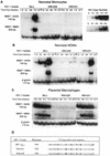
Comparison of PR and LA HIV-1 RT in neonatal monocytes, MDMs, and PMs. The PCR utilized one 32P-labeled primer per reaction as described in Materials and Methods, and standards represent HIV-1 DNA copy number equivalents amplified from 8E5 cell DNA. Primer pairs M667-AA55 and M667-M661 were described by Zack et al. (78) and amplified HIV-1 DNA, indicating RT initiation and completion (or near completion), respectively. Controls for de novo RT were treated for 2 h with 10 μM AZT prior to HIV infection, which was performed in the continued presence of the nucleoside analog. AZT-treated cells were harvested for DNA at day 17 postinfection. Input DNA was controlled by using β-globin specific primers (59), and the results shown are representative of three experiments. (A) Day 1 neonatal monocytes; (B) day 4 neonatal MDMs; (C) day 7 PMs; (D) comparison of HIV-1Ba-L, WM-628, and WM-631 phenotypes and V3 loop amino acid sequences (amino acid substitutions are in boldface).
FIG. 6
FIG. 6
CD4 expression on neonatal monocytes and MDMs. Nonpermeabilized CD14-positive neonatal monocytes were prepared and analyzed on day 0 (prior to adherence) or day 1 (after overnight adherence) of culture, and MDMs were analyzed on day 4, by using the anti-Leu-3a–FITC (CD4) MAb as described in Materials and Methods. CD14-positive cells were gated by using the anti-Leu-M3–PE MAb, and results are representative of three experiments. Isotype control (open) and specific (shaded) histograms are shown.
FIG. 7
FIG. 7
RT-PCR analysis of chemokine receptor mRNA expression in neonatal monocytes, MDMs, and PMs. mRNA was prepared and analyzed as described in Materials and Methods. CCR-1, CCR-3, CCR-5, and CXCR-4 mRNA transcripts were amplified as 296-, 539-, 272-, and 381-bp products, respectively; the GAPDH product is shown at 191 bp. cDNA samples were amplified following RT reactions with or without reverse transcriptase. Representative results from three experiments are shown; 2% agarose was used.
FIG. 8
FIG. 8
Chemokine receptor expression by PMs. Day 7 PMs were labeled with the 12G5 MAb to CXCR-4 or the 3A9 MAb to CCR5 and compared with isotype control antibodies (open histograms) by flow cytometry. Cells were either permeabilized prior to staining (cytoplasmic labeling) or left untreated (surface labeling), as described in Materials and Methods, and data representative of three experiments are presented.
FIG. 9
FIG. 9
Chemokine receptor expression on differentiating neonatal monocytes. Nonpermeabilized neonatal monocytes were prepared for flow cytometry on day 1 or 7 of culture and labeled with anti-Leu-M3–PE (CD14), 12G5 (CXCR-4), or 3A9 (CCR-5). Representative data are presented. Fluorescence-activated cell sorter analyses for CXCR-4 and CCR-5 were gated on the CD14-positive fraction of the cell populations and compared with results for isotype control antibodies (open histograms).

Similar articles

Cited by

References

    1. Alkhatib G, Combadiere C, Broder C C, Feng Y, Kennedy P E, Murphy P M, Berger E A. CC CKR5: a RANTES, MIP-1α, MIP-1β receptor as a fusion cofactor for macrophage-tropic HIV-1. Science. 1996;272:1955–1958. - PubMed
    1. Bleul C C, Wu L, Hoxie J A, Springer T A, Mackay C R. The HIV coreceptors CXCR4 and CCR5 are differentially expressed and regulated on human T lymphocytes. Proc Natl Acad Sci USA. 1997;94:1925–1930. - PMC - PubMed
    1. Chandwani S, Greco M, Mittal K, Antoine C, Kraninski K, Borkowsky W. Pathology and human immunodeficiency virus expression in placentas of seropositive women. J Infect Dis. 1991;163:1134–1138. - PubMed
    1. Chang J, Li S, Naif H, Cunningham A L. The magnitude of HIV replication in monocytes and macrophages is influenced by environmental conditions, viral strain, and host cells. J Leukocyte Biol. 1994;56:230–235. - PubMed
    1. Chang J, Naif H M, Li S, Sullivan J S, Randle C M, Cunningham A L. Twin studies demonstrate a host cell genetic effect on productive human immunodeficiency virus infection of human monocytes and macrophages in vitro. J Virol. 1996;70:7792–7803. - PMC - PubMed

Publication types

Substances