. 1998 Feb;72(2):1394-402.
doi: 10.1128/JVI.72.2.1394-1402.1998.

Nucleotide sequence, genome organization, and transcription map of bovine adenovirus type 3

Affiliations

Nucleotide sequence, genome organization, and transcription map of bovine adenovirus type 3

P S Reddy et al. J Virol. 1998 Feb.

Abstract

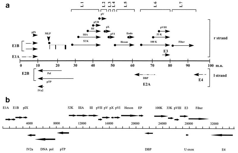
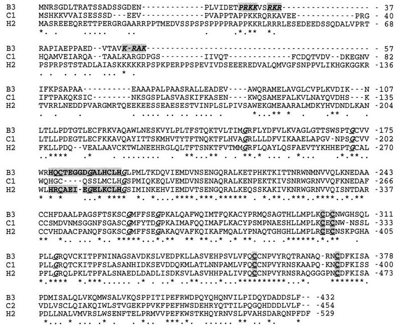
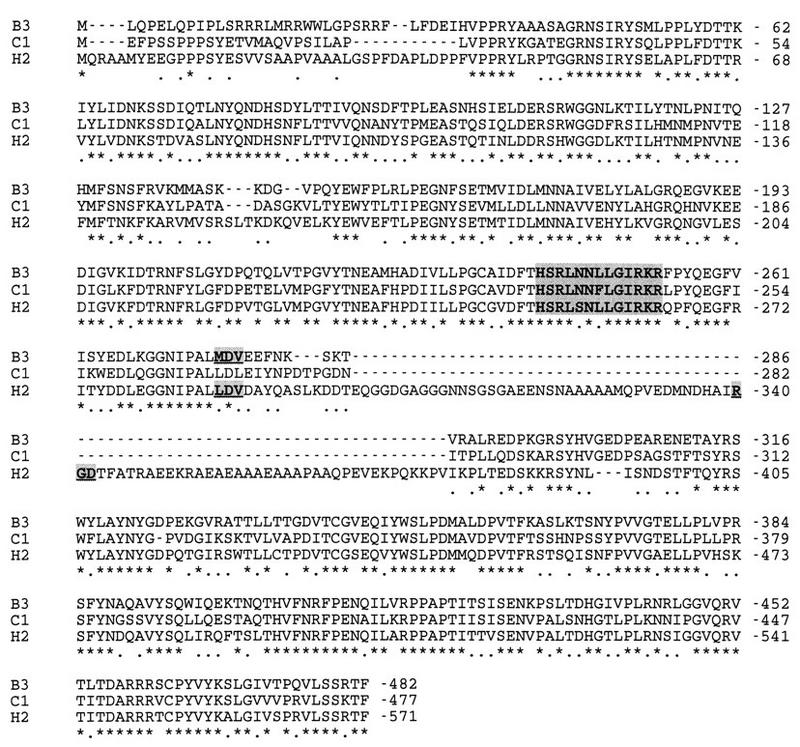
The complete DNA sequence of bovine adenovirus type 3 is reported here. The size of the genome is 34,446 bp in length with a G+C content of 54%. All the genes of the early and late regions are present in the expected locations of the genome. However, the late-region genes are organized into seven families, instead of five as they are in human adenovirus type 2. The deduced amino acid sequences of open reading frames (ORFs) in the late regions and early region 2 (E2) and for IVa2 show higher degrees of homology, whereas the predicted amino acid sequences of ORFs in the E1, E3, and E4 regions and the pIX, fiber, and 33,000-molecular-weight nonstructural proteins show little or no homology with the corresponding proteins of other adenoviruses. In addition, the penton base protein lacks the integrin binding motif, RGD, but has an LDV motif instead of an MDV motif. Interestingly, as in other animal adenoviruses, the virus-associated RNA genes appear to be absent from their usual location. Sequence analysis of cDNA clones representing the early- and late-region genes identified splice acceptor and splice donor sites, polyadenylation signals and polyadenylation sites, and tripartite leader sequences.


Figures

FIG. 1
FIG. 1
Transcription map and genome organization of BAV-3. (a) Summary of transcriptional analysis of BAV-3. The schematic diagram of the BAV-3 genome (34,446 bp) is divided into 100 m.u. Arrows above and below the central line represent mRNAs from the r and l strands, respectively. Solid lines represent sequences found in mRNA, broken lines indicate introns, and arrowheads represent poly(A) sites and show the direction of transcription. Each mRNA in L1 to L7 has a TPL spliced to its 5′ terminus (•). Detailed structures of transcripts in E1A and E1B (50), E3 (31) and E4 (38) have been described elsewhere. (b) Summary of the genomic organization of BAV-3. Genes are named as homologs of HAV-2. Arrows show the positions of the ORFs for the indicated proteins.
FIG. 2
FIG. 2
Multiple-sequence alignment of DBP homologs. The amino acid sequences of BAV-3 (B3), CAV-1 (C1), and HAV-2 (H2) DBPs were aligned as indicated. Identical and conserved amino acid residues are indicated by asterisks and dots, respectively. Dashes indicate gaps. Conserved Cys residues that are involved in metal binding are shown in bold, underlined, and shaded. NLSs are shown in bold italics and shaded. Conserved glycine residues are shown in bold italics and underlined. The sequence motif HXCX8CXH is shown in bold and shaded. The alignment was done with the CLUSTAL program by using default parameters (open gap cost and unit gap cost = 10). The same parameters were used for the alignment of protein sequences in Fig. 3.
FIG. 3
FIG. 3
Multiple-sequence alignment of penton base protein homologs. The amino acid sequences of BAV-3 (B3), CAV-1 (C1), and HAV-2 (H2) penton base proteins were aligned as described in the legend to Fig. 2. Identical and conserved amino acids are indicated by asterisks and dots, respectively. Dashes indicate gaps. RGD, LDV, MDV motifs are shown in bold, underlined, and shaded. The putative fiber-interacting domain is shown in bold and shaded.

References

    1. Anderson C W, Young M E, Flint S J. Characterization of the adenovirus 2 virion protein, Mu. Virology. 1989;172:506–512. - PubMed
    1. Anderson K P, Klessig D F. Altered mRNA splicing in monkey cells abortively infected with human adenovirus may be responsible for inefficient synthesis of the virion fiber polypeptide. Proc Natl Acad Sci USA. 1984;81:4023–4027. - PMC - PubMed
    1. Athappilly F K, Murali R, Rux J J, Cai Z P, Burnett R M. The refined crystal structure of hexon, the major coat protein of adenovirus type 2 at 2.9 angstrom resolution. J Mol Biol. 1994;242:430–455. - PubMed
    1. Bailey A, Mautner V. Phylogenetic relationships among adenovirus serotypes. Virology. 1994;205:438–452. - PubMed
    1. Bartha A. Proposal for subgrouping of bovine adenoviruses. Acta Vet. 1969;19:319–321. - PubMed

Publication types

Associated data

LinkOut - more resources