. 1998 Feb;72(2):1606-9.
doi: 10.1128/JVI.72.2.1606-1609.1998.

The spike protein of murine coronavirus mouse hepatitis virus strain A59 is not cleaved in primary glial cells and primary hepatocytes

Affiliations

The spike protein of murine coronavirus mouse hepatitis virus strain A59 is not cleaved in primary glial cells and primary hepatocytes

S T Hingley et al. J Virol. 1998 Feb.

Abstract

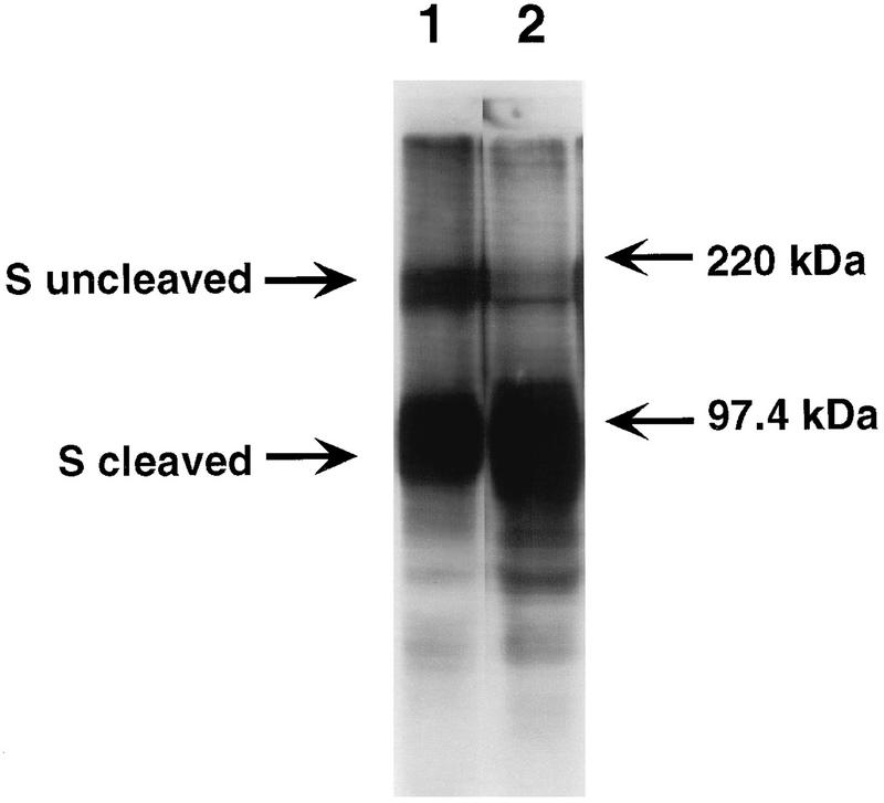
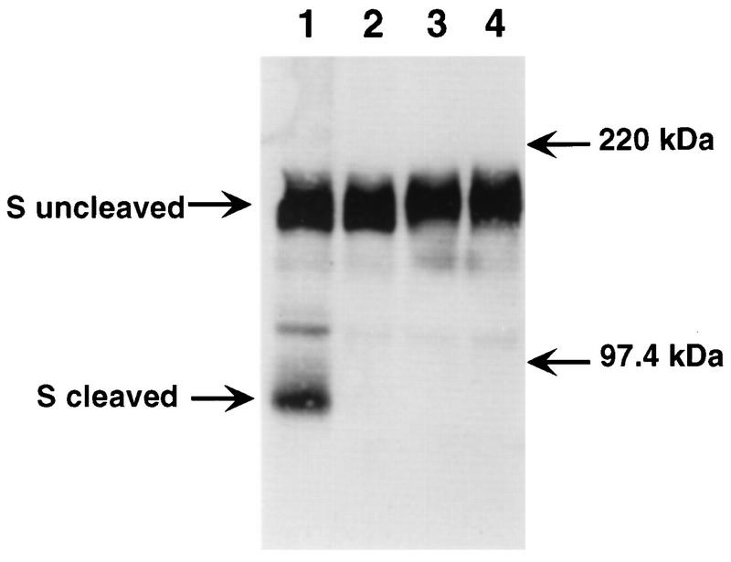
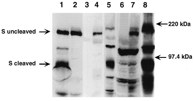
Mouse hepatitis virus strain A59 (MHV-A59) produces meningoencephalitis and severe hepatitis during acute infection. Infection of primary cells derived from the central nervous system (CNS) and liver was examined to analyze the interaction of virus with individual cell types derived from the two principal sites of viral replication in vivo. In glial cell cultures derived from C57BL/6 mice, MHV-A59 produces a productive but nonlytic infection, with no evidence of cell-to-cell fusion. In contrast, in continuously cultured cells, this virus produces a lytic infection with extensive formation of syncytia. The observation of few and delayed syncytia following MHV-A59 infection of hepatocytes more closely resembles infection of glial cells than that of continuously cultured cell lines. For MHV-A59, lack of syncytium formation correlates with lack of cleavage of the fusion glycoprotein, or spike (S) protein. The absence of cell-to-cell fusion following infection of both primary cell types prompted us to examine the cleavage of the spike protein. Cleavage of S protein was below the level of detection by Western blot analysis in MHV-A59-infected hepatocytes and glial cells. Furthermore, no cleavage of this protein was detected in liver homogenates from C57BL/6 mice infected with MHV-A59. Thus, cleavage of the spike protein does not seem to be essential for entry and spread of the virus in vivo, as well as for replication in vitro.


Figures

FIG. 1
FIG. 1
Replication of MHV-A59 in primary hepatocyte cultures. Confluent monolayers of hepatocytes derived from C57BL/6 mice were infected with MHV-A59 at an MOI of 5 PFU/cell. Culture supernatants were removed, and cellular fractions were obtained by three consecutive rounds of freezing and thawing at various times postinfection. The viral titers of the cellular and supernatant fractions are shown. Each point represents the viral titer from a single 35-mm-diameter dish of MHV-A59-infected hepatocytes, while the lines are drawn between the means for each time point. Titers were determined by plaque assay on L2 cells.
FIG. 2
FIG. 2
Western blot analysis of lysates of infected fibroblasts. Proteins in lysates from MHV-A59-infected L2 cells (lane 1) and MHV-3-infected L2 cells (lane 2) were separated by electrophoresis in sodium dodecyl sulfate–8% polyacrylamide gel electrophoresis gel, transferred to nitrocellulose, and visualized by chemiluminescence (Amersham) with a polyclonal goat anti-S protein antibody (obtained from K. Holmes) as the primary antibody to detect cleaved and uncleaved forms of S protein. The migration of the molecular mass markers is indicated.
FIG. 3
FIG. 3
Comparison of the cleavage of S protein in fibroblasts and in glial cells. Culture supernatants of MHV-A59- and C12-infected L2 cells (lanes 1 and 2, respectively) and MHV-A59- and MHV-3-infected glial cells (lanes 3 and 4, respectively) were analyzed by sodium dodecyl sulfate–8% polyacrylamide gel electrophoresis and Western blotting as described in the legend to Fig. 2. C12 is a variant of MHV-A59 with an uncleaved S protein (6) and is included here as a negative control for cleavage. The migration of the molecular mass markers is indicated.
FIG. 4
FIG. 4
Western blot analysis of S protein present in hepatocytes and liver lysates. Culture supernatants of MHV-A59- and C12-infected L2 cells (lanes 1 and 2, respectively), cell lysates of uninfected, MHV-A59-infected, and MHV-3-infected hepatocytes (lanes 3, 4, and 5, respectively), and liver homogenates of uninfected, MHV-A59-infected, and MHV-3-infected mice (lanes 6, 7, and 8, respectively) were analyzed by sodium dodecyl sulfate–8% polyacrylamide gel electrophoresis and Western blotting as described in the legend to Fig. 2. Lanes 1 and 2 were included as positive and negative controls for cleavage, respectively. The migration of the molecular mass markers is indicated.

Similar articles

Cited by

References

    1. Bos E C W, Heijnen L, Luytjes W, Spaan W J M. Mutational analysis of the murine coronavirus spike protein: effect on cell to cell fusion. Virology. 1996;214:453–463. - PMC - PubMed
    1. Cavanagh D. The coronavirus surface glycoprotein. In: Siddell S G, editor. The Coronaviridae. New York, N.Y: Plenum Press; 1995. pp. 73–113.
    1. Fleming J O, Trousdale M D, El-Zaatari F A K, Stohlman S A, Weiner L P. Pathogenicity of antigenic variants of murine coronavirus JHM selected with monoclonal antibodies. J Virol. 1986;58:869–875. - PMC - PubMed
    1. Frana M F, Behnke J N, Sturman L S, Holmes K V. Proteolytic cleavage of the E2 glycoprotein of murine coronavirus: host-dependent differences in proteolytic cleavage and cell fusion. J Virol. 1985;56:912–920. - PMC - PubMed
    1. Gallagher T M, Escarmis C, Buchmeier M J. Alteration of pH dependence of coronavirus-induced cell fusion: effect of mutations in the spike glycoprotein. J Virol. 1991;65:1916–1928. - PMC - PubMed

Publication types

Substances

LinkOut - more resources