. 1998 Feb;18(2):694-702.
doi: 10.1128/MCB.18.2.694.

Different cis-acting elements are involved in the regulation of TRP1 and TRP2 promoter activities by cyclic AMP: pivotal role of M boxes (GTCATGTGCT) and of microphthalmia

Affiliations

Different cis-acting elements are involved in the regulation of TRP1 and TRP2 promoter activities by cyclic AMP: pivotal role of M boxes (GTCATGTGCT) and of microphthalmia

C Bertolotto et al. Mol Cell Biol. 1998 Feb.

Abstract

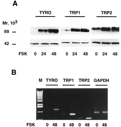
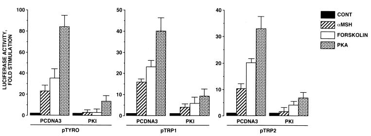
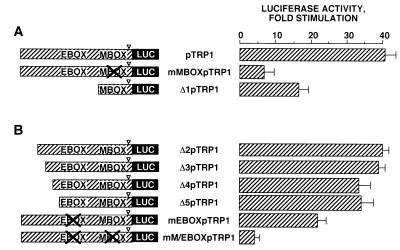
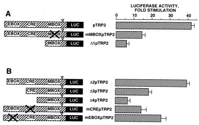
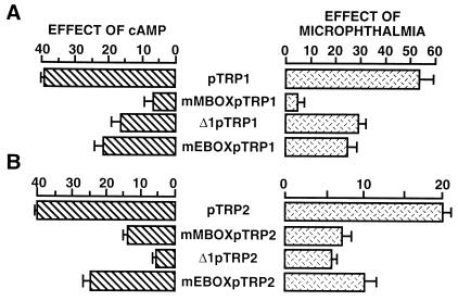
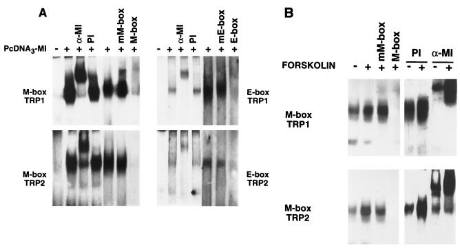
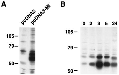
In melanocytes and in melanoma cells, cyclic AMP (cAMP)-elevating agents stimulate melanogenesis and increase the transcription of tyrosinase, the rate-limiting enzyme in melanin synthesis. However, two other enzymes, tyrosinase-related protein 1 (TRP1) and TRP2, are required for a normal melanization process leading to eumelanin synthesis. In B16 melanoma cells, we demonstrated that stimulation of melanogenesis by cAMP-elevating agents results in an increase in tyrosinase, TRP1, and TRP2 expression. cAMP, through a cAMP-dependent protein kinase pathway, stimulates TRP1 and TRP2 promoter activities in both B16 mouse melanoma cells and normal human melanocytes. Regulation of the TRP1 and TRP2 promoters by cAMP involves a M box and an E box. Further, a classical cAMP response element-like motif participates in the cAMP responsiveness of the TRP2 promoter, demonstrating that the TRP2 gene is subjected to different regulatory processes, which could account for its different expression patterns during embryonic development or under specific physiological and pathological conditions. We also found that microphthalmia, a basic helix-loop-helix transcription factor, strongly stimulates the transcriptional activities of the TRP1 and TRP2 promoters, mainly through binding to the M boxes. Additionally, we demonstrated that cAMP increases microphthalmia expression and thereby its binding to TRP1 and TRP2 M boxes. These convergent and compelling results disclose at least a part of the molecular mechanism involved in the regulation of melanogenic gene expression by cAMP and emphasize the pivotal role of microphthalmia in this process.


Figures

FIG. 1
FIG. 1
Forskolin treatment increases tyrosinase, TRP1, and TRP2 gene expression in B16 mouse melanoma cells. (A) Thirty micrograms of proteins from control cells and cells treated for 24 or 48 h with 20 μM forskolin (FSK) was subjected to Western blot analysis using antibodies PEP7 for tyrosinase detection, B8G3 for TRP1, and PEP8 for TRP2 (upper panel). The detection of the 44-kDa ERK1 protein ensured even loading of lanes (lower panel). (B) Ten micrograms of total RNA from control B16 cells or cells treated for 48 h with 20 μM forskolin was reverse transcribed. The resulting cDNAs were subjected to 28-cycle PCRs using specific primers that gave amplified products of 1,191 bp for tyrosinase (TYRO), 784 bp for TRP1, and 518 bp for TRP2. A control of PCR amplification with specific primers of GAPDH transcript showed a 983-bp fragment. PCR products were electrophoresed on a 1% agarose gel and stained with ethidium bromide before UV light visualization. DNA molecular weight markers (lane M), from bottom to top: 510, 1,018, 1,636, 2,036, 3,054, and 4,072 bp.
FIG. 2
FIG. 2
cAMP-elevating agents and PKA stimulate tyrosinase, TRP1, and TRP2 promoter activities in B16 cells: reversal of these effects by PKI. B16 cells were transfected with 0.3 μg of luciferase reporter plasmid pTyro, pTRP1, or pTRP2 and 0.05 μg of pCMVβGAL. In control (CONT), α-MSH, and forskolin conditions, 0.2 μg of empty pCDNA3 or 0.1 μg of empty pCDNA3 plus 0.1 μg of pCDNA3 encoding PKI was cotransfected with reporter plasmids. Cells were treated for 48 h with 20 μM forskolin or 1 μM α-MSH. To study the effects of PKA expression, B16 cells were transfected with 0.3 μg of luciferase reporter plasmid and 0.1 μg of pCDNA3 encoding PKA plus 0.1 μg of empty pCDNA3 or 0.1 μg of pCDNA3 encoding PKA plus 0.1 μg of pCDNA3 encoding PKI. Luciferase activity was normalized to β-galactosidase activity, and the results were expressed as fold stimulation of the basal luciferase activity from unstimulated cells. Data are means ± standard errors of five experiments performed in triplicate.
FIG. 3
FIG. 3
Regulation of TRP1 transcriptional activity by PKA is mediated by the M box (−44 to −33) and the E box (−238 to −233). B16 cells were transfected with 0.3 μg of pTRP1, mMBOXpTRP1, and Δ1pTRP1 (A) or Δ2pTRP1, Δ3pTRP1, Δ4pTRP1, Δ5pTRP1, mEBOXpTRP1, and mM/EBOXpTRP1 (B), 0.2 μg of pCDNA3, empty or encoding PKA, and 0.05 μg of pCMVβGAL. After 48 h, luciferase activity was assayed as described in the text and normalized to β-galactosidase activity. ▿, TATA-box position. Results are expressed as fold stimulation of the basal luciferase activity from unstimulated cells. Data are means ± standard errors of five experiments performed in triplicate.
FIG. 4
FIG. 4
Regulation of TRP2 transcriptional activity by PKA involved the M box (−129 to −135), the CRE-like motif (−239 to −232), and the E box (−346 to −340). B16 cells were transfected with 0.3 μg of pTRP2, mMBOXpTRP2, and Δ1pTRP2 (A) or Δ2pTRP2, Δ3pTRP2, Δ4pTRP2, mCREpTRP2, and mEBOXpTRP2 (B), 0.2 μg of pCDNA3, empty or encoding PKA, and 0.05 μg of pCMVβGAL. After 48 h, luciferase activity was assayed as described in the text and normalized to β-galactosidase activity. ▿, TATA-box position. Results are expressed as fold stimulation of the basal luciferase activity from unstimulated cells. Data are means ± standard errors of five experiments performed in triplicate.
FIG. 5
FIG. 5
The effects of microphthalmia on TRP1 and TRP2 promoter constructs correlate with their cAMP responsiveness. Histograms on the left represent the cAMP responsiveness of the different TRP1 (A) and TRP2 (B) constructs as determined in Fig. 3 and 4. To study the effects of microphthalmia on these different constructs, B16 cells were cotransfected with 0.3 μg of the reporter plasmid and 0.05 μg of pCMVβGAL plus 0.04 μg of empty pCDNA3 or pCDNA3 encoding microphthalmia. Reporter plasmids were pTRP1, mMBOXpTRP1, Δ1pTRP1, and mEBOXpTRP1 (A) and pTRP2, mMBOXpTRP2, Δ1pTRP2, and mEBOXpTRP2 (B). After 48 h, luciferase activity was assayed as previously described and normalized to β-galactosidase activity. Results are expressed as fold stimulation of the luciferase activity from cells transfected with empty pCDNA3. Data are means ± standard error of five experiments performed in triplicate.
FIG. 6
FIG. 6
Microphthalmia binds to TRP1 and TRP2 M and E boxes. (A) TRP1 M box (−44 to −33), TRP1 E box (−238 to −233), TRP2 M box (−129 to −135), and TRP2 E box (−346 to −340) were used as probes. Gel shift assays were performed in an in vitro transcription-translation reaction using empty pCDNA3 or pCDNA3 encoding microphthalmia (pCDNA3-MI). For competition experiments, unlabeled homologous and mutated oligonucleotides were added in 50-fold excess. (B) B16 nuclear extracts from control cells (−) or cells treated with forskolin for 6 h (+) were incubated with labeled TRP1 M box and TRP2 M box. Where indicated, reactions were carried out in the presence of preimmune serum (PI) or a specific antibody directed against the C terminus of the microphthalmia (α-MI). For competition experiments, unlabeled homologous and mutated oligonucleotides were added in 50-fold excess. Autoradiograms were exposed for 15 h at −80°C, except for competition experiments with TRP1 or TRP2 E box, which were exposed for 48 h.
FIG. 7
FIG. 7
cAMP increases microphthalmia expression in B16 mouse melanoma cells. B16 melanoma cells and NIH 3T3 cells nontransfected or transfected with microphthalmia were labeled with [35S]methionine-cysteine mix for 30 h. (A) NIH 3T3 cells were solubilized and immunoprecipitated with antimicrophthalmia antibody. (B) B16 melanoma cells were exposed to 20 μM forskolin for the indicated time, and then solubilized proteins were immunoprecipitated with antimicrophthalmia antibody. The immune complexes were analyzed by gel electrophoresis and autoradiography. Molecular masses, indicated on the left, are expressed in kilodaltons.

References

    1. Abdel-Malek Z, Swope V B, Amornsiripanitch N, Nordlund J J. In vitro modulation of proliferation and melanization of S91 melanoma cells by prostaglandins. Cancer Res. 1987;47:3141–3146. - PubMed
    1. Abdel-Malek Z, Swope V B, Suzuki I, Akcali C, Harriger M D, Boyce S T, Urabe K, Hearing V. Mitogenic and melanogenic stimulation of normal human melanocytes by melanotropic peptides. Proc Natl Acad Sci USA. 1995;92:1789–1793. - PMC - PubMed
    1. Barber J I, Townsend D, Olds D P A, King R A. Dopachrome oxydoreductase: a new enzyme in the pigment pathway. J Invest Dermatol. 1984;83:145–149. - PubMed
    1. Bentley N J, Eisen T, Goding C R. Melanocyte-specific expression of the human tyrosinase promoter: activation by the microphthalmia gene product and role of the initiator. Mol Cell Biol. 1994;14:7996–8006. - PMC - PubMed
    1. Bertolotto C, Bille K, Ortonne J P, Ballotti R. Regulation of tyrosinase gene expression by cAMP in B16 melanoma cells involves two CATGTG motifs surrounding the TATA box: implication of the microphthalmia gene product. J Cell Biol. 1996;134:747–755. - PMC - PubMed

Publication types

MeSH terms