. 1998 Feb;18(2):732-41.
doi: 10.1128/MCB.18.2.732.

Regulation of hypoxia-inducible mRNAs by the von Hippel-Lindau tumor suppressor protein requires binding to complexes containing elongins B/C and Cul2

Affiliations

Regulation of hypoxia-inducible mRNAs by the von Hippel-Lindau tumor suppressor protein requires binding to complexes containing elongins B/C and Cul2

K M Lonergan et al. Mol Cell Biol. 1998 Feb.

Abstract

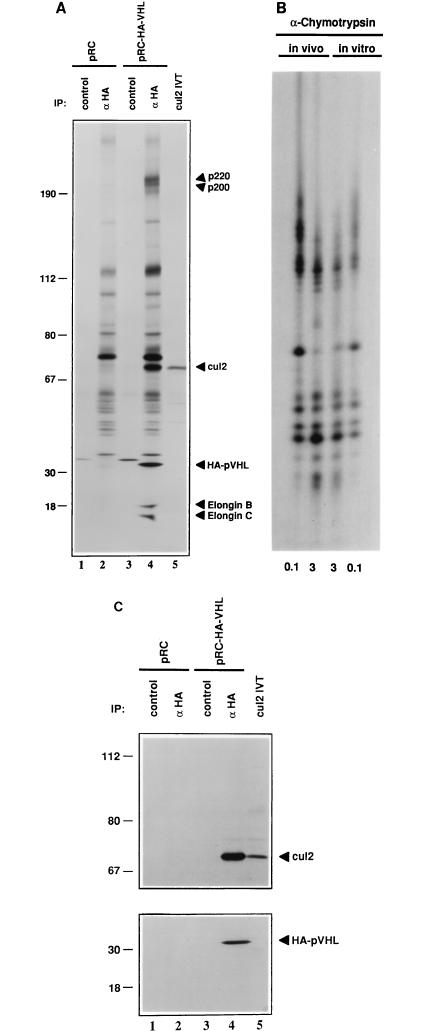
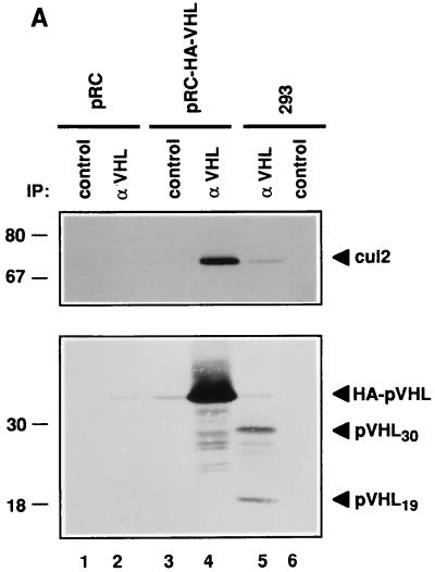
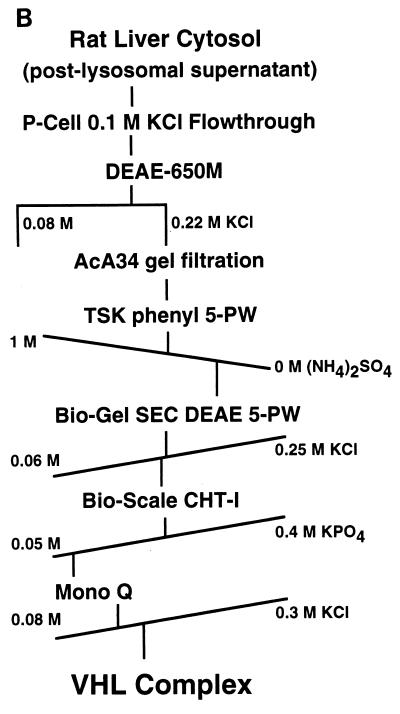
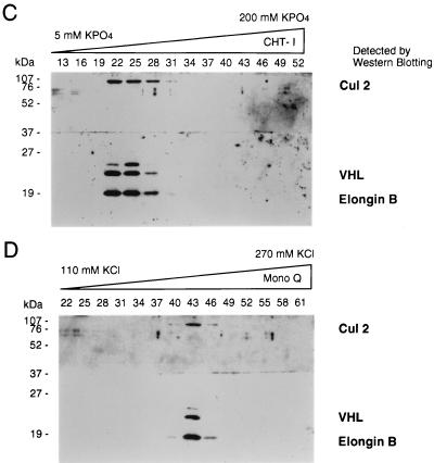
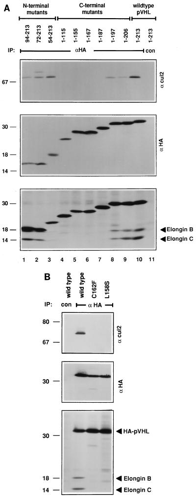
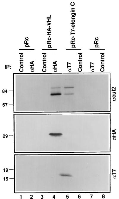
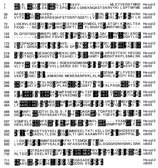
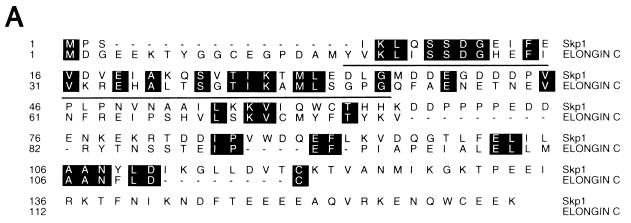
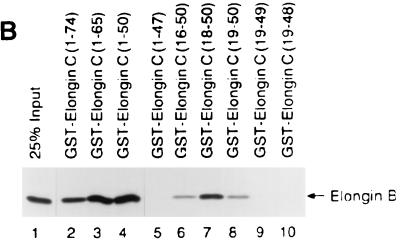
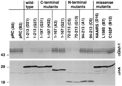
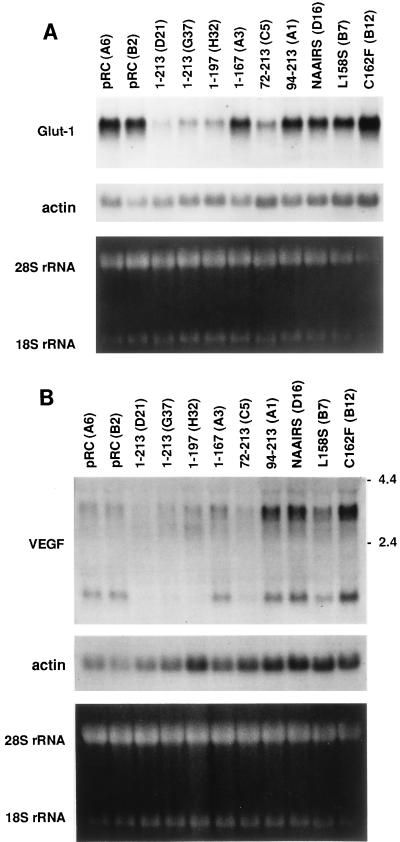
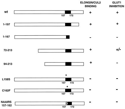
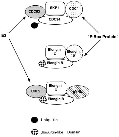
The von Hippel-Lindau tumor suppressor protein (pVHL) binds to elongins B and C and posttranscriptionally regulates the accumulation of hypoxia-inducible mRNAs under normoxic (21% O2) conditions. Here we report that pVHL binds, via elongin C, to the human homolog of the Caenorhabditis elegans Cul2 protein. Coimmunoprecipitation and chromatographic copurification data suggest that pVHL-Cul2 complexes exist in native cells. pVHL mutants that were unable to bind to complexes containing elongin C and Cul2 were likewise unable to inhibit the accumulation of hypoxia-inducible mRNAs. A model for the regulation of hypoxia-inducible mRNAs by pVHL is presented based on the apparent similarity of elongin C and Cul2 to Skp1 and Cdc53, respectively. These latter proteins form complexes that target specific proteins for ubiquitin-dependent proteolysis.


Figures

FIG. 1
FIG. 1
Coimmunoprecipitation of Cul2 with ectopically produced pVHL. (A and C) VHL (−/−) renal carcinoma cells stably transfected with a plasmid encoding HA-tagged pVHL (lanes 3 and 4) or with the backbone expression plasmid (lanes 1 and 2) were metabolically labelled with 35S, lysed, and immunoprecipitated (IP) with control or anti-HA antibody (αHA) as indicated. Bound proteins (lanes 1 to 4) as well as 35S-labelled Cul2 in vitro translate (IVT) (lane 5) were resolved by SDS-polyacrylamide gel electrophoresis and detected by autoradiography (A), anti-Cul2 immunoblot analysis (C, upper panel), or anti-HA immunoblot analysis (C, lower panel). Numbers at left are in kilodaltons. (B) The 35S-labelled Cul2 bands in panel A, lanes 4 and 5 (in vivo and in vitro, respectively), were excised and digested with 0.1 and 3 μg of α-chymotrypsin as indicated. Proteolytic fragments were resolved in an SDS–20% polyacrylamide gel and detected by autoradiography.
FIG. 2
FIG. 2
Copurification of Cul2 and endogenous pVHL. (A) 293 [VHL (+/+)] human embryonic kidney cells (lanes 5 and 6) and VHL (−/−) renal carcinoma cells stably transfected with a plasmid encoding HA-tagged pVHL (lanes 3 and 4) or with the backbone expression plasmid (lanes 1 and 2) were lysed and immunoprecipitated (IP) with anti-VHL (αVHL) or control antibody as indicated. Bound proteins were detected by anti-Cul2 (upper panel) or anti-VHL (lower panel) immunoblot analysis. Numbers at left are in kilodaltons. (B) Scheme for biochemical purification of pVHL complexes. P-cell, phosphocellulose P11. (C and D) Cochromatography of Cul2 with pVHL-elongin complexes. Aliquots of column fractions from the final two steps (hydroxylapatite Bio-Scale CHT-I high-pressure liquid chromatography and Mono Q chromatography [C and D, respectively]) were resolved by SDS–12% polyacrylamide gel electrophoresis and transferred to a polyvinylidene difluoride membrane. The top half of the membrane was immunoblotted with rabbit anti-Cul2 sera, and the bottom half was immunoblotted with a cocktail containing an anti-VHL monoclonal antibody and rabbit polyclonal anti-elongin B sera. Fraction numbers are shown above the lanes.
FIG. 2
FIG. 2
Copurification of Cul2 and endogenous pVHL. (A) 293 [VHL (+/+)] human embryonic kidney cells (lanes 5 and 6) and VHL (−/−) renal carcinoma cells stably transfected with a plasmid encoding HA-tagged pVHL (lanes 3 and 4) or with the backbone expression plasmid (lanes 1 and 2) were lysed and immunoprecipitated (IP) with anti-VHL (αVHL) or control antibody as indicated. Bound proteins were detected by anti-Cul2 (upper panel) or anti-VHL (lower panel) immunoblot analysis. Numbers at left are in kilodaltons. (B) Scheme for biochemical purification of pVHL complexes. P-cell, phosphocellulose P11. (C and D) Cochromatography of Cul2 with pVHL-elongin complexes. Aliquots of column fractions from the final two steps (hydroxylapatite Bio-Scale CHT-I high-pressure liquid chromatography and Mono Q chromatography [C and D, respectively]) were resolved by SDS–12% polyacrylamide gel electrophoresis and transferred to a polyvinylidene difluoride membrane. The top half of the membrane was immunoblotted with rabbit anti-Cul2 sera, and the bottom half was immunoblotted with a cocktail containing an anti-VHL monoclonal antibody and rabbit polyclonal anti-elongin B sera. Fraction numbers are shown above the lanes.
FIG. 3
FIG. 3
Binding of Cul2 and elongins to pVHL mutants. (A) Deletion mutants; (B) missense mutants. VHL (−/−) renal carcinoma cells ectopically producing the indicated HA-tagged pVHL species were metabolically labelled with 35S, lysed, and immunoprecipitated (IP) with anti-HA (αHA) or control (con) antibody as indicated. Bound proteins were resolved by SDS-polyacrylamide gel electrophoresis and detected by anti-Cul2 immunoblot analysis (upper panel), anti-HA immunoblot analysis (middle panel), or autoradiography (lower panel). The apparent decrease in the binding of elongins B and C to pVHL(54-213) in panel A was not a reproducible finding. Numbers at left are in kilodaltons.
FIG. 4
FIG. 4
Cul2 binds to elongin C in the absence of pVHL. VHL (−/−) renal carcinoma cells stably transfected with a plasmid encoding HA-tagged pVHL (lanes 3 and 4), T7 epitope-tagged elongin C (lanes 5 and 6), or the backbone expression plasmid (lanes 1, 2, 7, and 8) were lysed and immunoprecipitated (IP) with anti-HA (αHA), anti-T7 (αT7), or control antibody as indicated. Bound proteins were resolved by SDS-polyacrylamide gel electrophoresis and immunoblotted with anti-Cul2 polyclonal (upper panel), anti-HA monoclonal (middle panel), or anti-T7 monoclonal (lower panel) antibodies. Numbers at left are in kilodaltons.
FIG. 5
FIG. 5
Similarity of human Cul2 to S. cerevisiae Cdc53 protein. Identical amino acid residues are shaded with black.
FIG. 6
FIG. 6
The region of elongin C that is most similar to Skp1 is an elongin B-binding domain. (A) Similarity of elongin C to Skp1. Identical amino acid residues are shaded with black. Elongin C residues 18 to 50 are underlined. (B) Binding of radiolabelled elongin B to the indicated GST-elongin C fusion proteins (lanes 2 to 10). Bound proteins were resolved by SDS-polyacrylamide gel electrophoresis and detected by fluorography. Lane 1 contained 25% of the elongin B present in each binding reaction mixture.
FIG. 6
FIG. 6
The region of elongin C that is most similar to Skp1 is an elongin B-binding domain. (A) Similarity of elongin C to Skp1. Identical amino acid residues are shaded with black. Elongin C residues 18 to 50 are underlined. (B) Binding of radiolabelled elongin B to the indicated GST-elongin C fusion proteins (lanes 2 to 10). Bound proteins were resolved by SDS-polyacrylamide gel electrophoresis and detected by fluorography. Lane 1 contained 25% of the elongin B present in each binding reaction mixture.
FIG. 7
FIG. 7
Inhibition of Glut1 protein production by pVHL mutants. Whole-cell extracts (∼50 μg of protein per lane, as determined by the Bradford method) prepared from VHL (−/−) renal carcinoma cells ectopically producing the indicated HA-tagged pVHL species were resolved by SDS-polyacrylamide gel electrophoresis and immunoblotted with anti-Glut1 polyclonal (upper panel) or anti-HA monoclonal (lower panel) antibodies. Clone identifiers are indicated in parentheses.
FIG. 8
FIG. 8
Inhibition of hypoxia-inducible mRNA accumulation by pVHL mutants. Total RNA was isolated from VHL (−/−) renal carcinoma cells ectopically producing the indicated HA-tagged pVHL species and analyzed by Northern hybridization with radiolabelled probes specific for either the Glut1 mRNA (A) or the VEGF mRNA (B). Clone identifiers are indicated in parentheses. Comparable loading of RNA was confirmed by ethidium bromide staining of the rRNAs as well as by rehybridization of the filters to a radiolabelled actin probe. The presence of multiple VEGF mRNA isoforms was noted previously by others (26, 36, 43).
FIG. 9
FIG. 9
Summary of elongin-Cul2 binding and Glut1 inhibition by pVHL mutants. For Glut1 inhibition, + indicates that all clones tested produced Glut1 at levels similar to those in cells producing wild-type (wt) pVHL, − indicates that all clones tested produced Glut1 at levels similar to those in parental VHL (−/−) cells, and +/− indicates that some clones produced Glut1 at levels similar to those in cells producing wild-type pVHL and that some clones produced Glut1 at levels similar to those in parental VHL (−/−) cells. The reason for the variation in Glut1 production among these clones is currently unknown. Shown in Fig. 7 and 8 are data for pVHL(72-213) clones that scored positive. Black boxes indicate minimal elongin B and C binding domains based on in vitro studies (22). Asterisks indicate sites of missense mutations. The hatched box indicates the site of an NAAIRS substitution mutation.
FIG. 10
FIG. 10
Both Skp1 and elongin C participate in the formation of multiprotein complexes. Cdc4 and elongin A contain F-box motifs. Cdc53 and Cul2 are putative E3 ubiquitin ligases. Cdc34 is a putative E2 ubiquitin-conjugating enzyme, and elongin B contains a ubiquitin-like domain.

References

    1. Aso T, Haque D, Barstead R, Conaway R, Conaway J. The inducible elongin A elongation activation domain: structure, function and interaction with elongin BC complex. EMBO J. 1996;15:101–110. - PMC - PubMed
    1. Bai C, Sen P, Hofmann K, Ma L, Goebl M, Harper J W, Elledge S J. SKP1 connects cell cycle regulators to the ubiquitin proteolysis machinery through a novel motif, the F-box. Cell. 1996;86:263–274. - PubMed
    1. Berse B, Brown L, Livingston V, Dvorak H, Senger D. Vascular permeability factor (vascular endothelial growth factor) gene is expressed differentially in normal tissues, macrophages, and tumors. Mol Biol Cell. 1992;3:211–220. - PMC - PubMed
    1. Conaway R, Reines D, Garrett K, Powell W, Conaway J. Purification of RNA polymerase II general transcription factors from rat liver. Methods Enzymol. 1996;273:194–207. - PMC - PubMed
    1. Conaway, R. C., J. W. Conaway, and W. G. Kaelin, Jr. Unpublished data.

Publication types

LinkOut - more resources