. 1998 Feb;18(2):827-38.
doi: 10.1128/MCB.18.2.827.

GTP hydrolysis is not important for Ypt1 GTPase function in vesicular transport

Affiliations

GTP hydrolysis is not important for Ypt1 GTPase function in vesicular transport

C J Richardson et al. Mol Cell Biol. 1998 Feb.

Abstract

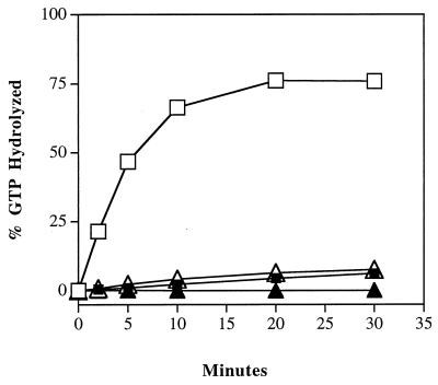
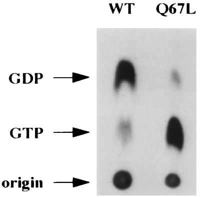
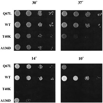
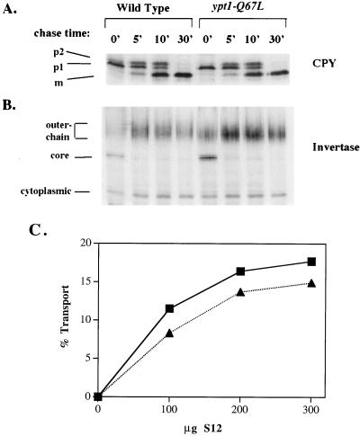
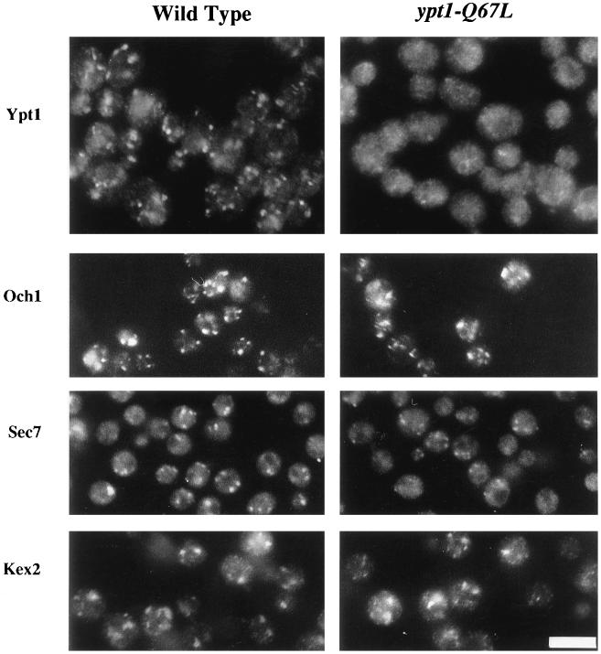
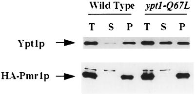
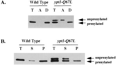
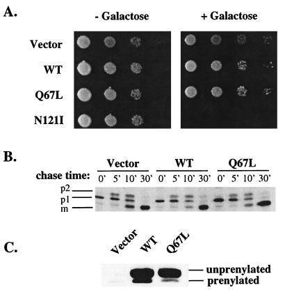
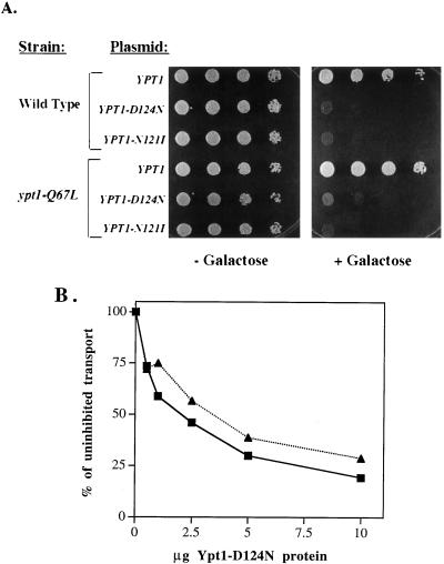
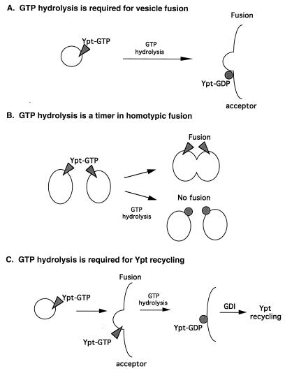
GTPases of the Ypt/Rab family play a key role in the regulation of vesicular transport. Their ability to cycle between the GTP- and the GDP-bound forms is thought to be crucial for their function. Conversion from the GTP- to the GDP-bound form is achieved by a weak endogenous GTPase activity, which can be stimulated by a GTPase-activating protein (GAP). Current models suggest that GTP hydrolysis and GAP activity are essential for vesicle fusion with the acceptor compartment or for timing membrane fusion. To test this idea, we inactivated the GTPase activity of Ypt1p by using the Q67L mutation, which targets a conserved residue that helps catalyze GTP hydrolysis in Ras. We demonstrate that the mutant Ypt1-Q67L protein is severely impaired in its ability to hydrolyze GTP both in the absence and in the presence of GAP and consequently is restricted mostly to the GTP-bound form. Surprisingly, a strain with ypt1-Q67L as the only YPT1 gene in the cell has no observable growth phenotypes at temperatures ranging from 14 to 37 degrees C. In addition, these mutant cells exhibit normal rates of secretion and normal membrane morphology as determined by electron microscopy. Furthermore, the ypt1-Q67L allele does not exhibit dominant phenotypes in cell growth and secretion when overexpressed. Together, these results lead us to suggest that, contrary to current models for Ypt/Rab function, GTP hydrolysis is not essential either for Ypt1p-mediated vesicular transport or as a timer to turn off Ypt1p-mediated membrane fusion but only for recycling of Ypt1p between compartments. Finally, the ypt1-Q67L allele, like the wild type, is inhibited by dominant nucleotide-free YPT1 mutations. Such mutations are thought to exert their dominant phenotype by sequestration of the guanine nucleotide exchange factor (GNEF). These results suggest that the function of Ypt1p in vesicular transport requires not only the GTP-bound form of the protein but also the interaction of Ypt1p with its GNEF.


Figures

FIG. 1
FIG. 1
Ypt1p-Q67L is defective in intrinsic and GAP-stimulated GTP hydrolysis. GTP hydrolysis was monitored by the charcoal binding assay. Wild-type (squares) and Ypt1p-Q67L (triangles) proteins were preloaded with [γ-32P]GTP for 15 min at 30°C. Unbound nucleotide was removed with two successive acrylamide spin columns. GTP hydrolysis assays were performed by incubating 2 nM preloaded Ypt1p with a P12 subcellular fraction (5 mg/ml) prepared from GPY60 cells (GAP-stimulated hydrolysis; open symbols) or without the P12 fraction (intrinsic hydrolysis; closed symbols) at 30°C. Aliquots were removed at the indicated time points and added to ice-cold activated charcoal to stop the reaction. The charcoal was pelleted, and an aliquot of the supernatant was removed and quantified by scintillation counting. The counts measured at time zero were subtracted as background. GTP binding was slightly less efficient for Ypt1p-Q67L, but hydrolysis rates were normalized for the amount of Ypt1p bound to GTP. Data shown are typical of three independent experiments.
FIG. 2
FIG. 2
Ypt1p-Q67L is predominantly bound to GTP in vivo. Wild-type (WT; NSY125) and ypt1-Q67L (NSY406) strains were grown to mid-logarithmic phase, spheroplasted, and labeled with [32P]orthophosphate for 1 h at 30°C. Spheroplasts were then lysed osmotically, and lysates were subjected to phase partitioning with 1% Triton X-114. Ypt1p was immunoprecipitated from the detergent phase with anti-Ypt1p antibodies for 2 h at 4°C. Associated nucleotides were released by heating in SDS and resolved by TLC on PEI-cellulose plates. Migration of nucleotides was determined by using unlabeled GDP and GTP standards which were visualized with UV light. Radiolabeled nucleotides were quantified by radioanalytic imaging. The results mentioned in the text are the averages from three independent experiments.
FIG. 3
FIG. 3
ypt1-Q67L mutant cells grow normally at temperatures ranging between 14 and 37°C. Tenfold serial dilutions of the ypt1-Q67L strain (NSY406) were spotted onto YPD plates and grown at the indicated temperatures. Wild type (WT; NSY125), ypt1-T40K (DBY1803), and ypt1-A136D (NSY222) strains were spotted as controls. ypt1-T40K and ypt1-A136D strains are shown to demonstrate that other mutations in YPT1 that deplete Ypt1p function do cause a growth phenotype and that the conditions used in the experiment are effective.
FIG. 4
FIG. 4
Secretory kinetics are normal in ypt1-Q67L mutant cells. (A) In vivo transport of CPY. Wild-type (NSY125) and ypt1-Q67L mutant (NSY406) cells were grown at 30°C in minimal medium without methionine to mid-logarithmic phase. Cells were then pulse-labeled with Tran35S-label for 6 min at 30°C. Samples were chased with excess unlabeled methionine and cysteine for the indicated times (minutes). CPY was immunoprecipitated with anti-CPY antibodies, and modified forms were separated on SDS–8% polyacrylamide gels. p1, ER form; p2, Golgi form; m, mature vacuolar form. (B) In vivo transport of invertase. A pulse-chase experiment was performed as described above for CPY except that invertase was derepressed by switching to low-glucose (0.1%) medium for 1 h at 30°C, and immunoprecipitation was done with anti-invertase antibodies. The constitutive cytoplasmic form, the core ER form, and Golgi outer chain forms are indicated. Rates of transport were determined by quantification of the different forms of CPY and invertase and found to be the same in wild-type and mutant cells. (C) In vitro ER-to-Golgi transport. Transport of 35S-labeled pro-α-factor was assayed in a cell-free reaction (30). Cell fractions were prepared from wild-type (NSY125; squares) or ypt1-Q67L (NSY406; triangles) cells. Permeabilized yeast cells serve as the donor compartment and were incubated with the indicated quantities of S12, the supernatant of a 12,000 × g spin that contributes the acceptor compartment as well as necessary soluble components. Percent transport was calculated as the percentage of ER-modified (core-glycosylated, concanavalin A-precipitable) α-factor that acquired Golgi-specific modifications (anti-α-1,6-mannose-precipitable counts per minute) in 90 min at 20°C. Data shown are typical of four independent experiments.
FIG. 5
FIG. 5
In ypt1-Q67L mutant cells, the immunofluorescence staining pattern of Ypt1p-Q67L is abnormal but other Golgi markers are normal. Yeast cells from wild-type (NSY125) or ypt1-Q67L (NSY406) strains were fixed and stained for fluorescence microscopy with affinity-purified anti-Ypt1p antibodies (1:500) or anti-Sec7p antibodies (1:500) as indicated. For visualization of Och1p and Kex2p, the same strains were transformed with plasmids expressing HA-tagged Och1p or Kex2p as indicated, fixed, and stained with anti-HA antibodies (1:1,000 dilution). Bar, 10 μm.
FIG. 6
FIG. 6
Mislocalization of mutant Ypt1p-Q67L to the cytosol as assessed by cell fractionation. Wild-type and ypt1-Q67L mutant cells were lysed with glass beads and centrifuged at 100,000 × g to generate supernatant (S) and pellet (P) fractions (T, total cell lysate). Proteins were resolved by SDS-PAGE, transferred to nylon membranes, and processed for Western blot analysis with affinity purified anti-Ypt1p antibodies (upper panel). For visualization of the Golgi marker Pmr1p, cells were transformed with a plasmid expressing HA-tagged Pmr1p. Cells were fractionated as above, and proteins were resolved by SDS-PAGE on 8% gels and processed for Western blot analysis with anti-HA antibodies (lower panel).
FIG. 7
FIG. 7
Mutant Ypt1p-Q67L is partially defective in prenylation. (A) A smaller fraction of the mutant Ypt1p-Q67L than of wild-type Ypt1p is prenylated, as determined by Triton X-114 phase partitioning and urea-acrylamide gradient gel electrophoresis. Wild-type (NSY125) and ypt1-Q67L mutant (NSY406) total cell lysates were subjected to phase partitioning with 1% Triton X-114. Total (T), aqueous (A), and detergent (D) phases were electrophoresed on 4 to 8 M urea–10 to 15% acrylamide gels and processed for Western blot analysis with anti-Ypt1p antibodies. Note that the aqueous phase contains all of the unprenylated form and the detergent phase contains all of the prenylated Ypt1p-Q67L. (B) Unprenylated and some prenylated mutant Ypt1p-Q67L is mislocalized to the cytoplasm (S100 fraction). Wild-type and ypt1-Q67L mutant cells were lysed with glass beads and centrifuged at 100,000 × g to generate supernatant (S) and pellet (P) fractions (T, total cell lysate). Proteins were resolved by SDS-PAGE on 4 to 8 M urea–10 to 15% acrylamide gradient gels, transferred to nylon membranes, and processed for Western blot analysis with affinity-purified anti-Ypt1p antibodies. The upper form of Ypt1p is unprenylated; the lower form is prenylated. Quantification indicates that there is the same amount of prenylated Ypt1p in wild-type and mutant strains. Data are typical of three independent experiments.
FIG. 8
FIG. 8
ypt1-Q67L is not dominant for growth or secretion when overexpressed. (A) Growth phenotypes. Wild-type YPT1 (WT; pNS326), ypt1-Q67L (pNS330), and YPT1-N121I (pNS327) were expressed from the galactose-inducible GAL10 promoter on a CEN URA-marked plasmid in the strain NSY125. Tenfold serial dilutions of cells were spotted onto SRaf-Ura or SRaf-Ura-plus-2% galactose plates and grown at 30°C. (B) CPY transport. The strains were grown overnight in SRaf-Ura minus methionine at 30°C to mid-logarithmic phase and then switched to inducing media (SRaf-Ura minus methionine plus 2% galactose) for 3 h at 30°C. Cells were harvested and pulse-labeled with Tran35S-label for 6 min at 30°C and chased for the indicated times (minutes). Cells were then lysed, and CPY was immunoprecipitated with anti-CPY antibodies and separated on SDS–8% polyacrylamide gels. p1, ER form; p2, Golgi form; m, mature vacuolar form. (C) Western blot analysis of Ypt1p expression. Cells used for the CPY assay were tested for Ypt1p expression. Ypt1p expression was induced with 2% galactose for 3 h at 30°C. Cells were lysed, and equivalent quantities of extract were run on 4 to 8 M urea–10 to 15% acrylamide SDS-containing gels. Proteins were transferred to nylon membranes and processed for Western blotting with anti-Ypt1p antibodies. The unprenylated and prenylated forms of Ypt1p are indicated.
FIG. 9
FIG. 9
Mutant Ypt1p-Q67L requires GNEF for function both in vivo and in vitro. (A) ypt1-Q67L cell growth is inhibited by expression of the dominant nucleotide-free alleles of YPT1. Wild-type (NSY125) and ypt1-Q67L (NSY406) cells were transformed with plasmids carrying the wild-type YPT1 gene (pNS326), YPT1-D124N (pNS317), or YPT1-N121I (pNS327) under the inducible GAL10 promoter. Yeast strains carrying these plasmids express Ypt1p when grown on medium containing galactose. Tenfold serial dilutions were spotted on SRaf-Ura medium without galactose (− Galactose) or with 2% galactose (+ Galactose). (B) In vitro transport in a ypt1-Q67L mutant cell reaction is inhibited by the nucleotide-free Ypt1p-D124N. In vitro transport cellular fractions from wild-type (squares) and ypt1-Q67L mutant (triangles) cells were prepared, and the reactions were performed as for Fig. 4C. Reactions were supplemented with the indicated quantities of purified mutant Ypt1p-D124N. Transport is expressed as percentage of uninhibited transport (reactions with no Ypt1p-D124N). Data shown are typical of four independent experiments.
FIG. 10
FIG. 10
Three models for the role of GTP hydrolysis in Ypt/Rab-mediated vesicular transport. (A) GTP hydrolysis is required for vesicle-membrane fusion (28, 60). (B) GTP hydrolysis is required to turn off Ypt/Rab-mediated homotypic membrane fusion (78). (C) GTP hydrolysis is required for Yptp recycling between membranes (this report). In this model, GTP hydrolysis is not required either for Yptp-mediated membrane fusion or for turning off Yptp function in heterotypic membrane fusion but rather is required for Yptp recycling between membranes.

References

    1. Alberts B, Bray D, Lewis J, Raff M, Roberts K, Watson J, editors. Molecular biology of the cell. New York, N.Y: Garland Publishing, Inc.; 1994. pp. 643–644.
    1. Alexandrov K, Horiuchi H, Steele-Mortimer O, Seabra M, Zerial M. Rab escort protein-1 is a multifunctional protein that accompanies newly prenylated rab proteins to their target membranes. EMBO J. 1994;13:5262–5273. - PMC - PubMed
    1. Antebi A, Fink G. The yeast Ca2+-ATPase homologue, PMRI, is required for normal Golgi function and localizes in a novel Golgi-like distribution. Mol Biol Cell. 1992;3:633–654. - PMC - PubMed
    1. Araki S, Kikuchi A, Hata Y, Isomura M, Takai Y. Regulation of reversible binding of smg p25A, a ras p21-like GTP-binding protein, to synaptic plasma membranes and vesicles by its specific regulatory protein, GDP dissociation inhibitor. J Biol Chem. 1990;256:13007–13015. - PubMed
    1. Bacon R A, Salminen A, Ruohola H, Novick P, Ferro-Novick S. The GTP-binding protein Ypt1 is required for transport in vitro: the Golgi apparatus is defective in ypt1 mutants. J Cell Biol. 1989;109:1015–1022. - PMC - PubMed

Publication types

MeSH terms